Anat 1 Ch 5 Osseous Tissue and Skeletal


- Slides: 22

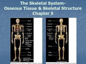
Anat 1 - Ch 5: Osseous Tissue and Skeletal Structure Skeletal system functions Bone structure – comparison of compact and spongy bone Bone development, growth and repair General classification of skeletal elements and bone markings

Bone Histology Osseous Tissue Specialized cells: 1. Osteocyte mature bone cell in lacuna 2. Osteoblast precursor of osteocyte responsible for osteogenesis (synthesizes organic components of matrix = osteoid)

3. Osteoprogenitor cell small number of cells still capable of mitosis – will produce osteoblasts 4. Osteoclast giant cell (>50 nuclei) many lysosomes capable of dissolving bony matrix Osteolysis and release of stored Ca 2+ and Phosphate

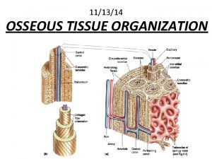
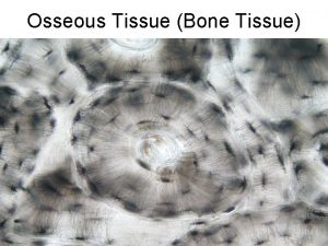
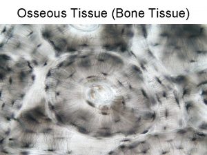
Osteon Fig 5. 2 Unit of dense bone structure. Concentric rings of mineral lamellae around the Haversian (central ) canals which are connected by Perforating or Volksmann canals. Lacuna (caves) hollows between lamella in which Osteocytes or bone cells are. Canaliculi - minute cracks which penetrate the mineral matrix and permit diffusion of nutrients, etc. between cells. Fig 5. 1

Cancellous Bone or spongy bone – does not show the above structuring Trabeculae = irregular thin plates Contains red bone marrow where blood cells are made Location?

Matrix = Protein - Crystal Mix 1. ~ 1/3 collagen fibers tough AND flexible 2. ~ 2/3 Ca(PO 4)2 3. ~ 2% cells tough

Bone Functions A. Support of body B. Mineral and fat storage C. Blood cell production D. Protection of internal organs E. Muscle attachment for movement Bone as an organ: Made up of what kinds of tissues?

Bone Coverings Periosteum (covers all bones) - fibrous outer layer - cellular inner layer Endosteum (inside bone) incomplete cellular layer matrix exposed purpose? Fig 5. 4

Bone Development & Growth Fetal ossification at 10 weeks 1. Intramembranous ossification = Dermal ossification 2. Endochondral ossification: A hyaline cartilage model is converted into bone.

Intramembranous Ossification Forms inside a membrane from an ossification center and grow peripherally Examples are cranial bones, mandible and clavicle Fig 5. 5

Endochondral Ossification Bones modeled first in hyaline cartilage. Blood vessels invade perichondrium and cause mineral precipitation forming a bone collar around the diaphysis. Lack of diffusion of nutrient supply leads to breakdown of cartilage center giving rise to the medullary cavity. Ossification centers form in the epiphyses leaving a cartilage band at the epiphyseal plate. Compare to Fig 5. 7

Bone Growth in Length at Metaphysis Reserve cartilage reproduces Chondrocytes enlarge and become more active metabolically, thus causing mineral to precipitate Eventually cartilage reserve ossifies creating the epiphyseal line indicating the end of bone growth

Appositional Growth Increase in Diameter Occurs as osteoclasts remodel bone from the medullary cavity and osteoblasts add bone to the outside

Bone Maintenance and Repair Skeleton is renewed/rebuilt every 5 years (= average; specifics depend on region) Bone is adaptable – stress vs. no stress on bone! Bone injury – classification of fractures

Match the Picture below to the fracture type AP view 1. ____Displaced, closed, simple Fracture Lateral view 2. ____Greenstick, closed fracture 3. ____Closed, simple transverse Fracture 4. ____Open fracture 5. ____Spiral, closed fracture 6. ____Comminuted closed fracture

Osteoporosis result of too little mineralization of bones for any of several reasons: 1. Loss of estrogen at menopause causes decreased efficiency of absorption of calcium and other minerals. 2. Deficiency of mineral in youth resulting in too little to begin with. 3. Imbalance of activity of osteoblasts and osteoclasts. 4. Occurs in men also but at an older age on average.

Bone Shapes All are covered with periosteum A. Long bone has a 1. diaphysis (shaft) and 2. proximal and distal epiphyses 3. medullary cavity lined with endosteum and filled with yellow bone marrow Examples? B. Short bone – no longer than it is wide e. g. : carpals

C. Flat bone – dense bone enclosing a thin spongy portion e. g. : cranial bones D. Irregular bones – no definable shape e. g. : vertebra and facial bones E. Sesamoid bone – “like a sesame seed” form inside a tendon Patella is only consistent example

Bone Markings (Surface Features) Frequently caused by stress on the bone from muscles or are present as articulations or passageways for other structures. "Insies" = indentations into bone 1. Facets: slight depressions about the size or shape of a finger mark 2. Fossae: larger and slightly deeper 3. Sulcus: groove as though made by dragging a spoon tip

4. Foramen: hole through a bone, usually with smooth edges through which blood vessels and nerves pass 5. Meatus: hole into, but not, through bone (e. g: ear hole) 6. fissure: irregular crack, usually between two bones, (earthquake type opening) 7. Sinus (antrum): cavity within bone, completely sealed space Be prepared to give one or two examples for each

"Outsies" include 1. Trochanter exists as only one pair (greater and 2. Tubercle much smaller projection from a bone, usually lesser) on the femur, very large projections for muscle attachment. somewhat rounded, muscle attachment. If the trochanter is the handle on a coffee mug, a tubercle is the pull tab on a soda can. 3. Condyle: knuckle like projection which articulates with 4. Epicondyle: prominence above a condyle 5. Tuberosity is a roughened result of muscle stress, another bone easier felt than seen, the texturing on a basketball. Fig 5 -14

 Bone cell
 Anat shenker-osorio
 Anat shahar
 Anat bremler-barr
 Wilyplus
 Digits of the foot
 Skeletal muscle
 Teased smooth muscle
 Skeletal muscle tissue structure
 N
N How is aerolar tissue different than aerenchyma tissue?
 Resective osseous surgery
 Positive architecture
 Fibro osseous tunnel
 Chorda tympani
 Osseous meatus
 Osseous coagulum
 Periapical granuloma vs abscess
 Osseous coagulum definition
 Sarcoplasmic
 Characteristics of skeletal smooth and cardiac muscle
 Micro muscles
 Chapter 36 skeletal muscular and integumentary systems
