seminar 2 1 1 1 Periapical granuloma 2


 Well-defined with a corticated radiopaque line or zone")


 radiolucence • asymptomatic • under 20 years old")

 same— • well defined • unilocular radiolucency different— • large blood-filled")
 Same— • scalloping • asymptomatic different– • wide age range • multiple(about")
 same— • painless • scalloping different— • usually anterior mandible •")

 • 40~60 years old ; ♂>♀ • site:")


 Malignant salivary gland tumors")


")


 • unicystic type (")


- Slides: 57
牙放seminar第一組 2. 1. 1 -1 Periapical granuloma 2. 1. 2 -4 Nasopalatine duct cyst 2. 1. 1 -2 Radicular cyst 2. 1. 2 -5 Eosinophilic granuloma 2. 1. 1 -3 Surgical defect 2. 2 -1 Dental follicle 2. 1. 1 -4 Periapical abscess 2. 2 -2 Pericoronitis 2. 1. 1 -5 Osteomyelitis 2. 2 -3 Paradental cyst 2. 1. 2 -1 Cementoma 2. 2 -4 Dentigerous cyst 2. 1. 2 -2 Periodontitis 2. 2 -5 Muralameloblastoma 2. 1. 2 -3 Trauma bone cyst 2. 2 -6 Adenomatoid Odontogenic Tumor
2. 1. 1 -1 these are well-defined round radiolucences surround both root apexes and bifurcation of the tooth #36, measuring not more than 2 cm in diameter. The radiolucence between the roots may develop alveolar bone resorption
Periapical Granuloma • Clinical features: asymptomatic pain and sensitivity can develop • 病理特徵: Periapical Granuloma 的 granulation tissue 是由三個東西組成 : - 發炎組織 : lymphocyte 、PMN、plasma cell -血管 -Fibrous tissue : 在發炎組織和血管的外面 • 看不到epithelium lining , 沒有表皮包圍 , 只有granulation tissue 。
2. 1. 1 -2 there is a well-defined unilocular round shaped circumrooted radiolucence with a corticated margin over the tooth #23 between the adjacent teeth. The radiopaque border is continuous with the lamina dura of the associated tooth.
Radicular cyst • 外圍是non-keratinized stratified squamous epithelium • 有rete process增生 • hyaline body • foamy cells • fibrous wall會有 heavy deposits of cholesterol crystals
Periapical granuloma 和 Radicular cyst的不同點 : • periapical granuloma 比 cyst常見 • periapical cyst 比較常發 生在 upper jaw bone. • X-ray 片上可以看到完整 的lamina dura • Granuloma 沒有治療就會 演發成radicular cyst • Radicular cyst發生在 nonvital tooth
2. 1. 1 -3
2. 1. 1 -4 There is ill-defined unilocular shaped radiolucence without a corticated margin on periapcial area of tooth 14, and with a periodontal pocket on the distal side. This maybe a combination syndrome of both endo. and perio. with progression, the abscess may extend through the medullary spaces away from the apical area resulting in osteomyelitis, or it may perforate the cortex and spread diffusely through the overlying soft tissue
Apical absess • arise as the initial periapical pathosis or from an acute exacerbation (phoenix abscess) of a chronic periapical inflammatory lesion. • In the early stage, the periapical periodontal ligament fibers may exhibit acute inflammation but no frank abscess formation. • best termed acute apical periodontitis.
Clinical Features • painful tender swelling of varying size and position • tenderness to pressure in buccal sulcus • fever and malaise • erythema and possibly draining sinus, intraoral or extraoral • unresponsive to thermal and electrical stimuli • positive percussion test
Differential diagnosis • Periapical granuloma: 根尖處牙周膜肥厚(raiolucency) Well-defined with a corticated radiopaque line or zone of sclerotic bone. • Radicular cyst: 與periapical granuloma類似,無法以radiolucency區別。 • Osteomyelitis: poor-defined “moth-eaten” radiolucency
2. 1. 1 -5
Osteomyelitis, periapical • Osteomyelitis就是骨髓的發炎 • 好發位置在posterior body of mandible,上顎是很少見的 • 其重要的特徵就是會有sequestra的產生。 • 分為 acute以及chronic
2. 1. 2 -1 There is a well-defined monolocular round shaped radiolucence without a corticated margin at the apical area of both mandibular central incisors(tooth 24, 25)extending from the mesial aspect of tooth 26 to the periapical area of tooth 24, measuring approximately 1 cm in diameter. the adjacent teeth are typically vital and not resorbed with an intact periapical ligament space.
Cemetoma , periapical cemento -osseous dysplasia , stage 1 • Middle-aged adults (typically black women ) • Monolocular , often multiple • Early stage : radiolucent , not corticated • Intermediate stage : radiopacity within the apical radiolucencies • Late stage : densely radiopaque but surrounded by a thin radiolucent line • Traumatic ( solitary ) bone cyst • Radicular cyst • Periapical granuloma
2. 1. 2 -2 • There is a continuous irregular radiolucence with a poor defined margin along the apical area of the right maxillary posterior teeth (tooth 2, 3, 4 and 5), and a well-defined round shaped radiolucence without a corticated margin at the periapical area of tooth 4 , with a superior margin at the apex os the root and a inferior margin near the midle one-third of the root, measuring approximately 1 cm in diameter. Severe bone destruction can be observed around the roots of the teeth.
Periodontitis • Localized severe bone destruction around the roots of the teeth • Endo-perio lesion • Eosinophilic granuloma • Traumatic (solitary ) bone cyst • Periapical granuloma • Radicular cyst
2. 1. 2 -3 There is a well-defined monolocular scalloped-shape radiolucence with a cortical margin between the root of the 35 and 37 extending from the distal of 35 to the root tip of 37 squeezing along the root outline,measuring approximately 4 × 2 cm in diameter. The involved teeth from 35 to 37 are still alive with lamina dura.
Trauma bone cyst • well-defined (corticated) radiolucence • asymptomatic • under 20 years old • 60% male • common in the mandibular premolar and molar areas • margin along the root, not push • vital teeth • scallop(several teeth) • empty or fluid filled cavity
lateral periodontal cyst Same-- • male prefer • asymptomatic • corticated round radiolucency • along the lateral root surface • in alveolar bone Different- • old ages • often in mandibular premolar-canine-lateral incicor area(rarely in molars) • some are botryoid round lucency(botryoid odontogenic cyst)
aneurysmal bone cyst(ABC) same— • well defined • unilocular radiolucency different— • large blood-filled spaces • often described as "soap bubble" • teeth moved and roots resorption
odontogenic keratocyst(OKC) Same— • scalloping • asymptomatic different– • wide age range • multiple(about 10%) • teeth moved and roots resorption
glangular odontogenetic cyst(GOC) same— • painless • scalloping different— • usually anterior mandible • middle aged • teeth moved and roots resorption
2. 1. 2 -4 There is a well-defined monolocular round shaped radiolucence with a corticated margin at midline of anterior maxilla , measuring approximately 2 x 4 cm in diameter. Upper central incisors are separated apart.
Nasopalatine duct cyst (Incisive Canal Cyst) • 40~60 years old ; ♂>♀ • site: midline, anterior maxilla • Unilocular, round or oval, welldefined, well corticated (unless infected) • It may cause palatal expansions • smooth cortical border • arises from epithelial remnants of the nasopalatine duct
• usually present in the midline of the anterior maxilla near the incisive foramen • many are inflamed • pain, pressure, drainage and swelling can occur
Differential Diagnosis : • periapical granuloma • radicular cyst
2. 1. 2 -5 There is a round monocular radiolucence without corticated margin between tooth 44 and tooth 47, measurely approximately 3 X 5 cm in diameter, Destruction of the periodontal bone (loose teeth)without otherwise affecting the teeth (e. g. root resorption).
Eosinophilic Granuloma n. Benign proliferation of Langerhans cells. n. Usually adolescents and young adults. n. Localized or multiple lesions. n. In the jaws, more than 75% in mandible. n. Round, monolocular, not corticated. n. Destruction of the periodontal bone (loose teeth)without otherwise affecting the teeth
Periodontitis Radicular cyst Squamous cell carcinoma Metastatic tumors (ill defined) Malignant salivary gland tumors
2. 2 -1 * There is a well-defined, unilocular, round-shaped radiolucence with a wellcorticated margin above tooth 22, surrouing crown of an impacted tooth, extending from distal aspect of tooth 21 to mesial aspect of tooth 24 and from the half part of the tooth 22’s root to alveolar bone above tooth 22. The height of the radiolucence is about to its width. ☆ Dental follicle !!
2. 2 -2 This radiograph is part of a panoramic one. There is a radiolucence locating at distal aspect of tooth 38, extending from distal of the tooth 38’s crown to the ramus of mandibular bone, measuring about 1 cm. Its height is approximately the length of tooth 38’s crown. It’s well-defined, surrouned by nearby soft tissue (gingiva) and the crown of tooth 38.
Differential diagnosis: Paradental cyst: 同: 同樣是發生在第三下顎臼齒的遠心面外 側或facial aspect,與部份阻生的牙齒有 關 。 異: 它所形成的是cyst,要形成cyst有3要 素,分別是cavity,epithelium,wall。 而pericoronitis並非cyst。
2. 2 -3 There is a small well-defined unilocular oval shaped radiolucence with corticated margin in the distal of the tooth 48 extending from distal cervical margin of the tooth 48 down to inferior alveolar canal and up to the 1/4 ramus
Paradentalcyst 病理特徵 好發位置:mandibular third molar的lateral root surface、靠近cervical margin 致病原因:uncertain. involved by pericoronitis (usually lower 3 rd molar).
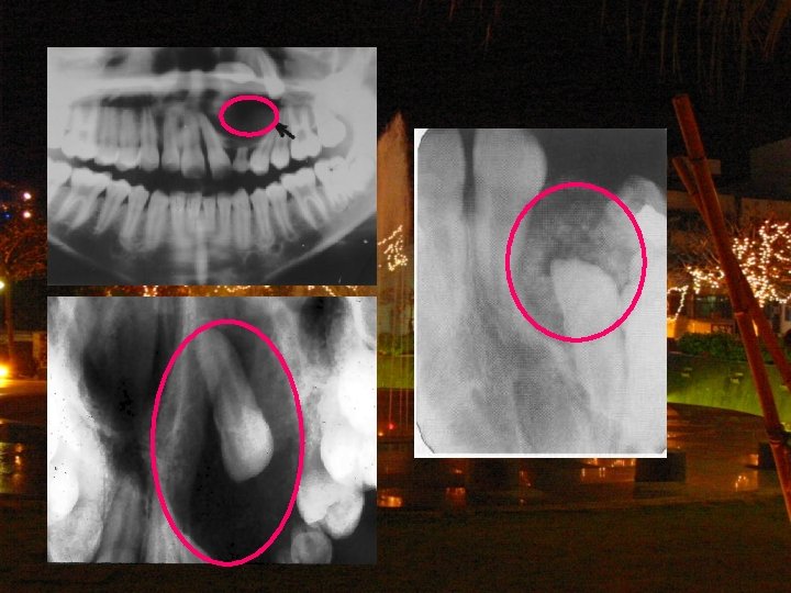
2. 2 -4 There is a well-defined unilocular oval shaped circumcoronal radiolucence with a corticated border over the submerged tooth 48 extending from retromolar area down to the mandibular angle, measuring approximately 1*2 cm in diameter.
Features of Dentigerous Cyst • Most common odontogenic cyst, next to radicular cyst. • Etiology • X-ray • Site • P’t ages: usually adolescents, 20~40 years old. • Eruption cyst • Effects on adjacent tooth • Treatment • Progonosis • Ameloblastic change (neoplastic transformation)
Differential diagnosis • Mural ameloblastoma
Differential diagnosis • Adenomatoid Odontogenic Tumor (AOT)
Differential diagnosis • Ameloblastic fibroma
Differential diagnosis • Paradental cyst
2. 2 -5 There is a well-defined unicystic irregular shaped pericoronal radiolucence without corticated margin associated with an unerupted 38 tooth in the cyst extending from distal aspect of the unerupted tooth 37 up to approximately 2/3 left ramus and from superior border of ramus down to left mandibular angle
Muralameloblastoma 病理特徵 ‧arises most commonly from a dentigerous cyst ‧most common site: Mandible posterior region ‧cause root resorption of the adjacent teeth ‧屬於unicystic ameloblastoma其中一種 tumor會侵入cystic wall(fibrous tissue),往外增 生,癒後最差
Ameloblastoma 臨床上ameloblastoma分成三大類: • conventional / multicystic type ( 86% ) • unicystic type ( 13% ) - luminal ameloblastoma - intraluminal ameloblastoma - mural (wall) ameloblastoma • Peripheral (extraosseous) type ( 1% )
Ameloblastoma • unicystic ameloblastoma 和 conventional ameloblastoma 的比較: conventional ameloblastoma Unicystic ameloblastoma Case比例 about 86% of all cases about 13% of all cases Shape multilocular radiolucence Unicystic radiolucence 發生位置 20% in maxilla 80% in mandible 56% in mandibular molar, ramus region 90% in ramus region 發生年齡 40、50歲的時候診斷出來,發生 18 -20歲 的平均年齡大約是 39、40歲左右 recurrent 50 -90% rate 10 -20%
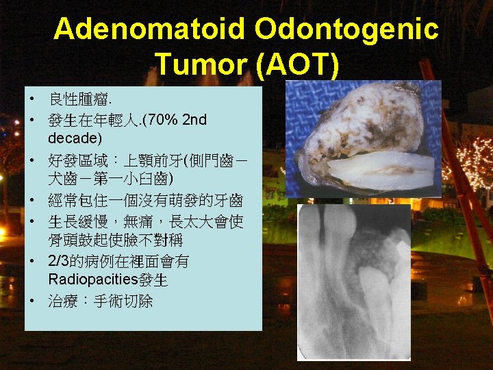
2. 2 -6 There is a well-defined unilocular oval shaped radiolucence with a corticated margin. It extend from apex of tooth 21 to apex of tooth 25 and from 3 mm below alveolar crest to nasal spine level, measuring approximately 3 x 3 cm in diameter. It causes tooth 21 and tooth 23 unerupted, tooth 21 malposition (root slants to mesisal) and tooth 24 slanted to distal.
Difference Disease • Dentigerous cyst • Dental follicle • Mural Ameloblastoma Full radiolucence的話只能用採樣確認
THE END