Microscopic Structure of Bone Osseous Tissue Another name

Microscopic Structure of Bone

Osseous Tissue Another name for bone tissue n Bone is a connective Tissue n n n Widely spread cells Matrix: n Water, Collagen Fibers, Mineral Salts

Calcification n Hardening of bone tissue by the deposition of mineral salts in the collagen fiber of the matrix

Hardness and Flexibility Hardness – Provided by the crystallized mineral salts n Flexibility – Provided by the collagen fibers n n Bones can resist being stretched or torn apart

Cells n There are 4 major types of cells found in osseous tissue n n Osteoblast Osteocyte Osteoprogenitor Osteoclast

Osteoblast Bone building cells n Synthesize and secrete collagen fibers and other organic components needed to build the matrix of the tissue n Osteoblasts surround themselves with matrix, become trapped in their secretions and become osteocytes n Do not undergo Mitosis n
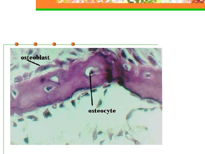

Osteocyte Mature Bone Cell n Main cells in bone tissue n n n Maintains daily metabolism Do not undergo mitosis

Osteoprogenitor “Bone stem cells” n These cells undergo mitosis then differentiate to form osteoblasts n

Osteoclasts HUGE cells formed from the fusion of as many as 50 monocytes n Concentrated in the endosteum n Release lysosomal enzymes and acids to digest the matrix n
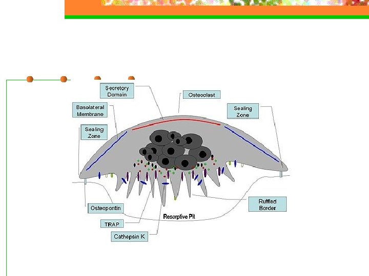

Reabsorption Breakdown of bone matrix n Part of the normal development, growth, repair and maintenance n

Categories of Bone Tissue Bone has many small spaces between the cells and matrix – it is not completely solid. n The category of tissue is based on the size and distribution of these spaces n About 80% of bone is compact bone; 20% is spongy bone n

Compact Bone Tissue Contains very few spaces n Forms the external layer of all bones and the diaphyses of long bones n Provides protection and support n Resists stress produced by weight and movement n

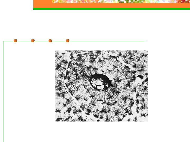
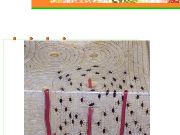
Osteon n Organizational Unit of Compact Bone

Perforating Canals n Transverse openings through which vessels from the periosteum penetrate the compact bone and eventually meet up with other vessels
 Canal Run longitudinally through bone n It is the center of the")
Central (Haversian) Canal Run longitudinally through bone n It is the center of the osteon n Contains blood vessels and nerves n

Concentric Lamellae n Rings of hard calcified matrix surrounding the central canal

Lacunae Means “little lake” n Small spaces between the lamellae n Contain osteocytes n

Canaliculi Minute canals that radiate off the lacunae in all directions. n Contain projections of the osteocytes n Connect lacunae creating a network throughout the compact bone to provide nutrients and oxygen to all the osteocytes and to get rid of waste n


Spongy Bone

Does not contain ostons n Made of trabeculae – an irregular network of thin columns of bone with many spaces in between n Trabeculae contain osteocytes within lacunae connected by canaliculi n

Spongy tissue makes up most of flat, short and irregular bones n Forms most of the epiphyses of long bones n Found in a narrow rim around the medullary cavity n

Spongy tissue is light – reducing the weight of the skeletal system n Red bone marrow is found in the spaces between trabeculae n n Hemopoiesis only occurs in the hip bones, ribs, sternum, vertebrae and epiphyses of long bones – where red bone marrow is found

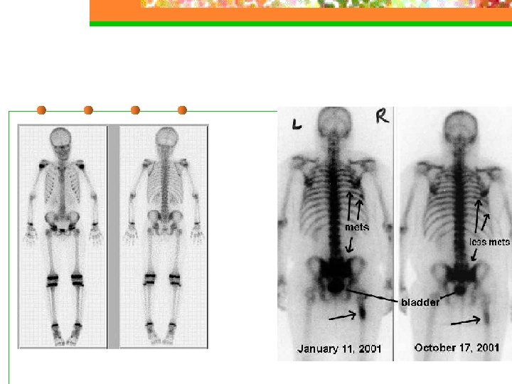
Bone Scan n n Radioactive Tracer is injected through an IV and absorbed by the bone A scanning device measures the amount of radiation emitted by the bones and translates the information into an x-ray Normal bones have a consistent gray color Darker/lighter areas indicate an abnormality n Ex - Bone cancer, abnormal healing, infections, arthritis

Checkpoint Questions – answer these in your notes… 1. 2. 3. 4. 5. 6. What kinds of tissue make up the skeletal system? How do red and yellow bone marrow differ in composition and function? What are the types of bones? Draw and label the parts of a typical long bone. What are the 4 types of bone cells? How are spongy and compact bone different in microscopic appearance, function and location?
- Slides: 30