Bone Quit Bone tissue or osseous tissue The

Bone Quit

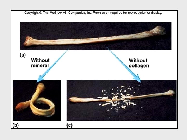
Bone tissue, or osseous tissue - The major structural and supportive connective tissue of the body - A rigid form of CT - Consists of matrix and cells - Matrix contains: Organic component 35% collagen fibres ( 95% collagen type I) Inorganic salts 65% #Calcium phosphate (58, 5%) (Hydroxyapatite) #Calcium carbonate (6, 5%) (responsible for hardness and rigidity)

Bones main functions: • Protection — Bones can serve to protect internal organs, such as the skull protecting the brain or the ribs protecting the heart and lungs. • Shape — Bones provide a frame to keep the body supported. • Blood production — The marrow, located within the medullary cavity of long bones and interstices of cancellous bone, produces blood cells in a process called haematopoiesis. • Mineral storage — Bones act as reserves of minerals important for the body, most notably calcium and phosphorus. • Fat Storage — The yellow bone marrow acts as a storage reserve of fatty acids • Movement — Bones, skeletal muscles, tendons, ligaments and joints function together to generate and transfer forces so that individual body parts or the whole body can be manipulated in three-dimensional space. The interaction between bone and muscle is studied in biomechanics. • Acid-base balance — Bone buffers the blood against excessive p. H changes by absorbing or releasing alkaline salts. • Detoxification — Bone tissues can also store heavy metals and other foreign elements, removing them from the blood and reducing their effects on other tissues. These can later be gradually released for excretion. • Sound transduction — Bones are important in the mechanical aspect of hearing.

Slide 1 Main menu Slide menu Quit
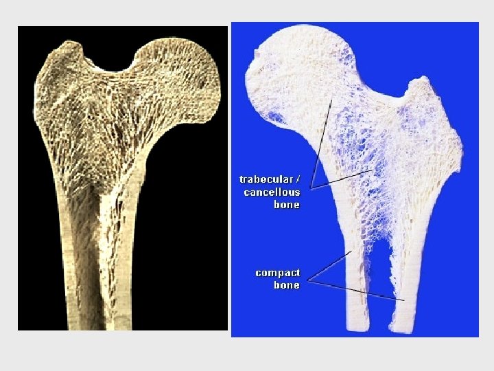
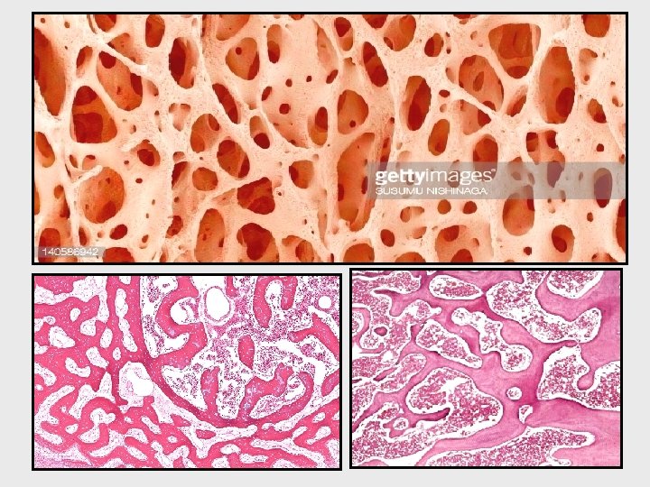

This is spongy or cancellous bone. This type of bone is found in the ends of long bones and between flat bones. The indicate trabeculae. The microscopic elements in spongy bone are the same as in compact bone. Slide 2 Main menu Slide menu Quit

Slide 3 Main menu Slide menu This is again spongy bone indicate trabeculae. The black dots are lacunae. Quit

Cancellous bone

Slide 4 Main menu Slide menu Quit
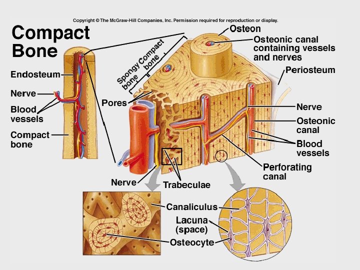

Haversian canal This is at Lamellae Lacuna Canaliculus Slide 5 Main menu Slide menu (Junqueira et al, 1986). section through a haversian system. The different parts of the haversian system has been annotated. The indicate an osteocyte with it’s processes that lie in the canaliculi. Around the osteocyte, in the lacuna is some bone fluid. Quit
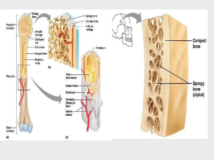
. Slide 6 Cortical bone Outer")
Inner circumferential lamellae Interstitial lamellae (Junqueira et al, 1986). Slide 6 Cortical bone Outer circumferential lamellae Haversian canal Main menu Slide menu Volkman’s canal Periosteum Endosteum This is a diagram of the structure of bone. Most of the elements are annotated. The = inner circumferential lamellae and the = the interstitial lamellae. The Endosteum lines the Volkman’s canals and the Haversian canals and is continuous with the deepest layer of the periosteum. Quit

Slide 7 Main menu Slide menu = the haversian canals while the = Volkman’s canal. Ground section in compact bone Quit

Slide 8 Main menu Slide menu Interstitial lamellae In this slide one can see a haversian system. The = interstitial lamellae that lie around the haversian system. Quit

This is a high powered view of bone. The = the canaliculi while the = a lacuna. In the lacuna lies an osteocyte with some bone fluid around it while the processes of the osteocyte lie in the canaliculi. Slide 9 Main menu Slide menu Quit

Decalcified section in compact bone

Slide 10 Main menu Slide menu Quit

Bone cells 1. Osteogenic cells 2. Osteoblasts 3. Osteocytes 4. Osteoclasts Osteogenic cells (osteoprogenitor cells) # spindle shape cell with oval nucleus and basophilic cytoplasm # essentially inactive osteoblasts which become active during bone growth and fracture (differentiate to osteoblasts) # present in periosteum and endosteum

Slide 11 Main menu Slide menu Quit
. Quit")
Slide 12 Main menu Slide menu (Junqueira et al, 1986). Quit

Slide 13 Main menu Slide menu Quit
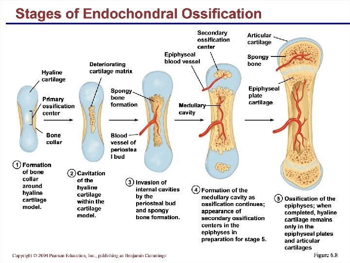
 Osteogenesis is the name given to the development of bone tissue. The")
Histogenesis (OSTEOGENESIS) Osteogenesis is the name given to the development of bone tissue. The first bone to develop is a form of spongy bone known as woven bone (immature bone or primary bone or bundle bone or non-lamellar bone). Woven bone is not usually found in people aged over 14 except for some specific locations including the vicinity of sutures of flat bones of the skull, in tooth sockets, and some tendon insertions. Woven bone also develops temporarily in cases of bone fracture and repair. Lamellar bone (Mature bone, Secondary bone) Most bone tissue is lamellar bone in which the tissue is well organized and regular.

Mature and immature bone Most of the compact and spongy bone from adult skeleton display the pattern of lamellar bone which classified as adult or mature bone (lamellar bone or secondary bone). On the other hand, the bony tissue initially deposited on the skeleton of developing fetus is called immature bone (woven bone or primary bone or nonlamellar bone or bundle bone). It differs from adult bone in several aspects • No organized lamellated appearance (called nonlamellar or woven bone) • It contains more cells per unit area than the mature bone • The cells tend to be randomly arranged • The matrix of immature bone has more ground substance • It forms more rapidly than mature bone • The mineral content is lower in immature bone • It is the major bone type in developing fetus while mature bone is the major type in adult (although in remodeling areas the immature bone is seen regularly) MB Mature Bone IB Immature Bone

Slide 14 Main menu Slide menu Quit

Slide 34 Main menu Slide menu Quit

Slide 35 Main menu Slide menu Quit
- Slides: 31