ALOPECIA Dr Ahmed AlHuchami 1 Hair Structure Anatomy

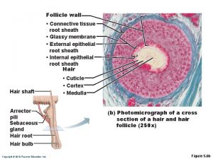
![Hair Structure [Anatomy of the Hair Follicle] 2 Hair Structure [Anatomy of the Hair Follicle] 2](https://slidetodoc.com/presentation_image/fd317f636ac90d749cef5c229dcc752f/image-2.jpg)


![Gray-patch tinea capitis presented as localized non scarring alopecia. [Alopecia, erythema and scaliness] Kerion Gray-patch tinea capitis presented as localized non scarring alopecia. [Alopecia, erythema and scaliness] Kerion](https://slidetodoc.com/presentation_image/fd317f636ac90d749cef5c229dcc752f/image-12.jpg)


![DLE Localized scarring alopecia (Red scalp with adherent scales) Localized scarring alopecia [DLE] 17 DLE Localized scarring alopecia (Red scalp with adherent scales) Localized scarring alopecia [DLE] 17](https://slidetodoc.com/presentation_image/fd317f636ac90d749cef5c229dcc752f/image-17.jpg)


- Slides: 56

ALOPECIA Dr. Ahmed Al-Huchami 1
![Hair Structure Anatomy of the Hair Follicle 2 Hair Structure [Anatomy of the Hair Follicle] 2](https://slidetodoc.com/presentation_image/fd317f636ac90d749cef5c229dcc752f/image-2.jpg)
Hair Structure [Anatomy of the Hair Follicle] 2

Classification of Hairs are classified into three main types: 1. Lanugo hairs: Fine long depigmented hairs covering the fetus, but shed about 1 month before birth. 2. Vellus hairs: Fine short unmedullated hairs, but coarses than lanugo hairs covering much of the body surface. They replace the lanugo hairs just before birth. 3. Terminal hairs: Long coarse medullated (pigmented) hairs replacing vellous hairs seen, for example, in the scalp, moustache, beard, chest, axillae, pubic region, trunk and the extremities. By age of adolescence, the terminal hairs cover the face and sexual area [secondary sexual characteristic]. Their growth is often influenced by circulating androgen levels. The lips, glans penis, labia minora, palms and soles remain free of hair follicles. 3

The Hair Cycle Each follicle passes, independently of its neighbours, through regular cycles of growth and shedding. There are three phases of follicular activity: 1. Anagen: The active phase of hair production. 2. Catagen: A short phase of conversion from active growth to the resting phase. Growth stops, and the end of the hair becomes clubshaped. 3. Telogen: A resting phase at the end of which the club hair is shed. * The duration of each of these stages varies from region to region. * The scalp contains an average of 100 000 hairs, anagen lasts for up to 5 years, catagen for about 2 weeks, and telogen for about 3 months. * As many as 100 -150 hairs may be shed from the normal scalp every day as a normal consequence of cycling. 4

Terminal hairs convert to vellus hairs in male pattern alopecia, and vellus to terminal hairs in hirsutism. 5

The Hair Cycle * The proportion of hairs in the growing and resting stages can be estimated by looking at plucked hairs (a trichogram). * The length of hair is determined by the duration of anagen; e. g. the hairs of the eyebrows have shorter cycles than those of the scalp. * Each hair follicle goes through its growth cycles out of phase with its neighbours, so there is no moulting period. However, if many pass into the resting phase (telogen) at the same time, then a correspondingly large number will be shed 2– 3 months later and this what occurs in telogen effluvium. * Mongoloids have less facial and body hair than Mediterranean people who also have more hair than northern Europeans. * Knowledge of hair growth phases is important in studying the patterns and types of hair fall e. g. anagen effluvium and telogen effluvium. 6

v a n A e g a r e p l a c s 7

STAGES OF HAIR GROWTH ANAGEN CATAGEN Growing stage Broom-shaped end Long and wider diameter Covered with IRS* 85% - 2 - 5 years * Transient stage Club-shaped end 1% - 2 weeks TELOGEN Resting stage Club-shaped end Short and thin diameter Not covered with IRS 14% - 3 months IRS: Inner Root Sheath 8

CLASSIFICATION OF ALOPECIA LOCALIZED SCARRING DIFFUSE NON-SCARRING 9

ü One convenient classification of alopecia is to divide it into localized and diffuse types. It is also important to decide whether the hair follicles have been replaced by scar tissue or not. The presence of any disease of the skin itself should also be noted. ü Localized A. is characterized by well-defined patchy areas of hair loss, while diffuse hair loss involves the whole scalp. ü Non-scarring A. has better prognosis than scarring A. as the hair usually grow again after a time because the hair roots are preserved. Hair loss in scarring A. is permanent [no hope for cure] due to damaged hair roots by the scarring process. If the follicular openings can no longer be seen with a lens, regrowth of hair cannot be expected. 10

CAUSES OF LOCALIZED NON-SCARRING ALOPECIA 1. Alopecia areata 2. Androgenetic alopecia (Early stages) 3. Tinea capitis (early treatment may prevent scarring) 4. Pyogenic infections e. g. boils or folliculitis. 5. Moth-eaten alopecia (Secondary syphilis) 6. Traumatic alopecia: a) Trichotillomania [Hair pulling habit] b) Traction alopecia c) Neonatal frictional alopecia (Occipital) 6. CTDs (e. g. SLE: early non-scarring alopecia but later may become scarring hair loss) 7. Epidermal cyst (pressure effect on hair follicles may cause hair loss) 11
![Graypatch tinea capitis presented as localized non scarring alopecia Alopecia erythema and scaliness Kerion Gray-patch tinea capitis presented as localized non scarring alopecia. [Alopecia, erythema and scaliness] Kerion](https://slidetodoc.com/presentation_image/fd317f636ac90d749cef5c229dcc752f/image-12.jpg)
Gray-patch tinea capitis presented as localized non scarring alopecia. [Alopecia, erythema and scaliness] Kerion [Inflammatory T. capitis] : A boggy mass with hair loss and pustules exuding pus from the hair follicle openings. If not treated , it will result in cause localized scarring alopecia. 12

Traumatic hair loss in a newborn “Frictional alopecia” alopecia 13

CAUSES OF LOCALIZED SCARRING ALOPECIA 1. Physical injury e. g. trauma and burn 2. Congenital aplasia cutis 3. Severe infections A. Fungal infections e. g. Kerion or Favus B. Bacterial infections e. g. Boils and Carbuncles C. Protozoal infestation (e. g. Cutaneous leishmaniasis) D. Viral infection e. g. Herpes zoster 5. Neoplasms: Cicatricial BCC, SCC, Metastatic Ca 6. Lichen planus (Lichenplanopilaris) 7. CTDs e. g. DLE and Morphea 8. Folliculitis decalvans 9. Sarcoidosis 10. Iatrogenic (IL-CS injections) 11. Idiopathic (Pseudopelade of Brocq) 14

Localized cicatricial alopecia (LP) 15

Localized scarring alopecia due to lichenplanopilaris 16
![DLE Localized scarring alopecia Red scalp with adherent scales Localized scarring alopecia DLE 17 DLE Localized scarring alopecia (Red scalp with adherent scales) Localized scarring alopecia [DLE] 17](https://slidetodoc.com/presentation_image/fd317f636ac90d749cef5c229dcc752f/image-17.jpg)
DLE Localized scarring alopecia (Red scalp with adherent scales) Localized scarring alopecia [DLE] 17

Localized scarring alopecia due to folliculitis decalvans 18

Localized scarring alopecia due to Pseudopelade of Brocq The term ‘pseudopelade’ pseudopelade is applied to a slowly progressive noninflamed type of scarring A. which leads to irregular areas of hair loss without any apparent preceding skin disease. 19

CAUSES OF DIFFUSE ALOPECIA 1. 2. 3. 4. 5. Genetically determined alopecia (Androgenitic alopecia) Diffuse alopecia areata (A. totalis and A. universalis) Physiological (Neonatal alopecia) Congenital (Ectodermal dysplasia) Endocrine disorders: Thyrotoxicosis, hypothyroidism, hypoparathyroidism and panhypopituitarism 6. Deficiency states (Kwashiorkor, Zinc deficiency, Iron deficiency) 7. Severe chronic illness (malnutrition, metabolic disturbances and emotional upset) e. g. uremia, lymphoma 8. Exfoliative skin disease e. g. erythroderma (stress may cause transient loss) 9. Hair shaft abnormalities (fragile hair) 10. Anagen effluvium 11. Teolgen effluvium 20

The cone-shaped incisors of hypohidrotic ectodermal dysplasia. Hypohidrotic ectodermal dysplasia (minimal hair & characteristic facies) 21

Alopecia areata ( ﺍﻟﺤﺎﺻﺔ ﺍﻟﺒﻘﻌﻴﺔ )ﺍﻟﺜﻌﻠﺒﺔ *AA is one of the commonest hair problems in dermatological daily practice. It affects about 1 -2% of the patients seen at outpatient skin clinics. * It is an acquired idiopathic disorder characterized by welldefined, single or multiple, non-cicatrical patches of hair loss. * It may affect any age group, although more common in young people (80% of cases occurs in 5 -40 years aged people). However, it may occur in infants, old people and during pregnancy. Both sexes are equally affected. * AA can involve any hairy area in the body (scalp, eyebrows, eyelashes, moustache, beard, axillary hair, pubic hair, body “truncal” hair and hair of the extremities). 22

* Patches of AA occur most commonly in the scalp and secondly in the beard area. * The skin in the involved area is normal i. e. no scarring, scaling or erythema. Close examination of the alopecic patches may show the tendency of hair at the periphery of these patches to be thinner proximally and thicker distally and look like an exclamation mark with the hair papilla is the tip of this exclamation mark [!]. Exclamation mark sign indicates active disease but not necessarily bad prognosis. * In some cases the hair loss may be so severe involving the entire scalp hair and in this case it is called alopecia totalis or even may involve the scalp with all other hairy areas and termed alopecia universalis. 23

An exclamation-mark hair 24

AA: Pathognomonic exclamation mark hairs seen around the edge of alopecic areas. They are broken off about 4 mm from the scalp, and are narrowed and less pigmented proximally. 25

A. universalis Extensive AA A A f o s e i t i r e v e s t n e r e f Dif AA in the beard 26

The characteristic uninflamed patch of alopecia areata Hair loss with normally looking scalp in AA 27

AA: Single patch in the scalp of an adult person 28

* The hair lost in AA may become grey in color on regrowth especially on spontaneous regrowth i. e. without treatment. AA usually dose not affect grey hair (only pigmented hair is involved by AA). *AA may involve the nails and up to 50% of patients may show fine pitting of the nails. Nail findings in AA may include: a) Nail pitting (fine and regular pits and may be called as Hammered brass pitting). b) Nail ridging c) Koilonychia (Spoon-shaped nails) d) Nail dystrophy e) Mottled lunulae 29

Aetiology of AA 1. Immunological factors: AA may be considered as an autoimmune disease for the following reasons: a) Association with other autoimmune disorders like vitiligo, Ass atopic dermatitis, Hashimoto’s thyroiditis , Addison’s disease, DM, Pernicious anemia …. . etc. b) Presence of circulating organ specific auto-antibodies e. g. antithyroid antibodies. c) Presence of circulating dysfunctioning T-lymphocytes. d) Histologically, T lymphocytes cluster like a swarm of bees around affected hair bulbs, having been attracted and made to divide by cytokines released from the dermal papilla. 2. Genetic factor: about 10 -25% of cases may be familial. 3. Psychological factor: Severe psychological upset probably acts as a precipitating or triggering factor. 30

Differential diagnosis of AA 1. Tinea capitis: the patches are not scaly and uninflamed and KOH hair examination is negative in AA in contrast to tinea capitis. 2. Lichen planus [the patches are inflamed with scarring]. 3. DLE [ also the patches are inflamed with scarring]. 4. Trichotillomania 5. Traction alopecia In both trichotillomania and traction alopecia broken hairs may be seen but true exclamation-mark hairs are absent. 6. Secondary syphilis can cause a ‘moth-eaten’ patchy hair loss. 31

Course & Prognosis of AA The prognosis (outcome) in AA in general is usually good as the recovery rate is high (> 90% of the cases recover within 6 months), but the relapse rate is also high so cure in AA is questionable, although some persons have remission for life, that is why the outcome in AA is unpredictable. Generally, in a first attack, regrowth is usual within few months. New hairs appear in the centre of the patches as fine pale down, and gradually regain their normal thickness and color, although the new hair may remain white in older patients. Subsequent episodes tend to be more extensive and regrowth is slower. Hair loss in some areas may coexist with regrowth in others. 32

Bad Prognostic Pointers (Signs) in AA 1. Unusually widespread alopecia especially alopecia totalis and alopecia universalis. 2. Early onset of the disease i. e. onset before puberty. 3. Multiplicity of the patches. 4. Chronicity (Recurrent cases). 5. Presence of nail changes. 6. Involvement of the scalp margin (Ophiasis), especially at the nape of the neck. 7. Familial AA [Positive F. H of AA]. 8. Association with atopy or Down’s syndrome. 33

Management of AA 1. Reassurance (not contagious with high rate of spontaneous recovery). The aim of therapy is to facilitate recovery. 2. Tranquillizers may be helpful at the start of therapy. 3. Topical sensitizing agents e. g. DNCB [Dinitrochlorobenzene]. 4. Topical irritants to stimulate hair growth such as topical phenol (5% solution), Dithranol (0. 1– 0. 25% ), Garlic, Plant extracts …. etc. are often used but with limited success. 5. Topical minoxidil 5% solution. Continue 34

Management of AA 6. Topical corticosteroids + Salicylic acid (Solution or cream) or as intralesional corticosteroids (triamcenolone 5 -10 mg/ml) especially for resistant cases. 7. PUVA [UVA + Psoralen] may be considered in resistant cases unresponsive to otherapies and in extensive cases like A. totalis and universalis. 8. Immunomodulators e. g. Oral zinc sulfate, BCG …. etc. may be tried in extensive or resistant cases. 9. Wigs may be necessary for extensive cases. The role of systemic corticosteroids in the management of AA is ? as they may induce hair growth but the problem is recurrence of alopecia on withdrawal of steroid may occur. 35

le p i lt u M A A f o s e h c t Pa Multiple & diffused patches of AA showing some hair growth on combination treatment with topical steroid and 5% Minoxidil solution 36

Trichotillomania or Hair-pulling habit is a minor comfort habit in children like nail-biting and lip-licking. It is a type of traumatic localized non-scarring alopecia not uncommonly seen in children and occurs more in girls than in boys. Affected individuals seldom have major psychiatric disorders, however some may have obsessive compulsive neurosis and when they are under the influence of psychological tension they develop and accustom the habit of twisting and pulling their hair resulting in localized non-scarring alopecia. The usual involved sites are the sides of the scalp or the frontovertical area. The hairs in the affected area are usually broken at different lengths from the scalp surface which is a characteristic feature of trichotillomania. 37

Trichotillomania Diagnosis can usually be made on the history, but some parents do not know what is going on. The bald areas do not show the exclamation-mark hairs of alopecia areata, or the scaling and inflammation of scalp ringworm. The patches are irregular in outline and hair loss is never complete Management includes reassurance, explanation to the parents or the patient that it is due to the habit of hair pulling, tranquilizers may be given and referral to psychiatrist may be necessary in some cases. Prognosis: Prognosis trichotillomania is usually of little consequence as the habit often goes away most quickly if it is ignored. However, more severe degrees of hair-pulling are occasionally seen in disturbed adolescents and in those with learning difficulties; then the outlook for full regrowth is less good, even with formal psychiatric help. 38

ia n a om l l i t ho c i r T irs a h en k o r b h t i w 39

Traction alopecia Clinical course Patients are often slow to accept that they are responsible for the hair loss, and notoriously slow to alter their cosmetic practices. Even if they do, regrowth is often disappointingly incomplete. Differential diagnosis The pattern of hair loss provides the main clue to the diagnosis and, if the possibility of traction alopecia is kept in mind, there is usually no difficulty. The absence of exclamation-mark hairs distinguishes it from alopecia areata, and of scaling from tinea capitis. Treatment Patients have to stop practices and procedures causing their hair loss. 40

Traction alopecia Cause Hair can be pulled out by several procedures intended to beautify, including hot-combing to straighten kinky hair, tight hairstyles such as a pony tail , rabbit’s ears or ‘corn rows’, and using hair rollers too often or too tightly. These will cause continuous traction on the hair follicles and may result in patchy hair loss at areas of maximum traction typically around the middle and sides of the scalp. Presentation The changes are usually seen in girls and young women. The pattern of hair loss is determined by the cosmetic procedure in use, hair being lost where there is maximal tug. Sometimes hair loss occurs mainly around the edge of the scalp-at the sides or at the front. The bald areas show short broken hairs, folliculitis and sometimes scarring. This is called marginal alopecia. 41

Traction alopecia caused by wearing the hair in a tight ponytail 42

Androgenetic Alopecia Synonyms Common baldness, Genetically-determined alopecia, Male pattern baldness. ü Androgenetic A. may be considered as a physiological event in the life of most men and many women. Pathology Bald scalp areas characterized pathologically by: 1. Reduction of blood vessels. 2. Reduction in pilo-sebaceus units. 3. Gradual decrease in the activity of anagen hair follicles: a. Decrease in size to a degree that become vellus hairs. b. The anagen phase will be reduced from 3 year to 1 year. So the number of telogen hairs will be increased in the involved area. 43

Aetiology and Pathogenesis üAlthough clearly familial, the exact mode of inheritance has not yet been clarified. In some families may run autosomal dominant inheritance. It occurs more in Caucasian and less in Japanese. It may occur in some animals like the chimpanzee. ü Male-pattern baldness is an androgen-dependent condition as androgen has an essential role in the pathogenesis both in males and females (eunuchoids & castrated males before puberty have no baldness). ü Normal androgen level in males is usually enough to produce baldness in genetically predisposed persons whereas in females normal androgen level causes baldness only in strongly predisposed women (less predisposed women need high androgen level to produce baldness). The female hormone, estrogen may have protective role in preventing common baldness. 44

Clinical Presentation P üOnset may start at puberty with gradual replacement of terminal hair by vellous hair especially in the fronto-vertical region of the scalp. Males have an earlier onset than females (15 -17 year versus >20 year). ü The severity of hair loss in androgenetic alopecia is variable as it a may be so mild and passes unnoticed or can be so prominent. üA common pattern of hair loss in men is the loss of hair first from the temples (temporal recessions) and then from the crown (vertical loss). Many men may have fronto-vertical loss or even complete baldness. In females, diffuse thinning of the fronto-vertical hair with preservation of the frontal line is the commonest pattern of hair loss. However , complete baldness in women is rare. Clinical course Hair loss is relentless, tending to follow the family pattern with some losing hair quickly and others more slowly. The diffuse pattern seen in women tends to progress slowly. 45

Androgenetic A. beginning in frontal area Male-pattern hair loss in a man Variations in male-pattern baldness 46

Male-pattern hair loss in a man Male-pattern hair loss in a woman 47

Treatment of common baldness 1. Surgery Surgical treatment may represent the only effective therapy for common baldness. Surgical procedures may include: a. Hair transplants: transplants Punch grafts or better Micrografts. The grafts are taken from the patient scalp (the sides of the scalp or from the occipital region). b. Scalp reduction + tissue expansion especially for small bald areas. 2. Medical therapy Till now there is no effective medical therapy in stopping the progression of baldness or restoring lost lair (hair regrowth). However medical therapies include: a. Topical application of minoxidil lotion may slow early hair loss and even stimulate new growth of hair in a few cases. Small and recently acquired patches respond best. When minoxidil treatment stops, the new hairs fall out after about 3 months. 48

b. Anti-androgens help some women with the diffuse type of androgenetic alopecia. Finasteride (Propecia), an inhibitor of human type II 5α-reductase, reduces serum and scalp skin levels of dihydrotestosterone in balding men. At the dosage of 1 mg/day, it may increase hair counts and so lead to a noticeable improvement in scalp hair. However, the beneficial effects slowly reverse once treatment has stopped. Finasteride is not indicated in women or children Side-effects are rare, but include decreased libido, erectile dysfunction and altered prostatespecific antigen levels. Other anti-androgens that can be used: Cyperoterone acetate (50 mg/day) Flutamide (125 -250 mg/day) Spironolactone (50 -100 mg/day) Cimetidine 3. Wigs are welcomed by some bald persons. 49

Telogen effluvium Definition Telogen effluvium is an early and excessive loss of normal club hairs from normal resting follicles in the scalp which results from traumatization of the normal hair by some stimulus such as surgery, parturition, fever and drugs which in short time precipitates the anagen phase into catagen and telogen phases. During this process the hair follicles are not diseased, and inflammation is absent. The usual time needed by the insult (stimulus) to cause hair shedding is about 2 -3 months but at times it may take closer to 6 months. Drugs causing T. effluvium may take shorter time to cause hair shedding usually 3 -5 weeks. 50

Cause of telogen effluvium: effluvium can be triggered by any severe illness or condition. These synchronize catagen so that, later on, large numbers of hairs are lost at the same time. The most common causes are: 1. Parturition (Postpartum telogen effluvium): it occurs 2 -6 months after delivery and lasts for 2 -6 moths and recover. Sudden stoppage of oral contraceptive pills may produce similar condition. Postnatal telogen effluvium occurs in the neonate with diffuse hair fall occurs from birth to 4 months, regrowth occurs by 6 months. 2. Fever Feve (Post febrile telogen effluvium): may be caused by any febrile illness. 3. Severe dieting: Kwashiorkor, starvation diet, crash diet and inappropriate weight reduction programs. Protein deprivation accompany these circumstances may result in telogen effluvium. 4. Severe stress (major surgery, hemorrhage, car accident, severe psychological upset …. . etc. ) 5. Hypothyroidism 6. Renal dialysis secondary to hypervitaminosis A 7. Drugs (Drug-induced telogen effluvium): A large group of drugs may be the cause, however the most common ones are: Bromocreptine, Captopril, Coumarin anticoagulants, Carbamazepine, Cimetidine, Danazol, Etretinate, Metoprolol, and Propranolol. 51

Post-febrile telogen hair loss “Telogen effluvium” 52

Presentation and course The hair fall occurs 2– 3 months after the provoking illness. It can be mild or severe. It is noted by the patient as “lots of hair coming out by roots”. Although the hair loss is diffuse only infrequently it causes clinically perceptible thinning of the hair since only rarely does it involve more than 50% of the hair. Regrowth, not always complete, usually occurs within a few months. Treatment There is no specific therapy for telogen effluvium but the patients can be reassured that their hair fall will be temporary and hair will grow again after few months. 53

Anagen effluvium is an excessive anagen hair loss. Causes: 1. Cancer chemotherapeutic agents (cytotoxic agents) Severe hair loss is seen with doxorubicin, the nitrosureas and cyclophosphamide. 2. Chemicals such as thallium and boron. The usual onset of anagen hair shedding is about 1 -2 months after administration of the causative agent. Up to 90% of hairs may be shed in anagen effluvium. It is to be noted that only growing (anagen) hairs are lost in anagen effluvium and with cessation of drug therapy, anagen follicles resume their normal activity within a few weeks; the process is entirely reversible. 54

Alopecia following chemotherapy for cancer 55

 Exclamation mark hairs
 Ahmed muhudiin ahmed
 Icd 10 eksim
 Alopecia
 Anthrophillic
 Alopecia sifilitica
 Oscar muñoz moreno arrones
 Trombocitopenia cuidados de enfermería
 Alopecia caracteristicas
 Fenastride
 Alopecia
 Alopecia
 Alopecia glen ellyn
 Dr colleen reisz
 Dark hair and blonde hair parents
 Tacheometric surveying formula
 The rebel wears fantastic clothes when others wear
 Hair grows in diagonal tubes called hair
 How to tell human hair from animal hair
 Heterozygous tabby x stripeless
 Label the structures of the hair follicle
 The two primary parts of hair are the hair fiber and the:
 Hair anatomy salon
 Sir syed ahmed khan father name
 Uv.khayyam
 Adel ahmed alalla
 United indian patriotic association
 Ahmed salman rushdie
 Ahmed salemi
 Salyaneli eyaletler şifre
 Dr mohammed ahmed
 Saad ahmed economics
 Ahmed mustapha
 Ideology of pakistan and sir syed ahmed khan
 Saad ahmed javed
 Dr mohammed ahmed
 Ahmed helmy uf
 Dr fawad randhawa
 Anna molka ahmed painting style
 Amina ahmed md
 Aida ahmed
 The hajji short story analysis
 Ali ahmed is a mathematics professor who tries to involve
 Ibrahim sammour
 Kulsum ahmed
 Ahmed miree
 Static compliance calculation
 Ahmed bawany academy school & college
 Glutamate oxaloacetate transaminase test
 Hezarfen ahmet çelebi mesleki ve teknik anadolu lisesi
 Dr. tahmeed ahmed
 Chikhaoui ahmed
 Shanzeh ahmed
 Dewan tanvir ahmed
 Amenities and facilities difference
 Ventsns
 Ahmed zarouf
