
TINEA CAPITIS Dr Immaculate Kariuki Consultant Paediatrician Nairobi
TINEA CAPITIS Dr. Immaculate Kariuki Consultant Paediatrician Nairobi, Kenya
Overview

TINEA INFECTIONS Superficial dermatophyte infections of keratinised tissue HAIR: Tinea capitis SKIN: Tinea cruris (groin & perineum) Tinea pedis (feet) Tinea manuum (hands) Tinea corporis (rest of body) NAIL: Tinea unguium
What is Tinea capitis? q Superficial fungal infection of the hair shafts & follicle and the skin of the scalp, eyebrows or eyelashes q Common superficial mycosis or dermatophytosis occurring predominantly in children q Affects the dead layers of skin and skin appendages with reduced ability to invade viable tissue except in context of immunosuppression q One of the water-related diseases (WHO) q Synonyms: Ringworm of the scalp
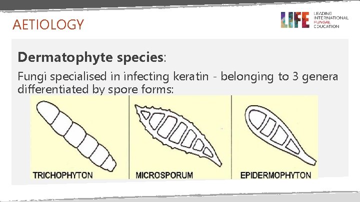
AETIOLOGY Dermatophyte species: Fungi specialised in infecting keratin - belonging to 3 genera differentiated by spore forms:
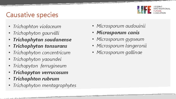
Causative species • • • Trichophton violaceum Trichophyton gourvilli Trichophyton soudanense Trichophyton tonsurans Trichophyton concentricum Trichophyton yaoundei Trichopyton ferrugineum Trichopyton verrucosum Trichophton rubrum Trichophyton mentagrophytes • • • Microsporum audouinii Microsporum canis Microsporum gypseum Microsporum langeronii Microsporum gallinae
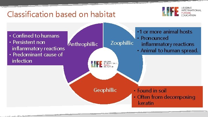
Classification based on habitat • Confined to humans • Persistent non Anthrophillic inflammatory reactions • Predominant cause of infection • 1 or more animal hosts • Pronounced Zoophillic inflammatory reactions • Animal to human spread. Geophillic • Found in soil • Often from decomposing keratin
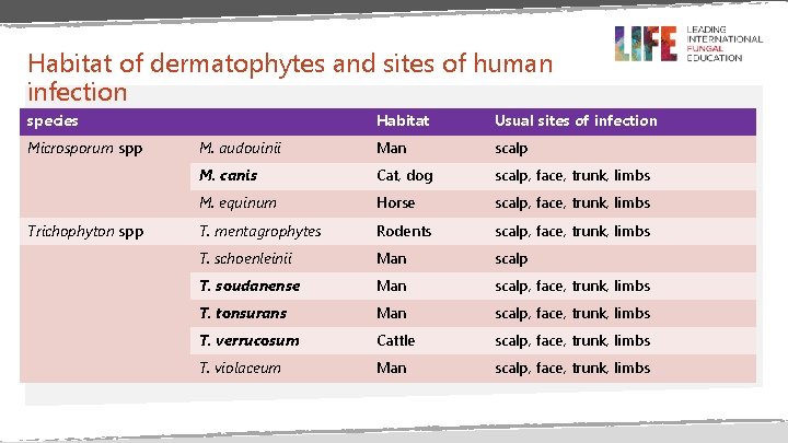
Habitat of dermatophytes and sites of human infection species Microsporum spp Trichophyton spp Habitat Usual sites of infection M. audouinii Man scalp M. canis Cat, dog scalp, face, trunk, limbs M. equinum Horse scalp, face, trunk, limbs T. mentagrophytes Rodents scalp, face, trunk, limbs T. schoenleinii Man scalp T. soudanense Man scalp, face, trunk, limbs T. tonsurans Man scalp, face, trunk, limbs T. verrucosum Cattle scalp, face, trunk, limbs T. violaceum Man scalp, face, trunk, limbs
Transmission • Direct Contact with organisms from: • Humans (anthrophillic organisms) • Animals (zoophillic organisms) • Soil (geophillic organisms) • Indirectly through fomites -hats, hair brushes etc.
Epidemiology
Intended Learning Objectives Describe the geographical variations in infection patterns Understand why Tinea capitis is a public health concern Describe the populations at higher risk of Tinea infections
EPIDEMIOLOGY • Common • Sporadic Vs. endemic • The prevalence of different types of dermatophytes varies between continents and from one country to another
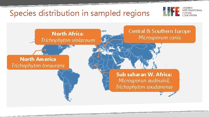
Species distribution in sampled regions North Africa: Trichophyton violaceum Central & Southern Europe Microsporum canis North America Trichophyton tonsurans Sub saharan W. Africa: Microsporun audouinii, Trichophyton soudanense
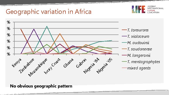
Geographic variation in Africa % % T. tonsurans % T. violaceum % M. audouinii % T. soudanense % No obvious geographic pattern ig 5 er ia '0 4 '8 N N ig er ia n bo Ga a Gh an t Iv or y Co as ue bi q M oz am bw ba Zi m Ke ny e a M. langeronii T. mentagrophytes mixed agents
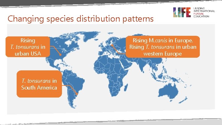
Changing species distribution patterns Rising T. tonsurans in urban USA T. tonsurans in South America Rising M. canis in Europe. Rising T. tonsurans in urban western Europe
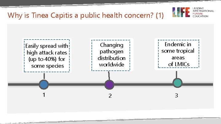
Why is Tinea Capitis a public health concern? (1) Easily spread with high attack rates (up to 40%) for some species 1 Changing pathogen distribution worldwide Endemic in some tropical areas of LMICs 2 3
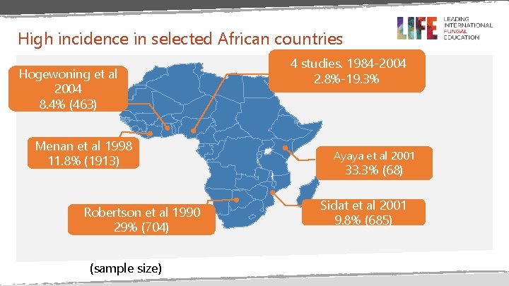
High incidence in selected African countries Hogewoning et al 2004 8. 4% (463) Menan et al 1998 11. 8% (1913) Robertson et al 1990 29% (704) (sample size) 4 studies. 1984 -2004 2. 8%-19. 3% Ayaya et al 2001 33. 3% (68) Sidat et al 2001 9. 8% (685)
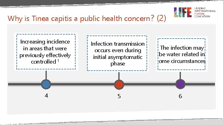
Why is Tinea capitis a public health concern ? (2) Increasing incidence in areas that were previously effectively controlled 1 Infection transmission occurs even during initial asymptomatic phase 4 5 The infection may be water related in ome circumstances 6
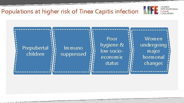
Populations at higher risk of Tinea Capitis infection Prepubertal children Immuno suppressed Poor hygiene & low socioeconomic status Women undergoing major hormonal changes
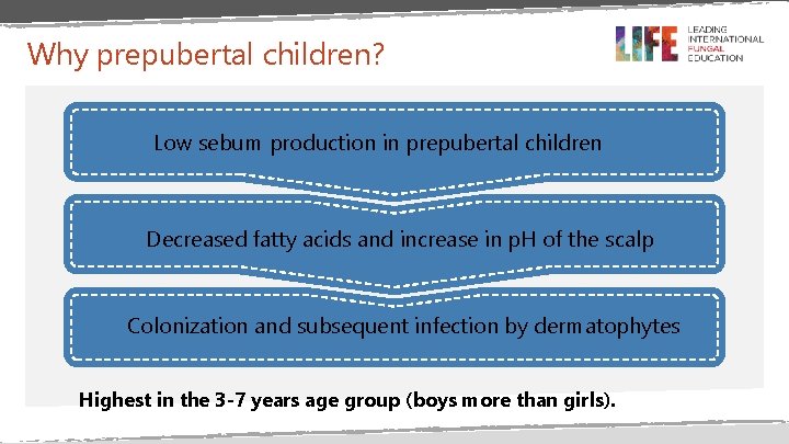
Why prepubertal children? Low sebum production in prepubertal children Decreased fatty acids and increase in p. H of the scalp Colonization and subsequent infection by dermatophytes Highest in the 3 -7 years age group (boys more than girls).
Immunosuppression leads to impaired hair shaft growth and strength leading to easier colonisation. • Diabetes mellitus • Prolonged steroid use • Cancer • Immunosuppressant drugs • Anaemia Not HIV - likely due to competitive colonisation with Malassezia
Women Ø Women under major hormonal changes including pregnancy and menopause. During these stages the hair shaft growth is altered leading to easier colonisation. Ø Other women Prolonged contact with prepubertal children? Contact with infected fomites due to frequent hairdresser visits?
Poor hygiene • Classified as a water-related disease by the World Health Organisation (WHO) • The spread of the disease is often related to insufficient clean water to wash the body or clothes • Low socio-economic status and poverty compounds this.
Summary • Tinea capitis causative organisms vary from one place to another with no obvious geographic pattern • Tinea capitis infections are a public health concern • Specific categories of the population – preburtal children and the immunosuppression are at higher risk of Tinea infections
MYCOLOGY
Structure of dermatophytes • All dermatophytes have a similar appearance in infected skin and skin appendages • The structure consists of septate hyphae, regular in width, branched and often fragmented into chains of arthrospores • In scalp and facial hair, the hyphae break up into arthrospores, which differ in terms of the size and pattern of infection
Patterns of infections Endothrix Fungal spores dwell within the hair shaft, the cortex remains intact Ectothrix Irregular fungal spores attach to the surface of the hair shaft, allowing for cuticle destruction Endothrix favosa Presence of hyphae and air bubbles within the hair shaft
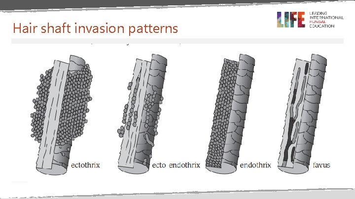
Hair shaft invasion patterns
Process of infection Adhesion of fungal cells to keratinocytes lasting 2 -3 hours after transfer from host Structural changes in organism Swelling of arthrospore and expression of extracellular fibrillar layer Hair shaft invasion via modified intercalary cells (penetrating organs), enzymes (proteases)
Dermatophyte antigens Ø Glycopeptides preferentially stimulate cell-mediated immunity Ø Keratinases - Enable skin invasion - Elicit delayed-type hypersensitivity (DTH) responses

Clinical Presentation
Intended Learning Objectives To be aware of what determines the clinical presentation To be familiar with the various presentations of the non inflammatory type To be aware of the various forms of the inflammatory presentation
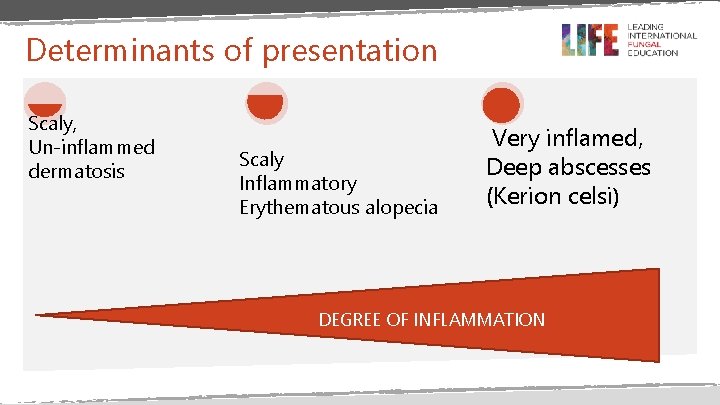
Determinants of presentation Scaly, Un-inflammed dermatosis Scaly Inflammatory Erythematous alopecia Very inflamed, Deep abscesses (Kerion celsi) DEGREE OF INFLAMMATION
Non-inflammatory presentation • Often due to anthrophillic dermatophytes with an ectothrix pattern of infection • Scaling, seborrheic form, well circumscribed patches of alopecia due to destroyed hair follicles • Diffuse alopecia
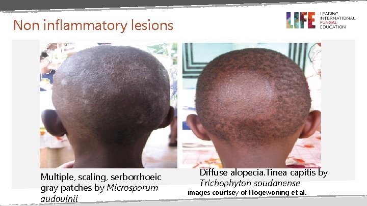
Non inflammatory lesions Multiple, scaling, serborrhoeic gray patches by Microsporum audouinii Diffuse alopecia. Tinea capitis by Trichophyton soudanense images courtsey of Hogewoning et al.
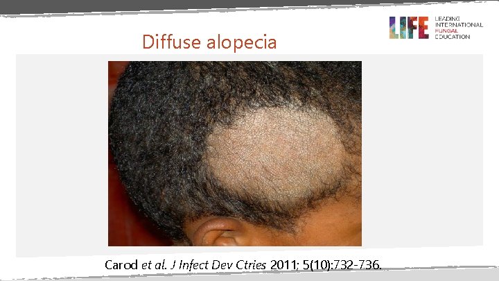
Diffuse alopecia Carod et al. J Infect Dev Ctries 2011; 5(10): 732 -736.
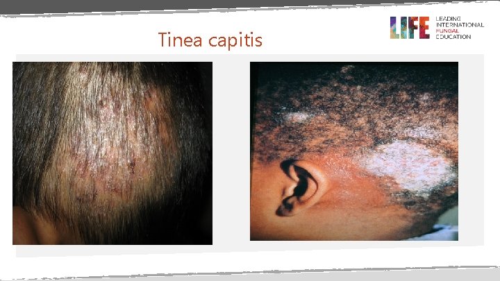
Tinea capitis
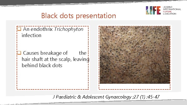
Black dots presentation q An endothrix Trichophyton infection q Causes breakage of the hair shaft at the scalp, leaving behind black dots J Paediatric & Adolescent Gynaecology ; 27 (1) : 45 -47
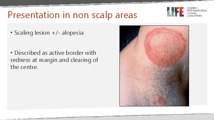
Presentation in non scalp areas • Scaling lesion +/- alopecia • Described as active border with redness at margin and clearing of the centre.
Inflammatory presentation
Determinants of inflammatory reaction • Host’s reactions to the metabolic products of the fungus • Virulence of the infecting strain or species • Anatomic location of the infection • Local environmental factors
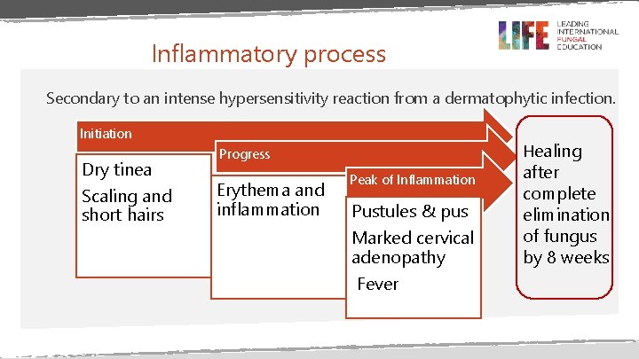
Inflammatory process Secondary to an intense hypersensitivity reaction from a dermatophytic infection. Initiation Dry tinea Scaling and short hairs Healing Progress Erythema and inflammation Peak of Inflammation Pustules & pus Marked cervical adenopathy Fever after complete elimination of fungus by 8 weeks
Dermatophytide reactions Also known as trichophytoid reactions Immunologic reactions (auto-eczematisation) associated with dermatophytoses • Localized or generalized • Occur at or after the height of infection and during treatment • Several studies have demonstrated a high rate of association of dermatophytide reactions and kerion • •
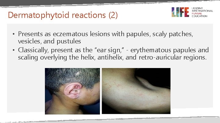
Dermatophytoid reactions (2) • Presents as eczematous lesions with papules, scaly patches, vesicles, and pustules • Classically, present as the “ear sign, ” - erythematous papules and scaling overlying the helix, antihelix, and retro-auricular regions.
Forms of inflammatory presentation All characterized by tender plaques covered with broken hairs and pustules • Pustular Patchy alopecia and scattered pustules or low-grade folliculitis • Favus Erythema around hair follicles and cicatricial alopecia. Eventually, scutula or yellow-crusted cup shape lesions form with hair loss and scarring May also involve the skin and nails • Mycetoma Nodular lesions overlying erythematous and scaly plaques, sinus tracts with purulent drainage & pseudo-alopecia Isa-Isa. et al Clinics in Dermatology 2010 ; 28 : 133 -136
KERION § Inflammatory sub-type of Tinea Capitis § Painful, crusty, matted, boggy scalp plaques or masses with drainage often confused with bacterial abscess § Often solitary, on the occipital area but there are reports of multiple lesions and unusual sites e. g. on vulva, beard, eyebrow § Usually infection by zoophilic dermatophytes - T. verrucosum, T. mentagrophytes § Lymphadenopathy is common § Complications: bacteria superinfection, permanent alopecia, circatrisation
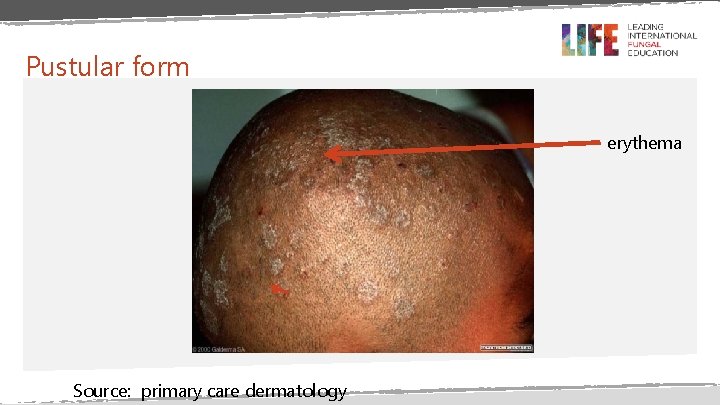
Pustular form erythema Source: primary care dermatology
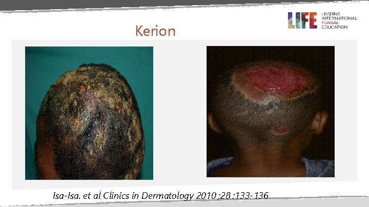
Kerion Isa-Isa. et al Clinics in Dermatology 2010 ; 28 : 133 -136
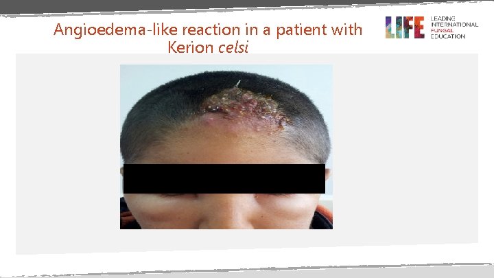
Angioedema-like reaction in a patient with Kerion celsi
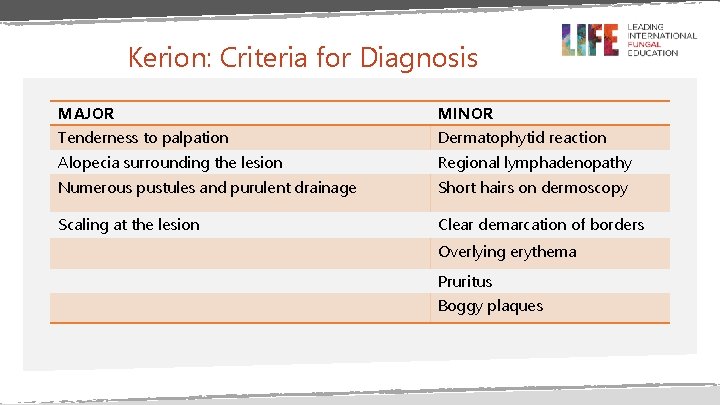
Kerion: Criteria for Diagnosis MAJOR MINOR Tenderness to palpation Dermatophytid reaction Alopecia surrounding the lesion Regional lymphadenopathy Numerous pustules and purulent drainage Short hairs on dermoscopy Scaling at the lesion Clear demarcation of borders Overlying erythema Pruritus Boggy plaques
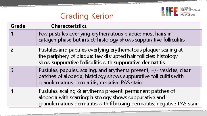
Grading Kerion Grade 1 Characteristics Few pustules overlying erythematous plaque; most hairs in catagen phase but intact; histology shows suppurative folliculitis 2 Pustules and papules overlying erythematous plaque; scaling at the periphery of plaque; few disrupted hair follicles; histology show suppurative folliculitis with suppurative dermatitis Pustules, papules, scaling, and erythema present; +/- vesicles; clear patches of alopecia; histology shows suppurative folliculitis with granulomatous dermatitis; negative PAS stain 3 4 Pustules, scaling & erythema present; permanent patches of alopecia with scarring; histology shows suppurative and granulomatous dermatitis with fibrosing dermatitis; negative PAS stain
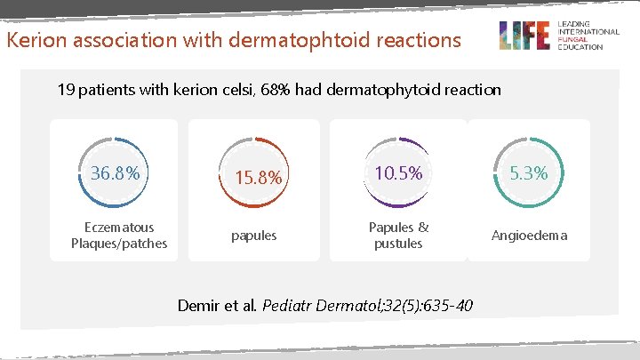
Kerion association with dermatophtoid reactions 19 patients with kerion celsi, 68% had dermatophytoid reaction 36. 8% Eczematous Plaques/patches 15. 8% papules 10. 5% 5. 3% Papules & pustules Angioedema Demir et al. Pediatr Dermatol; 32(5): 635 -40
DIAGNOSIS OF TINEA CAPITIS
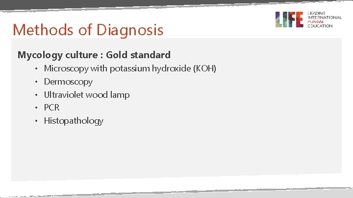
Methods of Diagnosis Mycology culture : Gold standard • • • Microscopy with potassium hydroxide (KOH) Dermoscopy Ultraviolet wood lamp PCR Histopathology
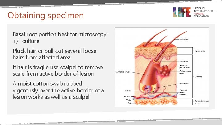
Obtaining specimen Basal root portion best for microscopy +/- culture Pluck hair or pull out several loose hairs from affected area If hair is fragile use scalpel to remove scale from active border of lesion A moist cotton swab rubbed vigorously over the active border of a lesion works as well as a scalpel
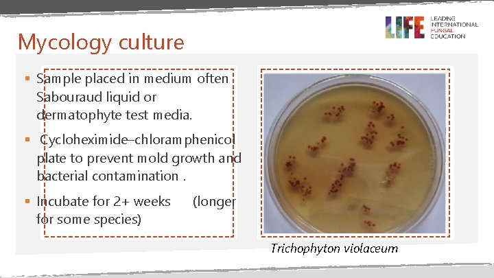
Mycology culture § Sample placed in medium often Sabouraud liquid or dermatophyte test media. § Cycloheximide–chloramphenicol plate to prevent mold growth and bacterial contamination. § Incubate for 2+ weeks for some species) (longer Trichophyton violaceum
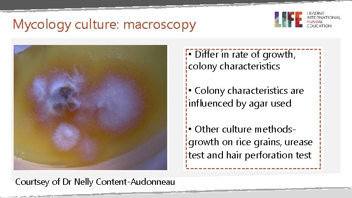
Mycology culture: macroscopy • Differ in rate of growth, colony characteristics • Colony characteristics are influenced by agar used • Other culture methodsgrowth on rice grains, urease test and hair perforation test Courtsey of Dr Nelly Content-Audonneau
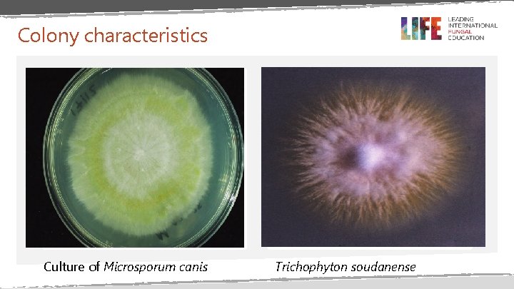
Colony characteristics Culture of Microsporum canis Trichophyton soudanense
Improving fungal culture yield for kerion May be necessary due to high false-negative culture rate with conventional methods of sampling, because sample may represent the inflammatory response • Gauze swabs • Moistened bacterial swab from the pustular area • Directly pressing the agar plate on to the kerion • Acytobrush which is sterile with softer bristles, improves sample quality and minimizes discomfort.
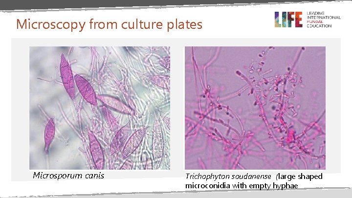
Microscopy from culture plates Microsporum canis Trichophyton soudanense (large shaped microconidia with empty hyphae
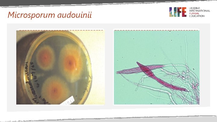
Microsporum audouinii
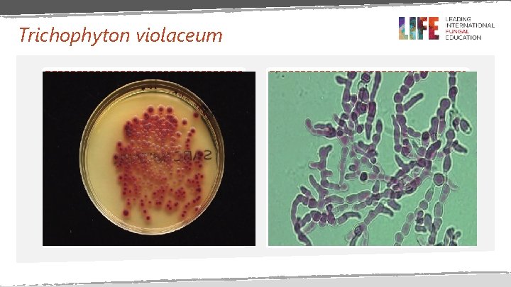
Trichophyton violaceum
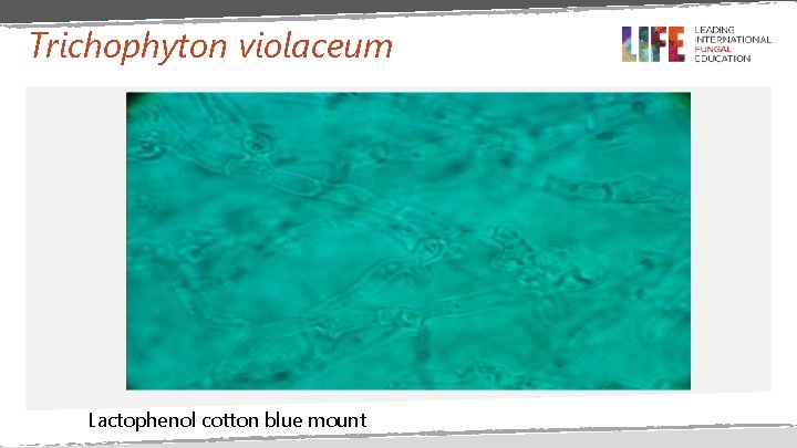
Trichophyton violaceum Lactophenol cotton blue mount
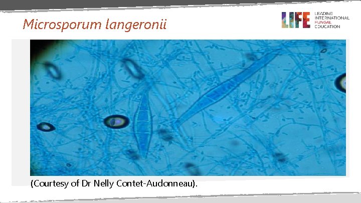
Microsporum langeronii (Courtesy of Dr Nelly Contet-Audonneau).
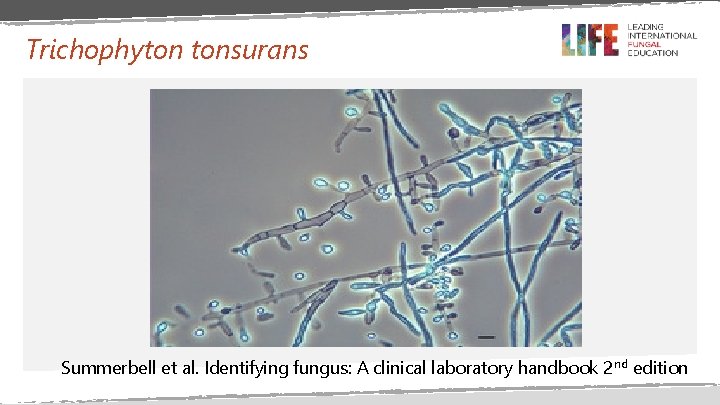
Trichophyton tonsurans Summerbell et al. Identifying fungus: A clinical laboratory handbook 2 nd edition
Microscopy with KOH: procedure Staining the specimen Transfer the scale, hair, or debris to a glass slide, and add a few drops of 10% to 20% KOH. Visualisation Examine under a microscope (× 400) with back-and-forth rotation of the focus knobs. Demonstrate septate branching hyphae or minute dermatophyte spores
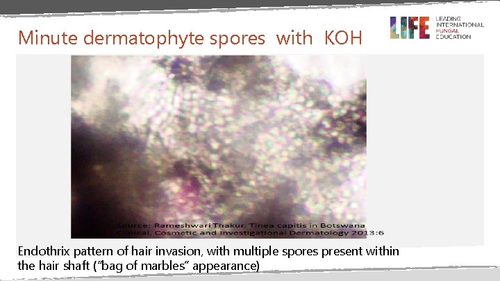
Minute dermatophyte spores with KOH Endothrix pattern of hair invasion, with multiple spores present within the hair shaft (“bag of marbles” appearance)
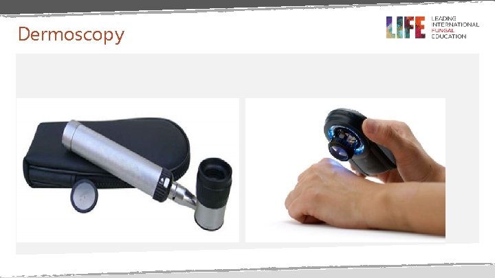
Dermoscopy
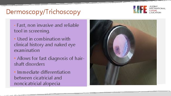
Dermoscopy/Trichoscopy • Fast, non invasive and reliable tool in screening. • Used in combination with clinical history and naked eye examination • Allows for fast diagnosis of hairshaft disorders • Immediate differentiation between cicatricial and noncicatricial alopecia
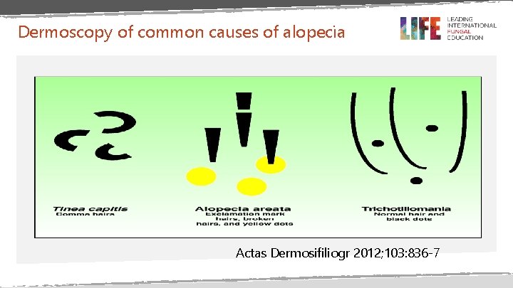
Dermoscopy of common causes of alopecia Actas Dermosifiliogr 2012; 103: 836 -7
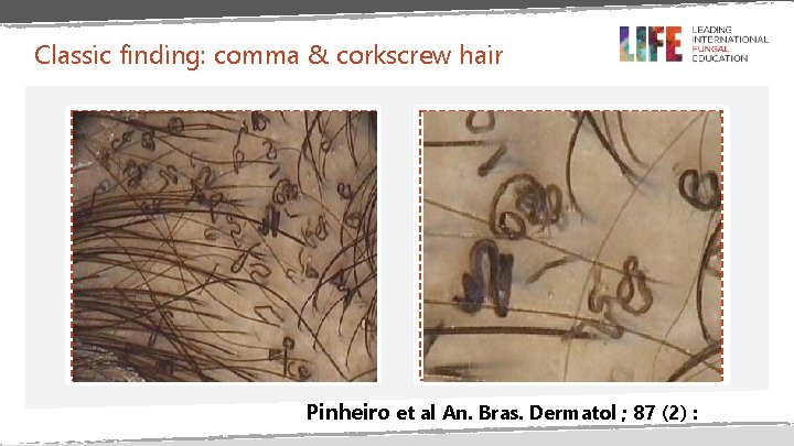
Classic finding: comma & corkscrew hair Pinheiro et al An. Bras. Dermatol ; 87 (2) :
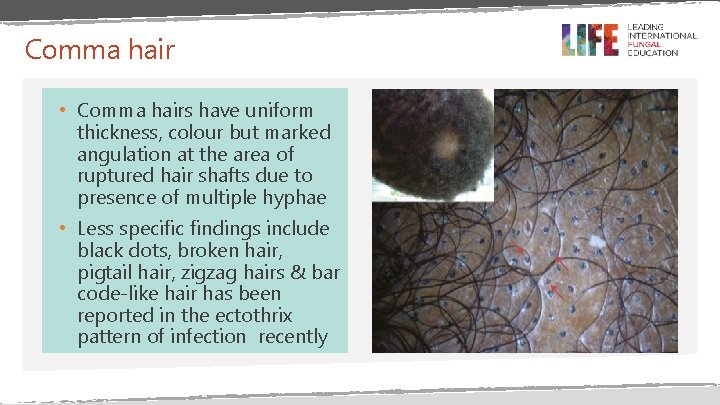
Comma hair • Comma hairs have uniform thickness, colour but marked angulation at the area of ruptured hair shafts due to presence of multiple hyphae • Less specific findings include black dots, broken hair, pigtail hair, zigzag hairs & bar code-like hair has been reported in the ectothrix pattern of infection recently
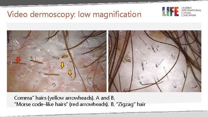
Video dermoscopy: low magnification Comma” hairs (yellow arrowheads). A and B, “Morse code–like hairs” (red arrowheads). B, “Zigzag” hair
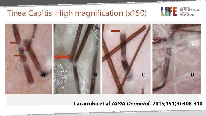
Tinea Capitis: High magnification (x 150) A B C D Lacarruba et al JAMA Dermatol. 2015; 151(3): 308 -310
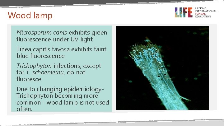
Wood lamp Microsporum canis exhibits green fluorescence under UV light Tinea capitis favosa exhibits faint blue fluorescence. Trichophyton infections, except for T. schoenleinii, do not fluoresce Due to changing epidemiology. Trichophyton becoming more common - wood lamp is not used often.
PCR • Real-time polymerase chain reaction (PCR) test or PCR reverse-line blot • A more recent method of diagnosis • Assays detect T. tonsurans ribosomal DNA in fluid used to ‘rinse’ the patient’s hairbrush. • Average test time is 5 hours.
HISTOPATHOLOGY: KERION • Reveals fungal spores surrounding the hair follicle, hyphae within the hair shaft and an inflammatory infiltrate in the dermis • Giant cell reaction may occur due to follicular destruction • Periodic acid-Schiff stain can be used to confirm diagnosis. • Kerion is characterized by neutrophilic and granulomatous infiltrates in early stages and fibrotic scar later • Biopsy for non inflammatory tinea capitis may be performed when culture and dermascopy are negative

TREATMENT
Systemic antifungals • The goal is to penetrate the hair shaft • Available options: • Griseofulvin • Newer agents: terbinafine, fluconazole and itraconazole, Newer antifungal agents are more concentrated in the hair & provide fungicidal concentrations after treatment is completed, allowing for shorter treatment duration and lower rates of re-infection • Topical agents are not very effective

GRISEOFULVIN Ø Oldest first-line treatment for tinea capitis (since 1958) Ø Dosage: 10 -15 mg/kg/day Ø Duration : 6 -8 weeks. Longer & higher doses for some infections Caution and contraindications Pregnant patients Lupus erythematosus Porphyria Severe liver disease Ø Reports of symptom resolution but failure to eradicate some Trichophyton spp in infected children have been made (continued spread even after treatment)
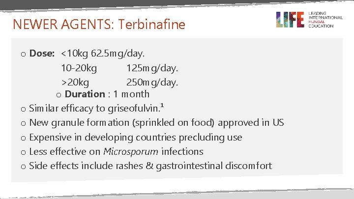
NEWER AGENTS: Terbinafine o Dose: <10 kg 62. 5 mg/day. 10 -20 kg 125 mg/day. >20 kg 250 mg/day. o Duration : 1 month o Similar efficacy to griseofulvin. ¹ o New granule formation (sprinkled on food) approved in US o Expensive in developing countries precluding use o Less effective on Microsporum infections o Side effects include rashes & gastrointestinal discomfort
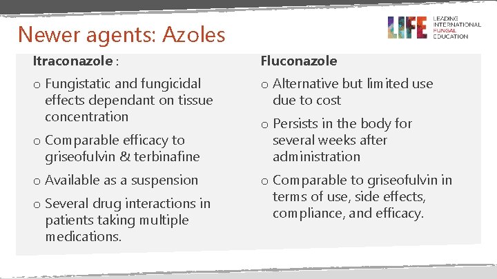
Newer agents: Azoles Itraconazole : Fluconazole o Fungistatic and fungicidal effects dependant on tissue concentration o Alternative but limited use due to cost o Comparable efficacy to griseofulvin & terbinafine o Available as a suspension o Several drug interactions in patients taking multiple medications. o Persists in the body for several weeks after administration o Comparable to griseofulvin in terms of use, side effects, compliance, and efficacy.
Systemic antifungal therapy for tinea capitis in children (2015) “With respect to complete cure (both cure of the infection and visible cure (i. e. fungal and clinical cure) low to moderate quality evidence suggests that newer treatments such as terbinafine, itraconazole and fluconazole are at least as good as griseofulvin, the usual treatment in children with tinea capitis caused by Trichophyton infections” Chen X et al Cochrane Database of Systematic Reviews 2016, Issue 5. Art. No. : CD 004685. DOI: 10. 1002/14651858. CD 004685. pub 3
Role of adjunctive Topical Agents - May reduce transmission if used within the first 2 weeks of infection by decreasing the carriage of viable spores. - Symptomatic improvement - less itching - Used for asymptomatic carriers with low spore loads. - Do not eradicate fungus - Applied to scalp & hair for 5 minutes then rinsed. - Applied 2 -3 times per week for 2 -4 weeks or until patient is mycological cured.
Topical agents used Ø Shampoos 1% or 2. 5% selenium sulphide 2% ketoconazole. 1– 2% zinc pyrithione 2. 5% povidone iodine shampoos (reported used in LMICs) Ø Topical squalamine, a natural aminosterol isolated from shark, has also been used
Additional management for inflammatory types v Prednisone During the first week to alleviate swelling and pain but no effects on scarring v Wet compresses to remove the crusts. May warrant inpatient care and improves healing v No role for surgical drainage on kerion
Follow up • Every 2 weeks after the first month of treatment to determine response. • Verify clearance of infection with culture. • For non-responders – increase dose or change medication • Identify source (human or animal) and screen contacts • Reporting to public health authorities
Primary prevention interventions • An adequate supply of water for personal washing and hygiene • Regular and thorough bathing with soap and water, with special attention to drying moist areas • Restrain from sharing hair brushes, combs, hats, hair accessories • Health education about how to prevent spread
Secondary prevention • The clothing and linen of infected persons should be frequently laundered in hot water to rid them of the fungus • Locate and treat source • Screen contacts
Screening and managing contacts • Who are the contacts? Home (siblings & parents) and school • How do we screen? - Careful physical inspection - Dermoscopy - Wood light for fluorescent types may be used - Fungal culture
Managing contacts Ø Asymptomatic carriers (positive fungal culture but no physical signs of infection ) i) If low fungal spores loads treat with antifungal shampoo ii) If high fungal spore loads treat with oral antifungals Ø Asymptomatic carriers of T. tonsurans irrespective of fungal load may be treated with oral antifungals.

Differential Diagnoses
Intended Leaning Objectives Identify disease entities with similar presentation to Tinea capitis To discuss distinct features of these conditions that may help in differentiating them from Tinea capitis infections
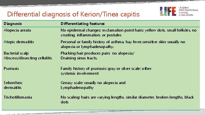
Differential diagnosis of Kerion/Tinea capitis Diagnosis Differentiating features Alopecia areata No epidermal changes; exclamation point hairs; yellow dots, small follicles, no crusting, inflammation, or pustules Atopic dermatitis Personal or family history of asthma, hay fever, sensitive skin; usually no alopecia or lymphadenopathy. Bacterial scalp Abscess/dissecting cellulitis Plucking hair produces pain; no alopecia/ Draining sinus tracts. Psoriasis Family history of psoriasis; gray or silver scale; other systemic involvement Seborrheic dermatitis Greasy scale; usually no alopecia and Lymphadenopathy Trichotillomania No scaling; hairs are varying lengths, similar diameter, broken lengths, black dots
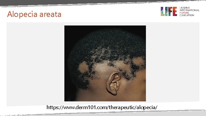
Alopecia areata https: //www. derm 101. com/therapeutic/alopecia/
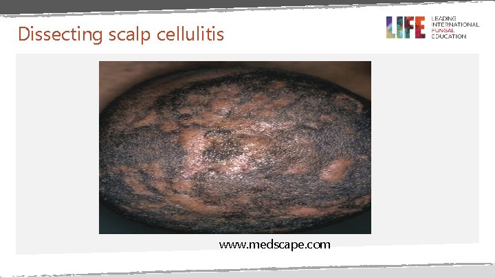
Dissecting scalp cellulitis www. medscape. com
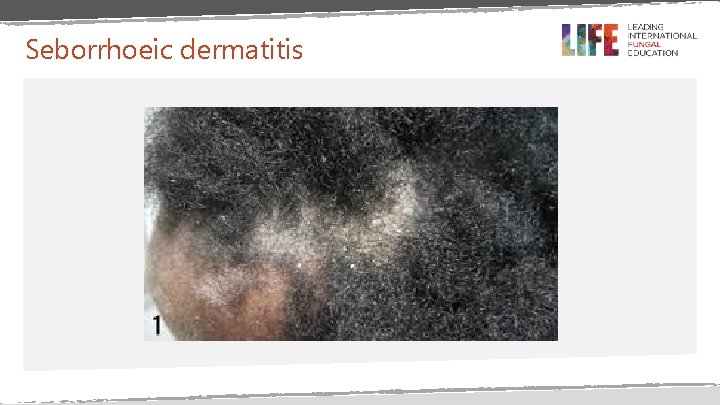
Seborrhoeic dermatitis
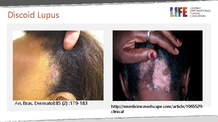
Discoid Lupus An. Bras. Dermatol; 85 (2) : 179 -183 http: //emedicine. medscape. com/article/1065529 clinical
END
- Slides: 99