Acute Inflammatory Dermatoses Literally thousands of inflammatory dermatoses


 is a common disorder of the skin characterized by localized")


- Slides: 29
Acute Inflammatory Dermatoses
• Literally thousands of inflammatory dermatoses have been described. • acute lesions last from days to weeks and are characterized by inflammatory infiltrates (usually composed of lymphocytes and macrophages rather than neutrophils), edema, and variable degrees of epidermal, vascular, or subcutaneous injury. • Chronic lesions, on the other hand, persist for months to years and are often associated with changes in epidermal growth (atrophy or hyperplasia) or dermal fibrosis.
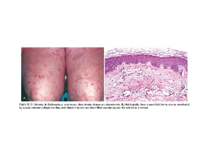
Urticaria • Urticaria (hives) is a common disorder of the skin characterized by localized mast cell degranulation and resultant dermal microvascular hyperpermeability. • This combination of effects produces pruritic edematous plaques called wheals. • Angioedema is closely related to urticaria and is characterized by edema of the deeper dermis and the subcutaneous fat.
• Urticaria most often occurs between ages 20 and 40, but all age groups are susceptible. • Individual lesions develop and fade within hours (usually less than 24 hours), and episodes may last for days or persist for months. • Sites of predilection for urticarial eruptions include any area exposed to pressure, such as the trunk, distal extremities, and ears. • Persistent episodes of urticaria may herald an underlying disease (e. g. , collagen vascular disorders, Hodgkin lymphoma), but in the majority of cases no underlying cause is identified.
• Pathogenesis: - Mast cell-dependent, Ig. E-dependent: Urticaria of this type follows exposure to many different antigens (pollens, foods, drugs, insect venom), and is an example of a localized immediate hypersensitivity (type I) reaction triggered by the binding of antigen to Ig. E antibodies that are attached to mast cells through Fc receptors. - Mast cell-dependent, Ig. E-independent: This subset results from substances that directly incite the degranulation of mast cells, such as opiates, certain antibiotics, curare, and radiographic contrast media. - Mast cell-independent, Ig. E-independent: These forms of urticaria are triggered by local factors that increase vascular permeability. One form is initiated by exposure to chemicals or drugs, such as aspirin, that inhibit cyclooxygenase and arachidonic acid production. The precise mechanism of aspirin-induced urticaria is unknown. A second form is hereditary angioneurotic edema, caused by an inherited deficiency of C 1 inhibitor that results in excessive activation of the early components of the complement system and production of vasoactive mediators.
• MORPHOLOGY: Lesions vary from small, pruritic papules to large edematous plaques. Individual lesions may coalesce to form annular, linear, or arciform configurations. The histologic features of urticaria may be very subtle. There is usually a sparse superficial perivenular infiltrate consisting of mononuclear cells and rare neutrophils. Eosinophils may also be present. Collagen bundles are more widely spaced than in normal skin, a result of superficial dermal edema. Superficial lymphatic channels are dilated due to increased absorption of edema fluid. There are no changes in the epidermis.
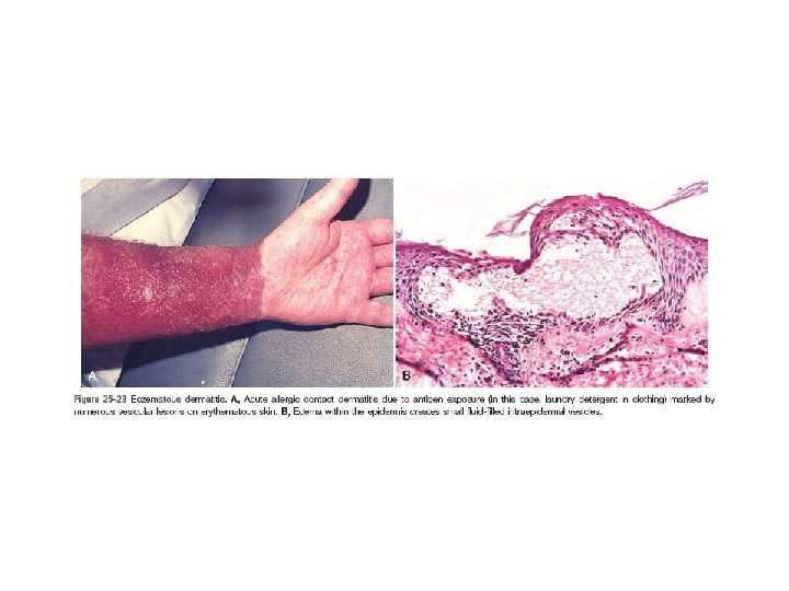
Acute Eczematous Dermatitis • Based on initiating factors, eczematous dermatitis can be subdivided into the following categories: (1) allergic contact dermatitis (2) atopic dermatitis (3) drug-related eczematous dermatitis, (4) photoeczematous dermatitis (5) primary irritant dermatitis.
• The causes of eczema are sometimes broadly separated into “inside” and “outside” types: disease resulting from external application of an antigen (e. g. , poison ivy) or a reaction to an internal circulating antigen (which may be derived from ingested food or a drug).
• Pathogenesis: Eczematous dermatitis typically results from T cell-mediated inflammatory reactions (type IV hypersensitivity). This has been well studied in dermatitis triggered by contact antigens (e. g. , uroshiol from poison ivy). It is believed that reactive chemicals introduced at the epidermal surface modify self proteins, acting as “haptens”, and these proteins become neoantigens. The antigens are taken up by Langerhans cells, which then migrate by way of dermal lymphatics to draining lymph nodes. Here the antigens are presented to naive CD 4+ T cells, which are activated and develop into effector and memory cells. On antigen reexposure, memory T cells expressing homing molecules migrate to skin sites of antigen localization. Here they release the cytokines and chemokines that recruit the numerous inflammatory cells characteristic of eczema. This process occurs within 24 hours.
• Langerhans cells within the epidermis play a central role in contact dermatitis, and understandably factors that affect Langerhans cell function impact the inflammatory reaction. • Chronic exposure to UV light is injurious to epidermal Langerhans cells and can prevent sensitization to contact antigens, although UV light can also alter antigens and generate forms that are more likely to induce sensitivity reactions.
• Treatment involves a search for offending substances that can be removed from the environment. • Topical steroids nonspecifically block the inflammatory response. • While such treatments are only palliative and do not cure, they are nevertheless helpful in interrupting acute exacerbations of eczema that can become self-perpetuating if unchecked.
• MORPHOLOGY: All types of eczematous dermatitis are characterized by red, papulovesicular, oozing, and crusted lesions that, if persistent, develop reactive acanthosis and hyperkeratosis that produce raised scaling plaques. Such lesions are prone to bacterial superinfection, which produces a yellow crust (impetiginization). With time, persistent lesions become less “wet” (fail to ooze or form vesicles) and become progressively (hyperkeratotic and acanthotic). Spongiosis characterizes acute eczematous dermatitis (edema seeps into the intercellular spaces of the epidermis). Mechanical shearing of intercellular attachment sites (desmosomes) and cell membranes by progressive accumulation of intercellular fluid may result in the formation of intraepidermal vesicles.
• During the earliest stages of eczematous dermatitis, there is a superficial, perivascular, lymphocytic infiltrate associated with papillary dermal edema and mast cell degranulation.
Erythema Multiforme • Erythema multiforme is an uncommon self-limited hypersensitivity reaction to certain infections and drugs. (1) infections such as herpes simplex, mycoplasmal infections, histoplasmosis, coccidioidomycosis, typhoid, and leprosy, among others (2) exposure to certain drugs (sulfonamides, penicillin, barbiturates, salicylates, hydantoins, and antimalarials) (3) cancer (carcinomas and lymphomas) (4) collagen vascular diseases (lupus erythematosus, dermatomyositis, and polyarteritis nodosa).
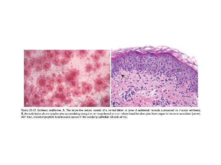
• Pathogenesis: Erythema multiforme is characterized by keratinocyte injury mediated by skin-homing CD 8+ cytotoxic T lymphocytes.
• MORPHOLOGY: Affected individuals present with a diverse array of lesions (hence the term multiforme), including macules, papules, vesicles, bullae, and characteristic targetoid (target-like) lesions. A febrile form associated with extensive involvement of the skin is called Stevens-Johnson syndrome, which is often (but not exclusively) seen in children. lesions involve not only the skin but also the lips and oral mucosa, conjunctiva, urethra, and genital and perianal areas. Secondary infection of involved areas due to loss of skin integrity may result in lifethreatening sepsis.
• Chronic Inflammatory Dermatoses: Psoriasis Seborrheic Dermatitis Lichen Planus
Acne Vulgaris • Disorder of Epidermal Appendages. • Virtually universal in the middle to late teenage years, acne vulgaris affects both males and females, although males tend to have more severe disease. • Acne is seen in all races but is usually milder in people of Asian descent. • It may be induced or exacerbated by drugs (corticosteroids, adrenocorticotropic hormone, testosterone, gonadotropins, contraceptives, trimethadione, iodides, and bromides), occupational exposures (cutting oils, chlorinated hydrocarbons, and coal tars), and conditions that favor occlusion of sebaceous glands, such as heavy clothing, cosmetics, and tropical climates. Some families seem to be particularly prone to acne, suggesting a hereditary component.
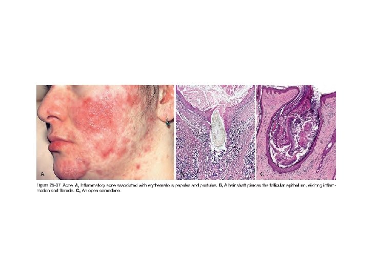
• Acne is divided into noninflammatory and inflammatory types, although both types may coexist. • Noninflammatory acne may take the form of open and closed comedones. Open comedones are small follicular papules containing a central black keratin plug. This color is the result of oxidation of melanin pigment (not dirt). Closed comedones are follicular papules without a visible central plug. Because the keratin plug is trapped beneath the epidermal surface, these lesions are potential sources of follicular rupture and inflammation.
• Pathogenesis: The pathogenesis of acne is incompletely understood and is likely multifactorial. At least four factors contribute to its development: (1) keratinization of the lower portion of the follicular infundibulum and development of a keratin plug that blocks outflow of sebum to the skin surface (2) hypertrophy of sebaceous glands during puberty under the influence of androgens (3) Lipase synthesizing bacteria (Propionibacterium acnes) colonizing the upper and midportion of the hair follicle, converting lipids within sebum to proinflammatory fatty acids (4) secondary inflammation of the involved follicle.
• MORPHOLOGY: - Inflammatory acne is marked by erythematous papules, nodules, and pustules. Severe variants result in sinus tract formation and dermal scarring. - Open comedones have large, patulous orifices, whereas those of closed comedones are identifiable only microscopically. - Variable infiltrates of lymphocytes and macrophages are present in and around affected follicles, and extensive acute inflammation accompanies follicular rupture. - Dermal abscesses may form in association with rupture and lead to scarring.
Panniculitis • Panniculitis is an inflammatory reaction in the subcutaneous adipose tissue that may preferentially affect (1) the lobules of fat or (2) the connective tissue that separates fat into lobules. Panniculitis often involves the lower legs. Erythema nodosum is the most common form and usually has a subacute presentation. A second somewhat distinctive form, erythema induratum.
• Erythema nodosum: - presents as poorly defined, tender, erythematous plaques and nodules that may be more readily palpated than seen. - Its occurrence is often associated with infections (β-hemolytic streptococcal infection, tuberculosis and, less commonly, coccidioidomycosis, histoplasmosis, and leprosy), drug administration (sulfonamides, oral contraceptives), sarcoidosis, inflammatory bowel disease, and certain malignant neoplasms, but many times a cause cannot be identified. - Fever and malaise may accompany the cutaneous signs. - It is considered to be caused by a delayed hypersensitivity reaction to microbial or drug related antigens. In some cases immune complexes have been implicated but in many cases the pathogenesis remains mysterious. - Over the course of weeks, lesions usually flatten and become bruiselike, leaving no residual clinical scars, while new lesions develop. - Biopsy of a deep wedge of tissue to generously sample the subcutis is usually required for histologic diagnosis.
• Erythema induratum: - is an uncommon type of panniculitis that affects primarily adolescents and menopausal women. - Although the cause is not known, most observers regard this as a primary vasculitis of deep vessels supplying the fat lobules of the subcutis; the associated vascular compromise leads to fat necrosis and inflammation. - Erythema induratum presents as an erythematous, slightly tender nodule that usually goes on to ulcerate. - Originally considered a hypersensitivity response to tuberculosis, erythema induratum today most commonly occurs without an associated underlying disease.
• MORPHOLOGY: - The histopathology of erythema nodosum is distinctive. In early lesions, the connective tissue septae are widened by edema, fibrin exudation, and neutrophilic infiltration. Later, infiltration by lymphocytes, histiocytes, multinucleated giant cells, and occasional eosinophils is associated with septal fibrosis. Vasculitis is not present. - In erythema induratum, on the other hand, granulomatous inflammation and zones of caseous necrosis involve the fat lobule. Early lesions show necrotizing vasculitis affecting small- to medium-sized arteries and veins in the deep dermis and subcutis.