
Acute Inflammatory Dermatoses Ghadeer Hayel M D Histopathologist

- Slides: 30
Acute Inflammatory Dermatoses Ghadeer Hayel M. D. Histopathologist Mar 8 th 2021
CONTENTS OF THIS LECTURE • • 1. 2. 3. Nomenclature of Skin Lesions Acute Inflammatory Dermatoses Urticaria Acute Eczematous Dermatitis Erythema Multiforme
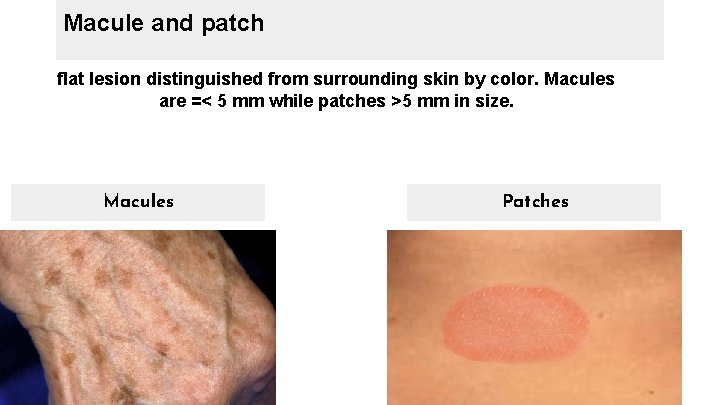
Macule and patch flat lesion distinguished from surrounding skin by color. Macules are =< 5 mm while patches >5 mm in size. Macules Patches
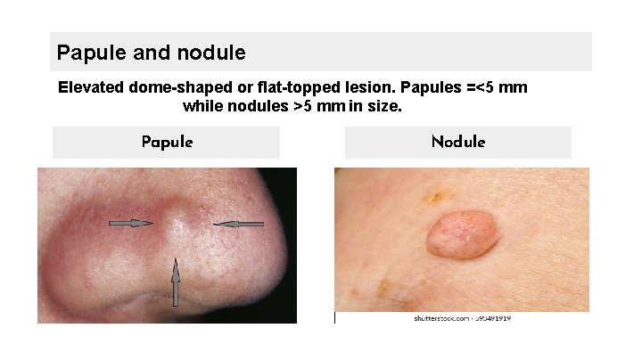
Papule and nodule Elevated dome-shaped or flat-topped lesion. Papules =<5 mm while nodules >5 mm in size. Papule Nodule
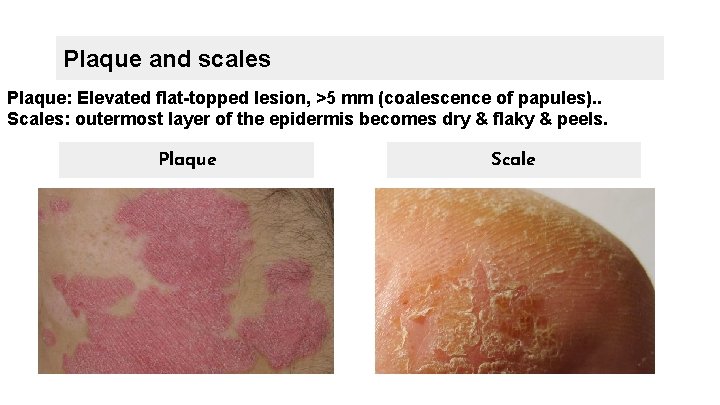
Plaque and scales Plaque: Elevated flat-topped lesion, >5 mm (coalescence of papules). . Scales: outermost layer of the epidermis becomes dry & flaky & peels. Plaque Scale
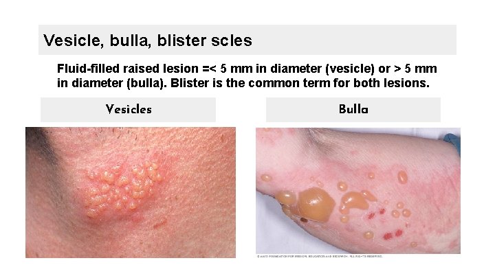
Vesicle, bulla, blister scles Fluid-filled raised lesion =< 5 mm in diameter (vesicle) or > 5 mm in diameter (bulla). Blister is the common term for both lesions. Vesicles Bulla
01. Urticaria a common disorder of the skin characterized by localized mast cell degranulation and resultant dermal microvascular hyperpermeability
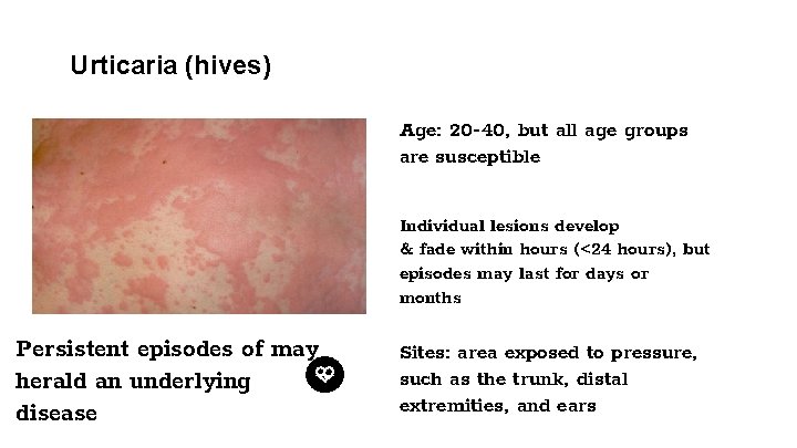
Urticaria (hives) Age: 20 -40, but all age groups are susceptible Individual lesions develop & fade within hours (<24 hours), but episodes may last for days or months Persistent episodes of may herald an underlying disease Sites: area exposed to pressure, such as the trunk, distal extremities, and ears
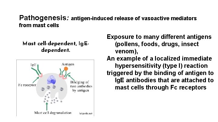
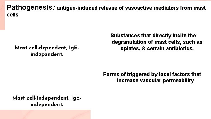
Pathogenesis: antigen-induced release of vasoactive mediators from mast cells Mast cell-dependent, Ig. Edependent. Exposure to many different antigens (pollens, foods, drugs, insect venom), An example of a localized immediate hypersensitivity (type I) reaction triggered by the binding of antigen to Ig. E antibodies that are attached to mast cells through Fc receptors
Pathogenesis: antigen-induced release of vasoactive mediators from mast cells Mast cell-dependent, Ig. Eindependent. Substances that directly incite the degranulation of mast cells, such as opiates, & certain antibiotics. Forms of triggered by local factors that increase vascular permeability. Mast cell-independent, Ig. Eindependent.
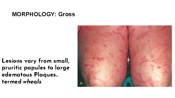
MORPHOLOGY: Gross Lesions vary from small, pruritic papules to large edematous Plaques. . termed wheals
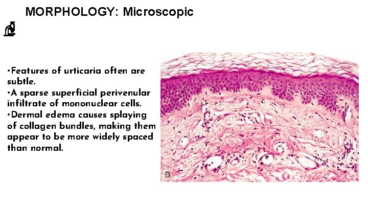
MORPHOLOGY: Microscopic • Features of urticaria often are subtle. • A sparse superficial perivenular infiltrate of mononuclear cells. • Dermal edema causes splaying of collagen bundles, making them appear to be more widely spaced than normal.
02. ● Acute Eczematous Dermatitis Greek word meaning “to boil over, ” One of the most common skin disorders
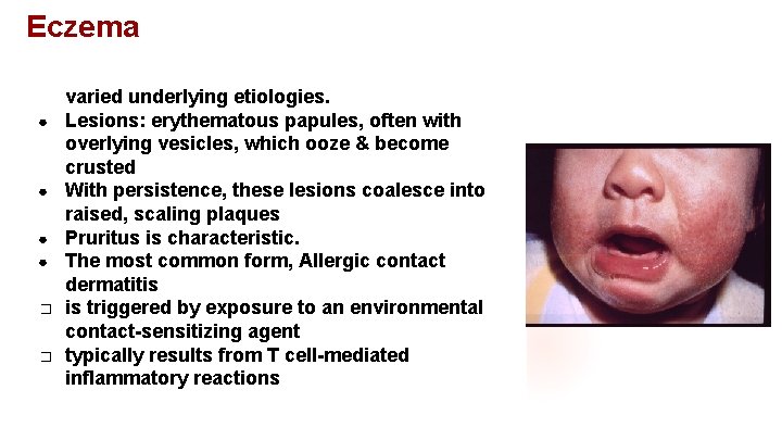
Eczema ● ● ● � � A clinical term for a number of conditions with varied underlying etiologies. Lesions: erythematous papules, often with overlying vesicles, which ooze & become crusted With persistence, these lesions coalesce into raised, scaling plaques Pruritus is characteristic. The most common form, Allergic contact dermatitis is triggered by exposure to an environmental contact-sensitizing agent typically results from T cell-mediated inflammatory reactions
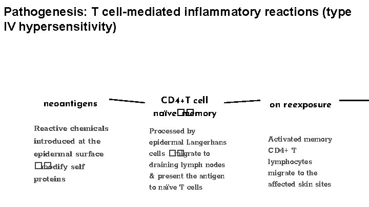
Pathogenesis: T cell-mediated inflammatory reactions (type IV hypersensitivity) neoantigens Reactive chemicals introduced at the epidermal surface �� modify self proteins CD 4+T cell naïve�� memory Processed by epidermal Langerhans cells �� migrate to draining lymph nodes & present the antigen to naïve T cells on reexposure Activated memory CD 4+ T lymphocytes migrate to the affected skin sites
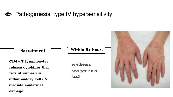
Pathogenesis: type IV hypersensitivity Recruitment CD 4+ T lymphocytes release cytokines that recruit numerous inflammatory cells & mediate epidermal damage Within 24 hours erythema and pruritus ﺍﻟﺤﻜﺔ
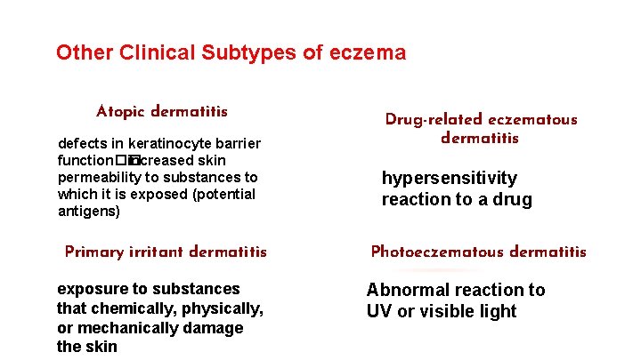
Other Clinical Subtypes of eczema Atopic dermatitis defects in keratinocyte barrier function�� increased skin permeability to substances to which it is exposed (potential antigens) Primary irritant dermatitis exposure to substances that chemically, physically, or mechanically damage the skin Drug-related eczematous dermatitis hypersensitivity reaction to a drug Photoeczematous dermatitis Abnormal reaction to UV or visible light
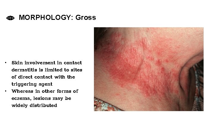
MORPHOLOGY: Gross • Skin involvement in contact dermatitis is limited to sites of direct contact with the triggering agent • Whereas in other forms of eczema, lesions may be widely distributed
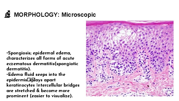
MORPHOLOGY: Microscopic • Spongiosis; epidermal edema, characterizes all forms of acute eczematous dermatitis(spongiotic dermatitis). • Edema fluid seeps into the epidermis�� splays apart keratinocytes Intercellular bridges are stretched & become more prominent (easier to visualize).
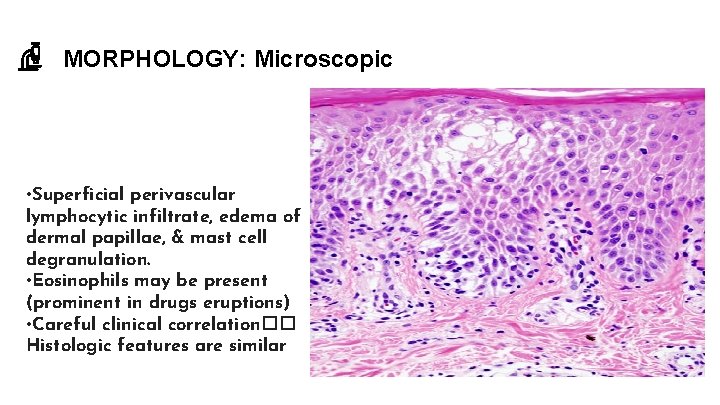
MORPHOLOGY: Microscopic • Superficial perivascular lymphocytic infiltrate, edema of dermal papillae, & mast cell degranulation. • Eosinophils may be present (prominent in drugs eruptions) • Careful clinical correlation�� Histologic features are similar
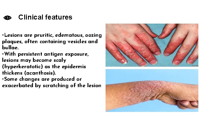
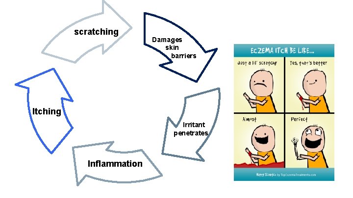
Clinical features • Lesions are pruritic, edematous, oozing plaques, often containing vesicles and bullae. • With persistent antigen exposure, lesions may become scaly (hyperkeratotic) as the epidermis thickens (acanthosis). • Some changes are produced or exacerbated by scratching of the lesion
scratching Damages skin barriers Itching Irritant penetrates Inflammation
Clinical features: Atopic dermatitis • Susceptibility to atopic dermatitis is often inherited; concordant in 80% of identical twins & 20% of fraternal twins. • It usually appears in early childhood & remits spontaneously as patients mature into adults. • Children with atopic dermatitis often have asthma & allergic rhinitis, termed the atopic triad. • Recent genetic studies have identified polymorphisms associated with increased risk in genes that encode proteins involved in keratinocyte barrier function, innate immunity, and T cell function.
03. Erythema Multiforme An uncommon, usually self-limited disorder that appears to be a hypersensitivity response to certain infections and drugs.
It affects individuals of any age & associated with the following conditions: Infections herpes simplex, mycoplasmal infections, histoplasmosis, coccidioidomycosis, typhoid, and leprosy Cancer Carcinomas & lymphomas Drugs sulfonamides, penicillin, barbiturates, salicylates, & antimalarials Collagen vascular diseases lupus erythematosus, dermatomyositis, & polyarteritis nodosa.
Pathogenesis • • Erythema multiforme is characterized by keratinocyte injury mediated by skin-homing CD 8+ cytotoxic T lymphocytes. The epidermal antigens that are recognized by the infiltrating T cells in erythema multiforme remain unknown.
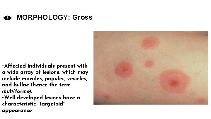
MORPHOLOGY: Gross • Affected individuals present with a wide array of lesions, which may include macules, papules, vesicles, and bullae (hence the term multiforme). • Well-developed lesions have a characteristic “targetoid” appearance
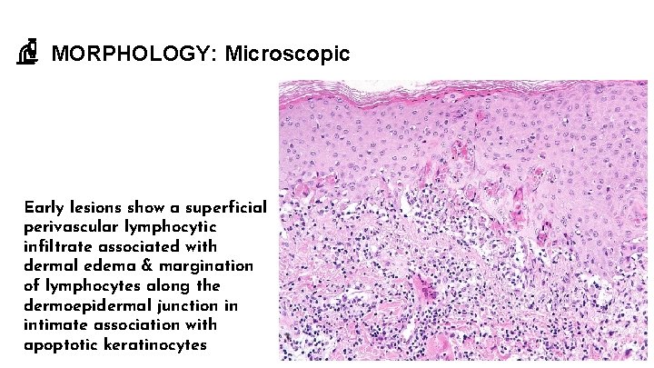
MORPHOLOGY: Microscopic Early lesions show a superficial perivascular lymphocytic infiltrate associated with dermal edema & margination of lymphocytes along the dermoepidermal junction in intimate association with apoptotic keratinocytes
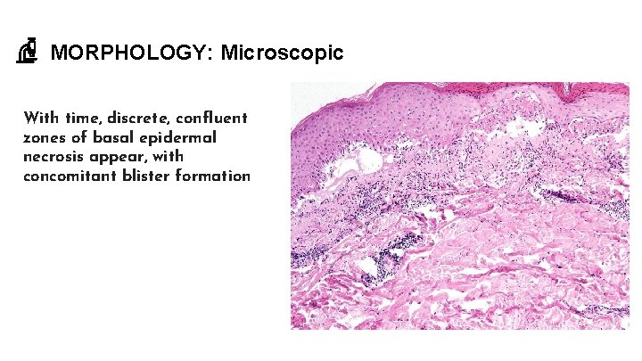
MORPHOLOGY: Microscopic With time, discrete, confluent zones of basal epidermal necrosis appear, with concomitant blister formation
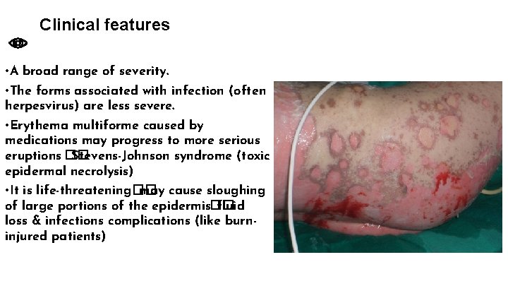
Clinical features • A broad range of severity. • The forms associated with infection (often herpesvirus) are less severe. • Erythema multiforme caused by medications may progress to more serious eruptions �� Stevens-Johnson syndrome (toxic epidermal necrolysis) • It is life-threatening�� may cause sloughing of large portions of the epidermis�� fluid loss & infections complications (like burninjured patients)