2 nd International Congress on Early Onset Scoliosis


 Sham (Screws placed and removed, grafts harvested and discarded) Instrumentation (Human")
 Randomized before the last non-retraceable operative step Craven 1994")
 Facet and Posterior Arthrodesis ready to Randomize")


 (ex vivo) Flexion-Compression Spinal column and portions of it Compression Body")
 Regression models Alpha < 0.")


 A-1 Baseline Flexion-compression stiffness (N/mm)")
 Control 376 ±")
")
")

 after Facet +")


")


 bone mineral")
 Implant only constructs (n=24) from similar")


 Negative: Quadripedal Positive: Biological system that responds to Wolff’s Law")

 All arthrodeses healed to fusion Less Stiff vs. More Stiff")

- Slides: 39
2 nd International Congress on Early Onset Scoliosis & Growing Spine November 7 -8, 2008: Montréal, Québec, CANADA BIOLOGICAL IMPLICATIONS OF INTRINSICALLY STABLE SPINAL INSTRUMENTATION Marc A. Asher, M. D. Kansas City, KS, USA Conflict of interest disclosure: I have received royalties, stock options, grant support, honoraria and travel expenses related to spinal implant research and development.
Acknowledgements Colleagues H. Clarke Anderson, MD William L. Carson, Ph. D E. James Harner, Ph. D Charles F. Heinig, MD Gopal Jayaraman, Ph. D Sue Min Lai, Ph. D Ralph Robinson, MD † Fellow James W. Hardacker, MD Residents Thomas G. Craven, MD Dale D. Dalenberg, MD Thomas R. Hunt, MD Hans Kioschos, MD K. Ronald Smith, MD Technician Richard Lark, BS
INTRODUCTION Background Spinal Arthrodesis First successful treatment for scoliosis Spinal instrumentation Required to reliably obtain fusion Intrinsically stable instrumentation Required to Control three dimensional deformity and Eliminate external immobilization
Possible negative consequences of intrinsically stable instrumentation on Fusion Bypassed bone and disk Unbypassed adjacent and periadjacent bone and disk
Purpose Experimentally quantify biological effects of stiff, pedicle screw anchored, posterior spinal instrumentation Hypotheses Anterior column growth can be controlled Potentially harmful biological consequences measurable Generally acceptably small Modifiable
METHODS Overview Summary Five published experiments One analysis in progress Institutional animal care committee approval obtained for each, and for pilot studies Experiments were designed to complement each other to gain maximum information
Animal Model Adult Mongrel Canines Up to one year survival Ten to 12 week old Walker hounds (Risser 0) Six months survival
Operation None (Control) Sham (Screws placed and removed, grafts harvested and discarded) Instrumentation (Human size on animals ½ to ¼ as heavy) Anchors Bicortical pedicle screws (6. 25 mm diameter) L 3 – L 5 anchors; L 4 bypassed (one L 1 – L 3 – L 5 anchors) Connections (Stiff) Longitudinal members Rods (6. 35 mm or 4. 76 mm diameter) or plates (human size) Transverse connection One with rods constructs Arthrodesis (Autogenous Iliac Crest Bone Graft) Facets only Facets and posterior
Experimental design (typical one shown) Randomized before the last non-retraceable operative step Craven 1994
Intra-operative Examples Instrumentation without Arthrodesis (Smith 1991) Facet and Posterior Arthrodesis ready to Randomize Rod Size [Asher (Hardacker) 2007]
Evaluations Radiographic in vivo and ex vivo Construct integrity
Bone mineral density Histomorphometric DEXA, in vivo and ex vivo Sham Implant without Arthrodesis
Biomechanical: Stiffness (N/mm) (ex vivo) Flexion-Compression Spinal column and portions of it Compression Body and Discs
Statistics Paired t test and Sign Rank Test (non-parametric) Regression models Alpha < 0. 05 Power Study group numbers most commonly 4 or 6 To prevent Type II error (false negative), 25 -30 (an unrealistic number) would be necessary
RESULTS I. Growing Spine A. Sagittal angulation C and I+F vs. F and S+F p <0. 01 Kioschos 1996
Control L 1 -L 5 posterior fusion Representative lateral lumbar spine radiographs of Walker Hounds [1012 weeks old (Risser 0) at baseline] at six months follow-up Kioschos 1996 L 1 -L 5 Instrumentation and fusion
B. Body and disk growth I + F v others p<0. 01 Kioschos 1996
II. Effects on the Mature Spine A. Arthrodesis (Fusion) A-1 Baseline Flexion-compression stiffness (N/mm) L 3 -5 Spinal Column L 3 -5 Implant Construct L 3 -5 Spinal Column + Implant Construct None 368 ± 45 [3] 376 ± 52 [5] Not applicable See below 4. 76 mm See Col. 4 152 ± 34 * [3] 154 ± 22 [5] 889 ± 125 [5] 283 ± 33 * [3] Not Done Implant Assembly 6. 35 mm Not Done * P < 0. 002 376 + 154 = 520 (<889) N/mm Post Inst. Resists column buckling [3] Craven 1994 [5] Asher 2007
A-2 Effect of sham operation on spinal column flexion-compression stiffness (N/mm) Control 376 ± 52* (n=4) Sham 6 months survival 12 months survival 6 & 12 mo. combined 588 ± 162 (n=4) 776 ± 160 (n=2) 648 ± 156* (n=6) *p = 0. 0167 Sham surgery limits flexion buckling (? scar) Control and sham (6 or 12 months survival) operated specimens had no posterior column stiffness Asher 2007
A-3 Effect of fusion type & survival time on spinal column flexion-compression stiffness (N/mm) Survival Impl ant Rod Diam eter 24 weeks Facet 6 months 12 months Facet + Posterior 4. 76 mm 1411 ± 361* (n=4)[3] 1147 ± 111‡ (n=4)[5] 6. 35 mm 727 ± 220* 1265 ± 252 (n=4)[3] * P <(n=4)[5] 0. 02 1594 ± 482‡ (n=4)[5] 1244 ± 250 (n=4)[5] ‡ P = 0. 0223 [3] Craven 1994 [5] Asher 2007
A-4 Effect of fusion type & survival time on posterior column flexion-compression stiffness (N/mm) Impl ant Rod Diam eter Survival 24 weeks 6 months Facet 12 months Facet + Posterior 4. 76 mm 192 ± 9* (n=4)[3] 164 ± 73‡ (n=4)[5] 462 ± 247‡ (n=4)[5] 6. 35 mm 112 ± 23* (n=4)[3] 153 ± 40 (n=4)[5] 280 ± 115 (n=4)[5] * P < 0. 003 ‡ P = 0. 0047 [3] Craven 1994 [5] Asher 2007
A-5 Effect of implant removal at 12 weeks, following baseline instrumentation and facet fusion, on spinal column flexion-compression stiffness (N/mm) at 24 weeks Implant Rod Diameter Survival ( 24 weeks) Implants Removed at 12 weeks Implants Retained all 24 weeks 4. 76 mm 1189 ± 436 1349 ± 233* 6. 35 mm 885 ± 226 799 ± 225* * P < 0. 05 Implant removal at 12 weeks does not result in stiffer facet fusions at 24 weeks. Craven 1994
A-6 Effect of retained implants on spinal column flexion-compression stiffness (N/mm) after Facet + Posterior Fusion Specimen With Implants Without Implants % Implant Contribution Control 4. 76 mm 6. 35 mm 6 Months 12 months 6 months 12 months 889 1336 1867 1739 1599 ± 125 ± 78 ± 579 ± 329 ± 249 376 1147* 1594* 1265 1244 ± 52 ± 111 ± 482 ± 250 57. 7 14. 3 27. 4 22. 3 ± 0. 7 ± 4. 0 ± 3. 6 ± 1. 2 ± 8. 5 * p = 0. 0233 All implant groups significantly stiffer than comparable control All with implant groups significantly stiffer than with implants removed Asher 2007
B. Effect of Instrumentation and Fusion on Bypassed Bone B-1 The effect of implant loosening in a non-fusion model Two non-fusion implant experiments were done. Without torque wrench tightening 1 of 6 constructs had loosening at 3 months and 4 of six constructs at 6 months. [Smith 1991] With torque wrench tightening none of the implant-implant connections were loose at 9 months while 20 of 56 (36%) pedicle screws were loose in bone to the extent that they could either be toggled or easily removed by hand. [Dalenberg 1993]
The effect of implant-bone interface loosening on bypassed bone mineral density in a non-fusion model Dalenberg 1993
B-2 The effect of implant removal compared to implant retained on bypassed (L 4) bone mineral density (BMD) following instrumentation and facet arthrodesis Neither retained group significantly less than baseline at 24 weeks Craven 1994
B-3 The effect of facet + posterior arthrodesis and retained implants on the bypassed (L-4) vertebral body compression stiffness (n=4) Specimen Control Bypassed 4900 L 4 Body 457 * ** ± 4. 76 mm Rods 6. 35 mm Rods 6 months 12 months 3244 2956 2828 3617 ± 534 ± 808 ± 887 ± 861 * ** P = 0. 0024 Interpretation: Similar to BMD studies that don’t show progressive bone loss from 6 to 12 months Asher 2007
C. Bypassed Disks The effect of facet + posterior arthrodesis and instrumentation on the compression stiffness (N/mm) of the L 3 -4 and L 4 -5 bypassed disks (n = 4 animals/ 8 disks) Control 4. 76 mm 6. 35 mm Specimen Bypassed Disks 6 months 12 months 1183 1386 1067 1459 1063 ± 138 ± 314 ± 218 ± 341 ± 307 ‡ ‡ * * * P = 0. 0162 ‡ P = 0. 0033 Interpretation: Bypassed disk stiffness appears to initially stiffen and then significantly loose stiffness Asher 2007
D. Unbypassed Disks The effect of facet + posterior arthrodesis and instrumentation on the compression stiffness (N/mm) of the L 1 -2, L 2 -3, and L 5 -6, L 6 -7 unbypassed disks (n = 4 animals/ 16 disks) Specimen Unbypassed Disks Control 4. 76 mm Rods 6. 35 mm Rods 6 months 12 months 1105 940 873 1039 904 ± 198 ± 166 ± 173 ± 211 ± 183 * ** ‡ Versus Control * P = 0. 0088; ** = 0. 0003; ‡ P = 0. 0015 Regression: Control and 4. 76 mm, P = 0. 0003; Control and 6. 35 mm, P = 0. 0056 Interpretation: 1. Unbypassed (adjacent and periadjacent disks) progressively loose stiffness 2. Not rod size (4. 76 mm vs. 6. 35 mm) related Asher 2007
E. Effect of Instrumentation without Arthrodesis on Unbypassed Bone Adjacent (L 6) bone mineral decrease ? 2 nd load transfer posterior ? Early adjacent vertebra compression fracture Smith 1991
III. Parameters Affecting Construct Stiffness (Carson, in preparation) Implant only constructs (n=24) from similar experiments compared [Craven 1994, Asher 2007] Flexion-compression stiffness showed wide-overlapping data spread 4. 76 mm rods: 120 N/mm to 275 N/mm 6. 35 mm rods: 240 N/mm to 570 N/mm More to stiffness than rod size!
Using strain-energy techniques the following equation was derived for implant construct flexion-compression stiffness, and measured stiffness values validated the equation Stiffness Ki is inversely related to flexibility fi
H 1 H 2 The stiffness of pedicle screw anchored, rigid/stiff connection rod constructs can be predicted using this formula.
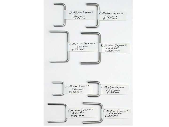
The following calculations are for stainless steel constructs. Two motion segments Screw length Rod length H 1 (mm) H 2 (mm) 4. 76 mm 6. 35 mm Thoracic 25 50 222 N/mm 433 N/mm Lumbar 45 70 53 N/mm 144 N/mm Anatomical Level Rod Diameter One motion segment Anatomical Level Screw length Rod Diameter H 1 (mm) H 2 (mm) 4. 76 mm 6. 35 mm Thoracic 25 25 344 N/mm 555 N/mm Lumbar 45 35 87 N/mm 154 N/mm
Discussion Limitations Canines (Healthy) Negative: Quadripedal Positive: Biological system that responds to Wolff’s Law Survival only from 6 months to one year Limited though significant (p=0. 002)implant stiffness differences: ~150 N/mm vs. ~300 N/mm Flexion-compression and compression mechanical testing Negative: Blind to other loading modes Positive: Antigravity axial load resistance critical
Conclusions Growing Spines Pedicle screw, stiff connection constructs restrict anterior column growth Mature spines Implants or sham surgery limit flexion compression buckling Without fusion Implant connections loosen unless properly tightened, then Implant – bone interface loosens Unbypassed bone, mild/significant bone mineral loss (3 and 6 months)
Mature Spines (with Fusion) All arthrodeses healed to fusion Less Stiff vs. More Stiff Constructs Fusion Increased stiffness Better stiffness maturation Bypassed bone Initially less bone mineral loss Later equalizing and stabilizing at ~ 90% baseline Bypassed disks Similar biphasic response Not significant increase then significant decrease Unbypassed disks Similar, significant progressive stiffness loss
Some Possible Clinical Correlations Limits need for concurrent anterior surgery Questions long term viability of instrumentation without fusion after maturity Temporary increase (? ) in risk of adjacent fracture may be due to posterior load shunting posterior from “sham” scar Suggests importance of an extensive arthrodesis May explain some post instrumentation removal fractures Remaining Questions Is residual implant stiffness detrimental to unbypassed disks Other Thank You