Urinary System Anatomy Paired kidneys Paired ureters Single


-glucose, amino acids,")


 • Secreted by the pituitary gland when sodium has been absorbed")
 • Has the opposite effect to ADH • When excess")
- Slides: 37
Urinary System
Anatomy: • Paired kidneys • Paired ureters • Single bladder • Single urethra
Function: • Rids the body of nitrogenous waste • Regulates blood chemistry
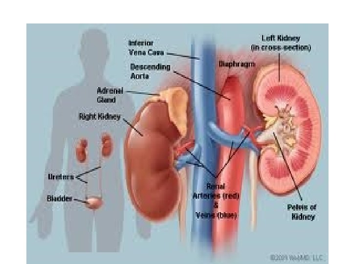
Kidneys • Location: • Superior lumbar • Against dorsal wall • Supported by fat cushions
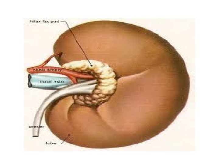
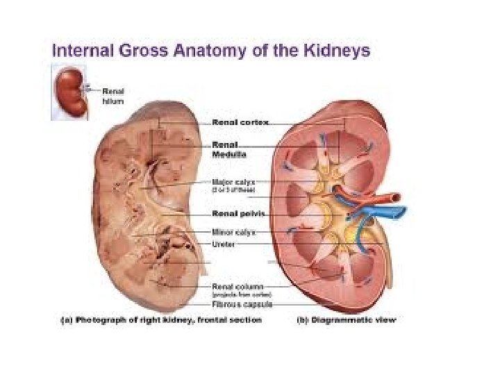
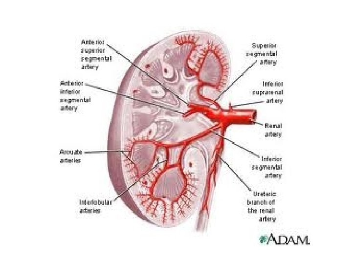
Outside Anatomy • Renal Capsule- fibrous, surrounds and protects the kidney • Medial Hilus- area where renal arteries, renal vein and ureter attach
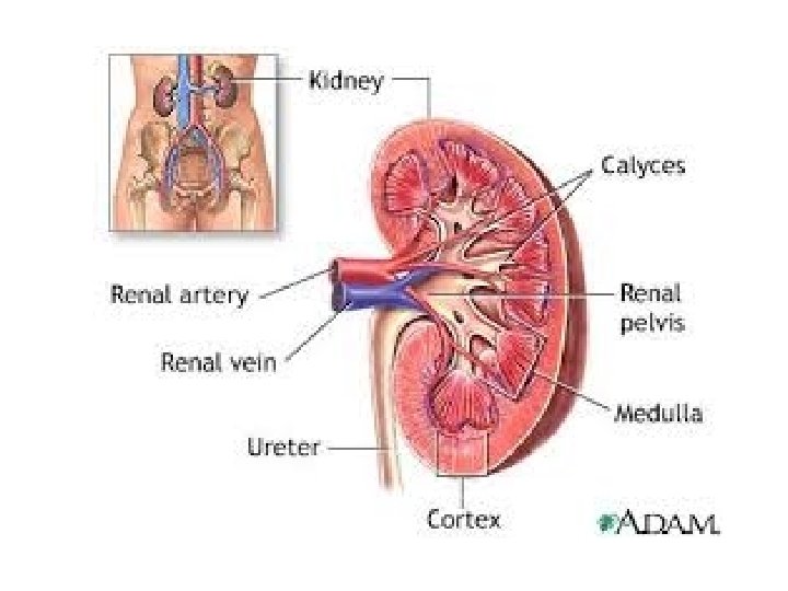
Inside anatomy • Renal Pelvis- flat basin like cavity – continuous with the ureter • Outer Cortex- lighter color outside margins, under the capsule
• Renal medulla- deeper, inner portion, darker color • Renal Pyramids- cone shaped, base faces cortex, apex (tip) faces inner most area • Calyces (calyx)- cup like extensions of the pelvis enclose the tip of the pyramids, collect urine that drains from the pyramids
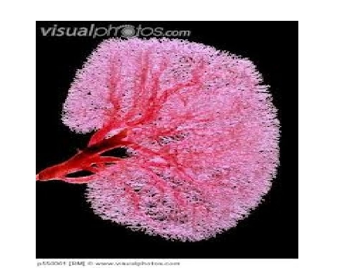
Blood Supply • The kidneys have a rich blood supply • ¼ of blood goes through per minute • Renal Arteries Interlobar arteries arcuate arteries interlobular arteries • Renal Veins interlobular veins arcuate veins interlobar veins
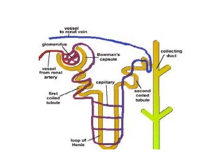
Nephron • Functional and structural unit of the kidney • 1 million nephrons per kidney • In charge of filtering, reabsorption, and excretion
Parts of the Nephron: • Glomerulus- a knot of capillaries • Renal Tubule- Bowman’s Capsule – cup shaped, completely encloses the glomerulus
The remaining tubule is about 5 cm long with 3 regions: -Proximal Convoluted Tubule (desending) -Loop of Henle -Distal Convoluted tubule (ascending)
Most of the nephron is located in the cortex, although the Loop of Henle dips down into the medulla
• The collecting tubules receive urine from many nephrons • Deliver urine to the calyces • Renal Pelvis Ureter Bladder Urethra out
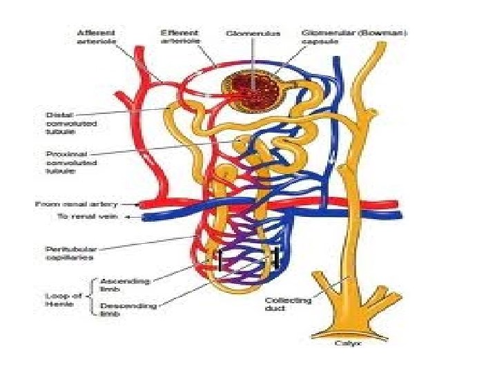
Blood Supply • The glomerulus is fed and drained by 2 arterioles • One feeds in = afferent arteriole • One drains out = efferent arteriole • The efferent continues to wrap around and cling to the tubules = peritubular bed
• The peritubular capillaries cling to the tubules and drains into the interlobular vein
• Blood pressure is high in the glomerulus- it is an arteriole going into capillaries • It forces fluid and solutes out of the blood • That drainage is captured in Bowman’s Capsule
-Red blood cells are too large to be squeezed out -Plasma is 92% water, 8% proteins, salts, sugar, oxygen, CO 2 , gases
Urine Formation • 4 Steps: Step 1: pressure filtrationoccurs in the glomelular capsule where things are squeezed out: glucose, amino acids, salts, plasma, and nitrogenous waste
What are nitrogenous wastes? • Urea- from amino acid metabolism • Creatinine- from muscle metabolism • Ammonia- from bacteria • Uric Acid- from breakdown of nucleotides
Other waste products: • Ketones from fat breakdown • Urobilinogen from breakdown of bilirubin (makes urine yellow) • Pressure forces water and waste out; large proteins and cells stay
Step 2 • Selective Re-absorption- takes some of the molecules (important ones)-glucose, amino acids, salts, water, and returns them to the blood by diffusion or active transport
Step 3 • Tubular Secretion- actively removes some waste that could not get squeezed out and moves it from the distal convoluted tubule (drugs)
Step 4 • Reabsorption of water- occurs all along, but finally in the nephron’s collecting tubes
Hemodialysis • Life saving technology, used when nephrons die • Based solely on diffusion
• Kidneys contribute to regulation of blood pressure by regulating blood volume
• Where salt goes water follows • Based on osmosis
• If salt is transported into blood: • Water follows • Blood volume increases • Blood pressure increases
Antidiuretic Hormone (ADH) • Secreted by the pituitary gland when sodium has been absorbed but not enough water has followed • Causes water to be absorbed • Less volume of urine is produced
Atrial Natriuretic Hormone (ANH) • Has the opposite effect to ADH • When excess volume/pressure goes through the heart, the stretch receptors in the heart detect the excess volume • The cells release ANH • Keeps sodium (and water) from being reabsorbed • Urine volume increases gets excreted