Uniting Nuclear Medicine with Ultrasound HHHoldorf What is


 was the first physician")


, especially")


 to")


- Slides: 27
Uniting Nuclear Medicine with Ultrasound HHHoldorf
What is Nuclear Medicine? Imaging tests that help in the diagnosis and treatment of medical conditions that are within the body Radioactive isotopes (tracers) are put into the body and the images are obtained after a certain amount of time
Nuclear Medicine Birthplace of nuclear medicine : Between the discovery of artificial radioactivity in 1934 and the production of radionuclide's by Oak Ridge National Laboratory for medicine related use in 1946 Origins of this medical idea date back to the mid 1920’s in Freiburg, Germany when George de Hevesy made experiments with radionuclide's administered in rats which displayed metabolic pathways of the substances which established the tracer principle
Pioneers From the Laboratory at the University of California at Berkeley Ernest Lawrence – invented a particle accelerator called the cyclotron in 1930.
Pioneers Continued…. John Lawrence - (the brother of Ernest Lawrence) was the first physician to treat a patient with a radioactive isotope of phosphate after the founding of the Donner Laboratory in 1936.
The Creation of Body Organ Imaging Benedict Cassen – Invented the radioisotope scanner used for thyroid imaging in 1951 at UCLA.
The Beginning of Medical Imaging Hal Anger – invented the Anger Camera, which uses gamma rays to detect tumors in 1953. During the 1970’s, the Anger Camera developed into PET and SPECT technology.
Official Recognition of Nuclear Medicine 1954 – The Society of Nuclear Medicine was created in Spokane, Washington. 1971 – Nuclear medicine was accepted as a medical field by the American Medical Association. 1972 – The American Board of Nuclear medicine was formed.
Nuclear Medicine’s Continued Development Gordon Brownell – created a detector device to utilize positron-electron destruction for organ imaging at MIT in 1953. David E. Kuhl and Colleagues in 1959 – invented the Mark 2 scanner, which is the forerunner of SPECT and CT scanners.
Nuclear Medicine - Improves 1973 - Elliot Lebowitz, Harold Atkins and colleagues created a more effective method for making thallium-201 for nuclear stress testing.
Improvement Continues 1974 - Michael E Phelps, Edward Hoffman and Michel M. Ter-Pogossian from Washington University constructed the Pett 3, which computes three dimensional images.
Nuclear Medicine continues to evolve 1984 - Michael J. Welch and John Katzenellenbogen from Washington University developed the PET radiotracer that images estrogen receptors in breast tumors.
Examples of More Recent Development 1990 – Steve Lamberts and Eric Krenning used somatostatin receptors binding radiotracers to Image endocrine tumors. 2001 - In the United States, a reported 16. 9 million nuclear medicine procedures were preformed. 2008 – Siemens Healthcare created the hybrid PET/MRI system. Currently, Nero Q version 36 software was approved by the FDA for the analysis amyloid uptake levels on regions of the brain.
How is the procedure performed? Radioactive material is usually given intravenously, however it’s sometimes given orally or inhaled as a gas This targets the specific body organs you are testing These radioactive compounds collect in that organ and gives off radiation and produces gamma rays The gamma camera detects these rays and the computer will produce the images of the organs you are looking at In many instances nuclear medicine images are superimposed with CT or MRI’s to produce specific views known as coregistration or fusion (the combing of the 2 different imaging techniques)
Procedure When it’s time to take the pictures, the camera or scanner rotates around the patient The patient will be asked to change positions between certain images while the camera is taking the pictures the patient must stay still
Experience during and after the procedure Most nuclear medicine procedures are painless and patients do not usually experience side affects However if injected patients may feel a cold sensation up the arm; when swallowed or inhaled it has little or no taste
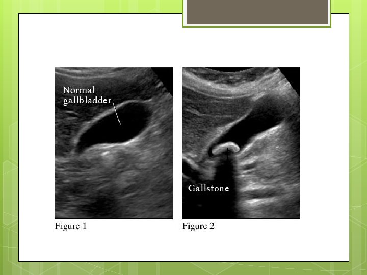
Common uses for Nuclear Medicine Heart Coronary artery disease, damage after a heart attack Lungs Respiratory and blood flow problems, detect lung transplant rejection Brain Abnormalities like seizures and memory loss Renal Detect urinary tract obstruction, evaluate kidneys for infection Cancer Stage and spread of cancer ( Thyroid Cancer) Gallbladder Gallstones, sludge
Examples of Radioisotopes used …. . Bismuth-213 -Used for targeted alpha therapy (TAT), especially cancers Iodine-131 - Widely used in treating thyroid cancer and in imaging the thyroid; also in diagnosis of abnormal liver function, renal (kidney) blood flow and urinary tract obstruction. Iron-59 - Used in studies of iron metabolism in the spleen
Case Study A 32 year old women has been complaining of twelve months history of recurrent abdominal pain. She stated the pain usually occurs at night and is located in the right upper quadrant. The pain she is experiencing is moderate to severe intensity and comes and goes for about an hour. The pain comes about once every 1 -2 months. There is no associated vomiting or diarrhea and the patient is well between attacks. The findings in the upper abdominal ultrasound is normal on two occasions with no evidence of gallstones. A hepatobillary scan was ordered. ( exam to determine the function of your gallbladder and sphincter of oddi dysfunction)
Images from Case Study – First Scan Figure 1: These are the images from the first part of the scan. The patient is injected with a radiolabelled bilirubin analogue with imaging performed over the upper abdomen. In normal fasting patients, there is uptake into the liver followed by excretion of tracer into the hepatic tracts, common bile duct, duodenum and gallbladder. By 60 minutes, maximal gallbladder filling is seen.
These are the images from the second part of the scan. At sixty minutes, an intravenous infusion of cholecystokinin (CCK) is administered with a further 30 minutes of imaging taking place. A Gallbladder Ejection fraction is then calculated. In normal patients, there is prompt gallbladder constriction with an ejection fraction of at least 35%. - Ejection Fraction identifies the % of bile that leaves the gallbladder with each contraction.
Scan findings The scan showed that there is a poor constriction of the gallbladder to CCK aggravation Normally gallbladders have an ejection fraction greater than 35% in response to CCK
Diagnosis The clinical diagnosis of chronic Cholecystitis is relatively direct with the patient complaining of symptoms of biliary colic with the ultrasound showing gallstones. Most of the time it is cured from a cholecystectomy It is also very common for patients that experience similar symptoms without gallstones( like this case study) with pain being caused by disturbed gallbladder function
The Role of Cholecystokinin The normal response to eating is for cholecystokinin (CCK) to be secreted from the stomach with the CCK causing dilation of the Sphincter of Oddi and gallbladder constriction. In these types of patients there is a paradoxical response to the CCK with pain resulting from a spasm from the gallbladder, Sphincter of Oddi or both Poor constriction of the gallbladder in response to CCK provocation with abnormal gallbladder ejection fraction
Compare and Contrast from the examination of the Gallbladder Ultrasound identifies if the gallbladder is inflamed If the gallbladder has sludge Measurements Nuclear Medicine Determines if the system being investigated is functioning or not Shows picture not the anatomy Shows bile leakage
Conclusion Patients with symptoms of biliary colic with no gallstones, an abnormal gallbladder ejection fraction in response to the CCK is a good indicator of gallbladder dysfunction and is advised that they will benefit from a cholecystectomy