The mammary gland nourishes the neonate Exocrine gland


 Glandular; secreting tissue = Parenchyma")


- Slides: 31
The mammary gland nourishes the neonate Exocrine gland; common to all mammals Function: nourish the neonate Food source: fat, protein, sugar (CHO), vitamins, minerals, water Protection: immunoglobulins (first Ab protection; absorbed via intestinal tract
The mammary gland is part the reproductive system The mammary gland is loosely considered part of the reproductive system: Serves a “reproductive function”; nourishment of the neonate = survival of species. Relies on same endocrine (hormonal) support for development and function. Example: gonadal steroids, prolactin, etc.
Endocrine Glands Affect Mammary Function
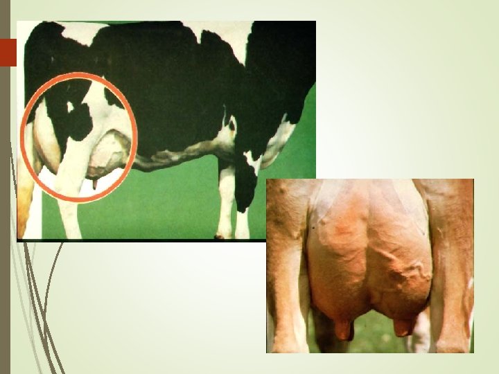
Mammary Gland Structure Udder consists of four separate glands A teat hangs from each quarter Bottom of teat closed by sphincter muscle known as streak canal Can have extra nonfunctional teats Called supernumerary teats Removed when calf is young Conformation of teats Vary in shape from cylindrical to conical Rear teats are usually shorter Each teat has one streak canal Teats should be moderately sized and located centrally on each quarter Sphincter in each teat should be tight enough to prevent leakage Teats are hairless
Mammary Gland Structure Support system = Stroma (connective tissue) Glandular; secreting tissue = Parenchyma Alveoli- secreting epithelial cells Duct system- lined by epithelial cells Lobules & lobes- clusters of alveolar tissue supported by connective tissue
Separate Mammary Glands. Quarters 60% 40%
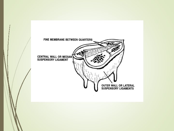
Mammary Gland Structure/Suspension Intermammary groove separates left and right halves of the udder Udder can weigh anywhere from 7 to 165 pounds May support up to 80 pounds of milk Rear quarters secrete 60% of the milk Udder continues to grow in size until cow is 6 years of age Well attached udder fits snugly against the abdominal wall in front and on the sides Extends high between thighs in rear 3 major supporting structures Skin Median suspensory ligament Lateral suspensory ligament
Mammary Gland Suspension Skin Minor role in support Median suspensory ligament Separates right and left halves of udder Connects udder to abdominal wall Lamellae Elastic tissue which responds to weight of milk in udder Lateral suspensory ligament Inflexible Surround the outer wall of udder Attached to prepubic and subpubic tendons Intermammary groove formed where lateral suspensory ligament and median suspensory ligament meets
Fig 29 -3. An illustrated view of the ligaments that permit udder suspension (Courtesy of Iowa State University)
Mammary Gland Support Medial suspensory ligament
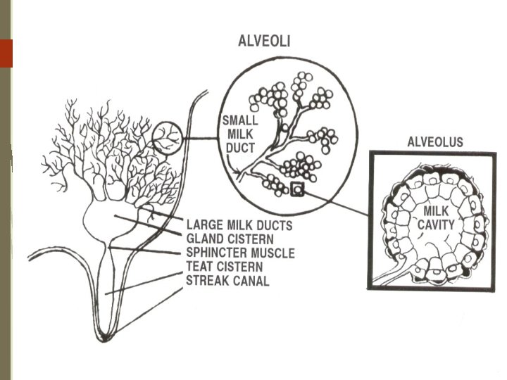
Internal Anatomy Streak canal Functions to keep milk in udder and bacteria out of udder Teat cistern Duct in teat with capacity of 30 -45 milliliters Separated from streak canal by folds of tissue called Furstenberg’s rosettes Gland cistern Separated from teat cistern by the cricoid fold Holds up to 400 milliliters of milk Collecting area for the mammary ducts From this branches the mammary ducts
Fig 29 -4. A dissected mammary gland showing the gland cistern, teat cistern and streak canal (Courtesy of Mark Kirkpatrick)
Alveoli and Duct System Alveoli is the basic milk producing unit Small bulb-shaped structure with hollow center Lined with epithelial cells that secrete milk Each cubic inch of udder tissue contains 1 million alveoli Each alveoli surrounded by network of capillaries and myoepithelial cell Contraction of myoepithelial cell stimulates milk ejection Groups of alveoli empty into a duct forming a unit called a lobule Several lobules create a lobe Ducts of lobe empty into a galatophore, which empties into the gland cistern Ducts provide storage area for milk and a means for transporting it outside Lined by two layers of epithelium Myoepithelial cells are arranged in longitudinal pattern Shorten to increase diameter to facilitate flow of milk
Alveoli and Duct System
Alveolar Products Alveolus: basic secretory unit; lined by epithelial cells which synthesize and/or secrete: lipid - triglycerides & free fatty acids (FFA) protein - caseins lactose - disaccharide; major CHO; osmoreactive molecule (draws water) minerals & vitamins - Ca, P, K; Vits. A, B, C, D water
Alveolar Structure Alveolar components & function: epithelial cells - milk synthesis & secretion lumen - collect milk components & water myoepithelial cells - milk ejection basement membrane - selective transfer terminal duct - milk transport out of alveoli capillary system - supply milk precursors and deliver hormones
Mammary Cell Function Alveolar milk component synthesis: RER > lipid, caseins Golgi apparatus > lactose (also packages lactose, caseins, minerals, water)
Circulation One gallon of milk requires 400 gallons of blood being passed through udder Ratio may increase in low producing cows Blood enters the udder through external pudic arteries Blood exiting udder from veins at the base of udder blood can travel through two routes Via external pudic veins Via subcutaneous abdominal veins
Fig 29 -6. Blood flow to and from the mammary gland determines milk producing capability of the cow (Courtesy of Iowa State University)
Mammary Venous Circle Cranial Mammary Vein
Mammary Vessels
Lymphatic System Lymph is clear, colorless contains less protein than blood plasma contains high [ ] of lymphocytes (WBC’s) which play a role in immune defense contains few RBC’s carries glucose, salts, fat (chylomicra from intestine) dissipates heat carrier of fibrinogen (clotting protein)
Lymphatic System Movement of lymph is passive: lymph moves through vessels by: 1. muscle movement (exercise, etc. ) 2. breathing 3. heart beat 4. tissue massage
Lymphatic System Helps regulate proper fluid balance within udder and combat infection Fluid drained from tissue only travels away from udder Blood capillary pressure Contraction of muscles surrounding the lymph vessels Valves that prevent backflow of lymph Mechanical action of breathing Lymph travels from udder to the thoracic duct and empties into blood system Flow rates of lymph depend on physiological status of the cow
Lymphatic System Fluid enters the lymph system through open-ended vessels called lacteals
Function of the Lymphatic System
Lymphatic System- Edema: low pressure, passive system fed by a high pressure vascular system! this situation results in pooling of interstitial fluid if evacuation of lymph is impaired tissue trauma; increased mammary blood flow at parturition Example:
Alleviating Mammary Edema Preparturient milking may be helpful store colostrum from healthy cows to feed calves Frequent milkout to reduce mammary pressure Diuretics, corticoids to reduce swelling • Mammary massage, icing –work fluid towards supramammary lymph nodes • Reduce salt intake • Don’t feed too much, too early before calving