Lactation the production of milk by the mammary

Lactation, the production of milk by the mammary gland, is a distinguishing characteristic of mammals, whose young at first feed solely on milk from their mothers. Mammary gland growth and development occur rapidly as the female reaches puberty. The ovarian hormones have a large effect on development. Estrogen is primarily responsible for duct and cistern growth, whereas progesterone stimulates growth of the alveoli. Growth hormone, adrenal corticoids, and prolactin are primarily responsible for the initiation of lactation. Lactation is maintained primarily through hormonal influence. Prolactin, thyroid hormones, adrenal hormones, and growth hormone are all important in the maintenance of lactation.

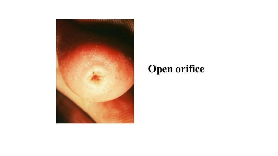
The Cow’s Udder External appearance; the four mammary glands of the dairy cow are grouped together in a structure called the udder. The udder should be reasonably large, possess a level floor, and be neatly attached both front and rear. The teats should be squarely placed, hang perpendicularly, and be of good size (teat length; mean 6 -7 cm). • The mammary gland serves two functions: 1) it provides nutrition to calf, and 2) it is a source of passive immunity to the calf. • The mammary gland is an exocrine gland located in the groin area. General structure The udder is composed of two principal types of tissue, secreting and connective. A limited amount of connective tissue is necessary for support of the glands. The desirable udder is one which contains a minimum amount of connective and fatty tissue and a maximum amount of secretory tissue. Internal structure The udder is divided into right and left halves by a heavy membrane which extends lengthwise of the body and helps to support the udder by its attachment to the abdominal wall. Front and rear quarters are separated by a very thin connective tissue membrane. The milk from each quarter can be removed only from the teat of that quarter. At the end of the teat is the opening through which the milk is removed. This opening is called the streak canal and is surrounded by a sphincter or circular muscle. This muscle normally keeps the stored milk from running out.

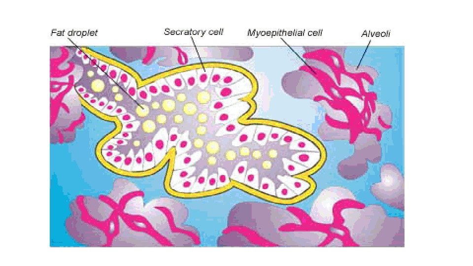
The importance of the suspensory attachments for the udder is indicated by the weight they have to carry. The secretory tissue of the mammary gland is composed of millions of grapelike structures called alveoli.

• The teat widens out into the cistern of the teat, which opens into the larger cistern of the gland. Leading into the cistern of the gland are 8 to 12 large milk ducts which branch into all parts of the gland carry the milk secreted down to the cistern. Each gland is divided into lobes, separated by connective tissue membranes which appear as white glistening bands between the orange-colored glandular tissues. The lobes are further divided into lobules, each made up of a large number of hollow spherical structures called alveoli. The cells lining the alveoli, called epithelial cells, first manufacture the milk from the constituents of the blood, then discharge the milk into a hollow cavity called the lumen. • It is estimated that 300 to 400 volumes of blood pass through the udder for each one volume of milk secreted. • On the average each of the front quarters produces about 20 per cent of the milk and each of the rear quarters, about 30 per cent. There is almost no difference in production between the right and left halves of the udder. • Secreting tissue is soft, pliable, and spongy to the touch, with no firm, fatty tissue or edema in the udder. A ‘’meaty’’ udder is a relatively poor producer.

Circulatory System; The arterial blood from the heart reaches the base of each half of the udder through an external pubic artery which comes through the inguinal canal. Each of these branches into two mammary arteries, the cranial, which supplies the fore-quarter, and the caudal the rear. These arteries branch to supply blood to the mammary glands. Blood from the capillaries collects in two large veins, one at the base of each half of the udder, which are joined together by a smaller vein in the rear. There are three routes by which blood can return to the heart. One is the so-called ‘’milk vein’’ or subcutaneous abdominal vein. The second is the external pubic vein which parallels the external pubic artery. The third route is the perineal vein which carries the blood through the pelvic arch. • The lymph vessels of the udder carry lymph upward and toward the rear of each half where it passes through the supramammary lymph glands. When the udder is congested at calving time, it is simply the accumulation of large quantities of lymph. • Milk let-down; as soon as one begins the milking process, either by hand or by machine, a small quantity of milk can be removed from the cistern and the larger ducts. The stimulus of milking causes the posterior lobe of the pituitary to discharge a hormone called ‘’oxytocin’’ into the blood stream. In about one minute it reaches the udder and causes a contraction of the smooth muscles in the udder forcing the milk from the lumina of the alveoli down the duct system into the cistern (great inflow of milk into the cistern). While the contractions continue, milk can be removed very rapidly from the udder. After about 8 minutes the gradual disappearance of the hormone permits the muscles to relax and, if milking has not been completed, the last milk cannot be removed. When cows are disturbed by barking dogs, shouting, or ill-treatment, they frequently will not ’’let down’’ their milk. This situation is believed to be due to the discharge of adrenalin from the adrenal glands when animals are excited. Adrenalin causes a relaxation of the smooth muscles of the udder and prevents the normal action of oxytocin upon the muscles. Later, when the adrenalin has been eliminated from the blood, the cow again can ’’let down’’ her milk upon the stimulus of milking.

Defective udders may be pendulous or pear-shaped, cut up between the quarters or halves, or may lack one or more quarters. The teats may be short or hard to milk.

ü Milk appears to be made by a combination of filtration of certain constituents from the blood stream, cell degeneration, and the synthesis of other constituents by means of true cell metabolism. ü Butterfat appears to be synthesized in the gland, apparently from neutral blood fat. ü Milk proteins appear to result partly from synthesis and partly from filtration. ü Lactose or milk sugar is not found in the blood. It appears to be synthesized from glycogen in the secreting cells of the gland. ü Vitamins, certain mineral salts, urea, and various flavoring compounds from the feed apparently pass from the blood to the milk without change. They do not seem to be essential ingredients in the synthesis of milk. Milk synthesis o After the removal of milk at milking time, the cycles of milk secretion and discharge are rapid, filling the lumina of the alveoli, the ducts and storage spaces of the duct system, and the gland cistern. With the continued secretion and discharge of milk there is a gradual rise in udder pressure. The cycles of secretion and discharge begin to slow down. o As the interval between milkings lengthens, the butterfat content of the total yield of milk is reduced. o The butterfat percentage of milk increases with decreasing production.

Milk is nature’s perfect food for infants. It is also a most important food for adults and is the most widely used of all foods. A further increase in the consumption of milk is to be desired, and the following facts must receive consideration: üMilk is very perishable üMilk that happens to contain disease organisms spreads the disease rapidly üMilk readily takes on bad flavors üSince milk is white, dirt is easily seen in it Milk of high sanitary quality, therefore, must be: v. Low in bacterial count v. Free from disease germs v. Of good flavor v. Free from dirt

The conditions that have a possible relation to the points noted: • Kind of barn • Kind of milk room or house • Health of cow • Health of attendants • Feeding route • Preparation of cow for milking • Preparation of milker • Kind of milk pail • Milking machine • Promptness in removing milk from barn • Straining or clarifying • Cooling methods • Method of cleaning utensils • Fly control Of these fourteen factors, all may effect the bacterial count, but the ones that usually have the greatest effect are health of cow, kind of pail, use of milking machine, cooling milk, and cleaning utensils, with particular emphasis on the last two.

• The milk room should have a concrete floor, smooth walls and ceiling, and should be kept strictly clean. • It is obvious that the all-important step in fly control is to practice sanitation in the barn and milk house and around the premises. • Good herd management also demands that a herd be kept free from disease. • Because the bacteria that cause most of the diseases spread by milk come from human beings, it is easy to see the great importance of making sure that everybody who milks the cows or handles the milk or utensils is in good health. • To make sure that the milk is free from feed flavors, it is desirable that nothing be fed just before or during milking. • Before milking, the udder and flanks of the cow should be throughly brushed; and just before milking each cow, the udder should be wiped with a cloth or paper towel moistened in warm water. • The milker should wear clean, and should wash his hands as often as they become soiled. • A small-mouth pail should be used. • The milking machine is used now almost universally, it affects the sanitary quality of milk. • Like the milking machine, unclean and unsterile utensils are the source of most of the bacteria that get into milk. • The milk should be removed from the barn after each cow is milked. • The fact that milk is to be strained is no excuse for letting dirt get into it. • The necessity of cooling milk to prevent bacterial growth.

Milking - Sucking of calf, - Hand-milking, - Milking by machine. Milking Systems Milking Parlors Hygiene and Udder Health Milking and Application High Quality Milk Production As the cow is prepared for milking by washing and drying the udder and teats, the nerves in the teats send a message to the pituitary gland located at the base of the brain. The milk letdown hormone, oxytocin, is released into the bloodstream -reaching the udder in about 1 minute. This hormone causes the muscle fibers surrounding the alveoli to contract and force the milk out into the ducts and udder cistern, making milking possible. Alveoli are the very tiny milk-secreting cells in the udder. The effect of oxytocin is diminished in about 7 minutes, so rapid milking is necessary if one is to obtain a complete emptying of the udder. When cows are mistreated or unduly disturbed or excited just before or during milking, adrenaline may be released into the bloodstream by the adrenal glands. This hormone counteracts the effects of oxytocin and prevents or stops milk letdown. This reaction cause incomplete milking and lead to udder problems and reduced milk yield.

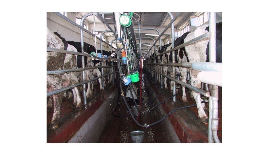
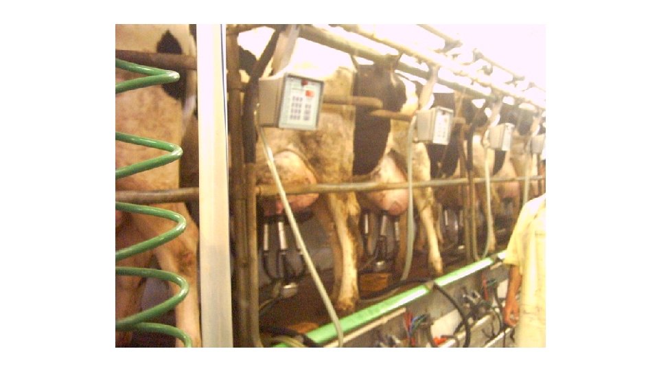
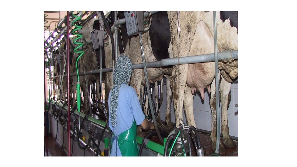
Milking Facilities Loose housing of dairy cows requires the use of a milking parlor or a conventional stall barn for housing the animals during the milking process. Milking parlor type, size and arrangement may vary widely depending on the several factors: Ø Size of herd Ø Ø Ø Number of persons to do milking Resources that can be invested Local sanitary regulations In general, milking facilities will include the following: ü A milking room, ü ü ü A milk room with space for a bulk tank, Room for utensil washing and storage, vacuum pump, and heating facilities (sometimes combined with milk room) Concentrate storage facilities. The milking room is generally arranged so that the cows will stand on a raised platform in order that they can be milked without the operator stooping. Milk generally is carried automatically through a sanitary pipeline to a bulk tank. The milking machine operates on a two-vacuum system: one vacuum is located inside the rubber liner and the other outside the rubber liner of the teat cup. There is a constant vacuum on the teat to remove the milk and keep the unit on the teat. The intermittent vacuum in the pulsation chamber causes the rubber liner to collapse around the teat. This assists blood and lymph to flow out of the distal teat into the upper part of the teat and udder. The milking parlor and its equipment must be kept sanitary.

Numerous nongenetic factors influence the milk and butterfat yield of cows; • • Age of the cow Length of the calving interval Length of the dry period Length of milking intervals Season of calving Nutrition and management etc. Causes of variation in the composition of milk The causes of variation in the concentration of milk constituents; ü Temporary variations from one milking to the next or from day to day ü Variations due to changes in the feeding, environmental temperature, or the health of the cow, ü Variations which are dependent on the stage of lactation or on the age of the cow, and ü Hereditary differences between breeds and between animals within the same breed.

The persistency of lactation Daily milk production typically increases during the first few weeks of lactation, peaks at approximately 4 -6 weeks, and then decreases over the next several weeks of the lactation period. Persistency of lactation measures how milk production is maintained over time. The lactation yield is determined to a large extent by the maximum yield and to a lesser extent by the persistency. Persistency is determined by calculating milk production of the current month as a percentage of the last month’s production. The persistency, or shape of the lactation curve, is influenced by various nongenetic factors, e. g. , the age of the cow, the length of the current calving interval, the length of the preceding dry period, the condition of the cow at time of calving, the level of nutrition during lactation, etc.

Decreased milk production during the lactation period is due primarily to a decreased number of active alveoli and less secretory tissue (epithelial cells) in the alveoli. These and other changes are associated with hormonal changes. When milking or suckling is stopped, the alveoli are distended and the capillaries are filled with blood. After a few days the secretory tissue becomes involuted (reduced in size and activity) and the lobes of the mammary gland consist primarily of ducts and connective tissue. The cow then becomes dry as milk secretion is not occurring. After the dry period (approximately 2 months) and when parturition approaches, hormones and other influences prepare the mammary gland to resume its secretion and production of milk.

Kow-Kant-Kick The heifer or cow that continues to kick or jump up and down during the milking process needs the restraint afforded by the Kow-Kant-Kick, Cattle Controller, rope, or tail hold.

Hand Milking Machine Milking

Portable Milking Machine

on Milking

Milk Cooling Tank

Variation of Fat, Protein and Lactose on Lactation

Udder Diseases • Oedema • Mastitis

Edema causes abnormal congestion at calving time and persists after cows have been fresh for a long time.

Mastitis • inflammation of the mammary gland udder tissue, • big problem in dairy cattle, • increase somatic cell count, • The disease costs to 200 $ per head, annually Identification Control Programmes

The only really satisfactory way to control mastitis is to prevent it by means of good management. Some of the most important management factors are; 1. Follow good milking procedures 2. Use a strip cup at every milking 3. Prevent udder injuries 4. Provide adequate bedding 5. Prevent exposure of cows to drafts and damp beds 6. Breed and select for desirable shaped udders 7. Follow a routine testing program under the supervision of a veterinarian 8. Treat in early stages of infection 9. Isolate or eliminate potential spreaders as quickly as possible

CMT

The teats should be uniformly placed at the corners of the udder floor. They should hang straight down from the udder floor and should be of average, convenient, size and shape. This is important because the teat and the udder size and shape are often associated with good milking qualities. Normal teat
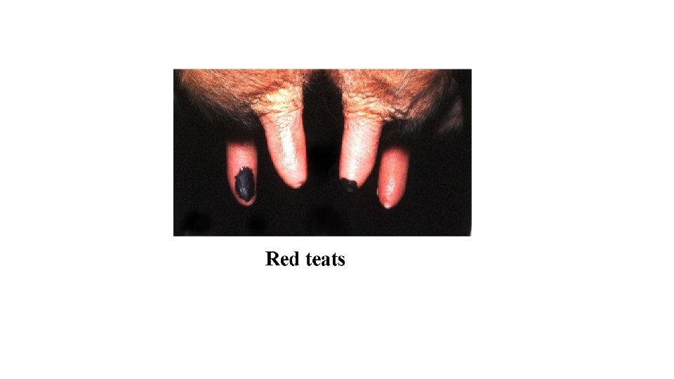
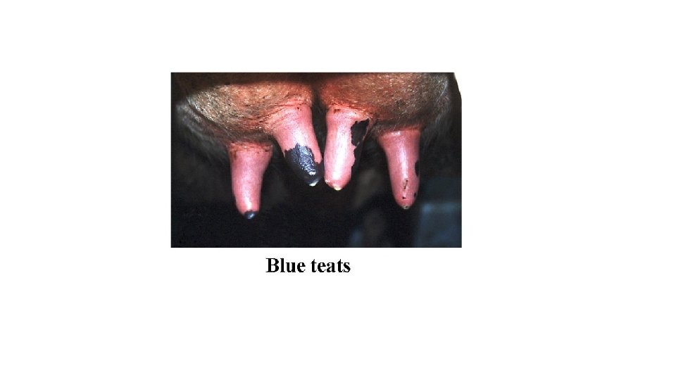

Petechia in teat

Milk yield, kg Lactation Curve Month of lactation

Factors Affecting Milk Yield Breed Age of the cow Dry period Estrous and Gestation Season of calving Live weight Feeding Climate Stage of lactation, Lactation length Parity, Milking frequency (time between milkings) Diseases, etc…

Standardization of Dairy Cattle Lactation-Production Records The lactation production of a cow results from the interplay of heredity and environment. For accuracy in selection it is important that the record reflect as precisely as predictable the cow’s genetic potential for milk or fat production. The actual records themselves may be poor indicators of breeding value, since so many environmental influences have a marked effect on a cow’s performance during a particular lactation. Some of the more important items which deserve consideration are length of the lactation period, number of times milked daily, age of the cow at freshening, length of the preceding dry period, and the season of freshening. For example: Factors for computing lactation records of over 305 days 305 -308 1. 00 309 -312 0. 99 313 -316 0. 98. . 361 -364 0. 86 365 0. 85 The same conversion factors are used for both milk and fat when a record is expressed on a 305 -day, 2 X, matureequivalent (M. E. ) basis.
- Slides: 38