MAMMARY TUMORS IN DOGS Originate in the mammary


- Slides: 30
MAMMARY TUMORS IN DOGS
Originate in the mammary gland Older female dogs and cats Being intact increases the occurrence Associated with teats and extend along the mammary chain
NINE YEAR OLD BITCH WITH MAMMARY TUMOR. Note deep emaciation in bitch
In dog thrice that of breast cancer in women Second most common tumors in dogs( after skin) Most common tumor in bitches Lifetime risk to develop mammary tumors • Intact bitches-25% • Males- 1%
Age of Spaying in bitches Risk of occurrence of mammary tumours vs. intact female Before first heat 0. 5% After just one heat cycle 8% Post second heat cycle 26%
Age of dogs affected 10 to 11 y for malignant tumours Body condition Obesity at 1 y Nutritional factors Eating red meat and high fat homemade diets
Breeds at increased risk: • Poodles • Brittany spaniel • English setter • Pointer • Fox Terrier • Boston Terrier • Cocker Spaniel • Lhasa Apso
• Approximately 50% of mammary tumours in dog – malignant Types of tumours Examples Benign Adenomas Fibroadenomas Benign mixed tumours Mesenchymal tumours Malignant Sarcoma Carcinosarcoma Inflammatory carcinoma Carcinomas
• Uniform grading system absent Criteria of classification Types Prognosis Blood vessel wall invasion With invasion Poor Without invasion Only slightly better Size Greater than 5 cm dia Increased chances of lymphnode metastasis Type Sarcoma and Carcinosarcomas better Inflammatory carcinoma Poor prognosis and will have metastasised by the time of diagnosis
Degree of nuclear differentiation Well differentiated 20% recurrence Moderately differentiated Poorly differentiated 80% recurrence
Sarcomas Osteosarcoma Fibrosarcoma Osteochondrosarcoma • Metastasis occurs to regional lymph nodes and lungs • Mixed malignant tumours have histological characteristics of mesenchymal and epithelial malignancy
Palpable mass underneath the skin of the abdomen A single mass or multiple masses The majority (greater than 65%) of mammary tumors develop in the fourth and fifth mammary glands
Benign tumors are often small, well-circumscribed, and firm Malignant tumors exhibit more aggressive behavior, such as: • rapid growth, • poorly defined borders, • fixation to skin or underlying tissue, and • inflammation or ulceration.
• An aggressive type of mammary tumor seen in dogs • Often painful with swollen mammary glands. • Swelling is often diffuse and can involve either a single mammary chain (i. e. , all glands on either the left or right side) or all mammary glands • Other signs include inappetence, weight loss, generalized weakness, and swelling of one or both hind legs
Appearance and location of tumour Thorough evaluation of all mammary glands, lymph nodes Auscultation of lungs Lameness examination FNA discouraged
Mast cell tumors Soft tissue sarcomas Epithelial inclusion cysts. Mastitis Dermatologic disease (i. e. , atopy or other allergies).
Thoracic radiographs Complete blood count Serum chemistry panel Urinalysis
Surgical excision is the treatment of choice Chest X rays taken prior to surgery Removal with wide margins Spaying recommended along with tumour excision Chemotherapy rarely used
Five pairs of mammary glands Lymphatic drainage by • Axillary • Superior inguinal • Inguinal • Sublumbar • Anterior mediastinal Lymphatic communication
Blood supply: • Perforating sternal branches of internal thoracic artery • Mammary branches of Epigastric arteries to Abdominal and inguinal glands
Extent of surgery is not associated with improved survival May have theoretical implications based on lymphatic drainage for invasive tumours
Simple Lumpectomy Mammectomy • Removal of just the tumour • Removal of a single gland, when tumour is in the centre of the gland, more than 1 cm dia, adherence to over or underlying skin or subcutis Regional Mastectomy • Removal of cranial (1 st to 3 rd) or caudal (3 rd to 5 th ) region segment of mammary chain, if consecutive glands are involved Radical Mastectomy • Removal of entire mammary chain if multiple masses preclude individual gland removal or regional mastectomy
Skin incised Blunt dissection of tumour with rim of normal tissue Malignancy revealed in histopathology Surgical margins found unclean A second more aggressive surgery indicated
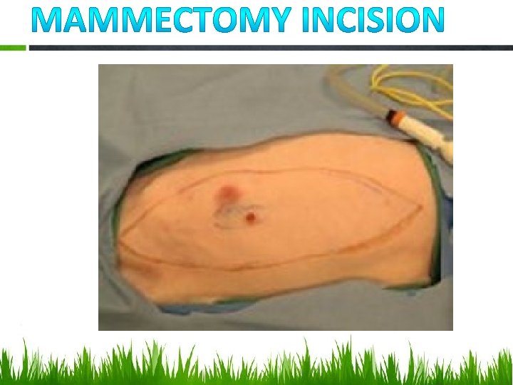
Elliptical incision around gland with 2 cm margins from the tumour Skin, subcutis and superficial layer of abdominal wall fascia removed if involved
Includes removal of the glands and their lymphatic drainage in one unit Procedure similar to mammectomy Elliptical incision being carried over the entire region concerned
Elliptical incision around the entire chain of mammary glands to be removed Caudal superficial epigastric artery and vein can be easily identified, isolated, ligated and divided. Ligature and electrocautery used to control haemorrhage Not indicated to improve the survival in dogs Tension free closure is essential to prevent dehiscence
Ovarohysterectomy performed along with tumour excision because: • To prevent pyometra • Nearly 50% of mammary tumours contain receptors for oestrogens If concurrent ovarohysterectomy is elected, done prior to tumour excision.
Advocated if one or more negative prognostic factors present Despite high frequency of mammary tumours in dogs, no protocol has been standardised Tamoxifen-use precluded in dogs due to high incidence of oestrogenic side effects Doxorubicin alone or in combination with Cyclophosphamide also used Radiation therapy not been reported widely for treating mammary gland malignancy in dogs
THANK YOU References: • Textbook of Small Animal Surgery by D. Slatter Edition 3 • Merck’s Veterinary Manual, Edition 9 • Clinical Textbook for Veterinary Technicians by Mc Currin and Bassert, Edition 6