
NEONATAL JAUNDICE Dr mirzarahimi CAUSES OF PHYSIOLOGIC HYPERBILIRUBINEMIA

NEONATAL JAUNDICE Dr. mirzarahimi

CAUSES OF PHYSIOLOGIC HYPERBILIRUBINEMIA 1 Definition: Full term infant 6 8 mg/l 00 ml by 3 days Premature infant 10 15 fig/100 ml by 5 days v. An increased bilirubin load : more than 8. 5 mg/kg/day ØIncreased RBCs volume/kg ØDecreased fetal RBCs survival 90 days ØIncreased early labeled bilirubin ØIncreased resorption of bilirubin from intestines

CAUSES OF PHYSIOLOGIC HYPERBILIRUBINEMIA 2 v Defective uptake of bilirubin from plasma ØDecreased y protein ØBinding of y and z proteins by other anions ØDecreased caloric intake v. Defective bilirubin conjugation and excretion Ølow levels of UDPGT ØInhibition of conjugation by maternal steroids
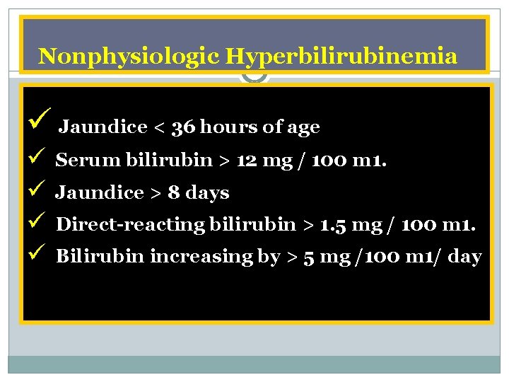
Nonphysiologic Hyperbilirubinemia ü Jaundice < 36 hours of age ü ü Serum bilirubin > 12 mg / 100 m 1. Jaundice > 8 days Direct reacting bilirubin > 1. 5 mg / 100 m 1. Bilirubin increasing by > 5 mg /100 m 1/ day

Causes For Neonatal Hyperbilirubinemia Overproduction 1 ØFetal maternal blood group incompatibility Rh, ABO, others ØHereditary spherocytosis , elliptocytosis, somatocytosis ØNonspherocytic hemolytic anemias ØG 6 PD deficiency and drug Pyruvate kinase deficiency ØOther red cell enzyme deficiencies Øa thalassemia ØB thalassemia ØVitamin K induced hemolysis

Causes For Neonatal Hyperbilirubinemia Overproduction 2 Extravascular blood : petechiae, hematomas pulmonary, cerebral, or occult hemorrhage Polycythemia: Maternal fetal or feto fetal transfusion Delayed clamping of the umbilical cord
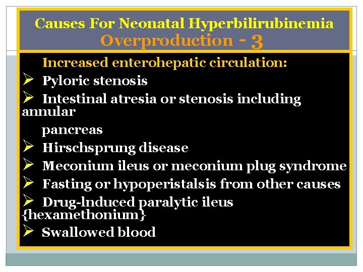
Causes For Neonatal Hyperbilirubinemia Overproduction 3 Increased enterohepatic circulation: Ø Pyloric stenosis Ø Intestinal atresia or stenosis including annular pancreas Ø Hirschsprung disease Ø Meconium ileus or meconium plug syndrome Ø Fasting or hypoperistalsis from other causes Ø Drug lnduced paralytic ileus {hexamethonium} Ø Swallowed blood

Causes For Neonatal Hyperbilirubinemia Undersecretion 1 Metabolic and endocrine conditions: Ø Familial nonhemolytic jaundice types 1 and 2 (Crigler Najjar syndrome) Ø Galactosemia Ø Hypothyroidism, Tyrosinosis, Hypermethioninemla Ø Drugs and hormones: Novobiocin, Pregnanediol Ø Certain breast milks Ø Lucey Driscoll syndrome Ø Infants of diabetic mothers Ø Prematurity Ø Hypopituitarism and anencephaly
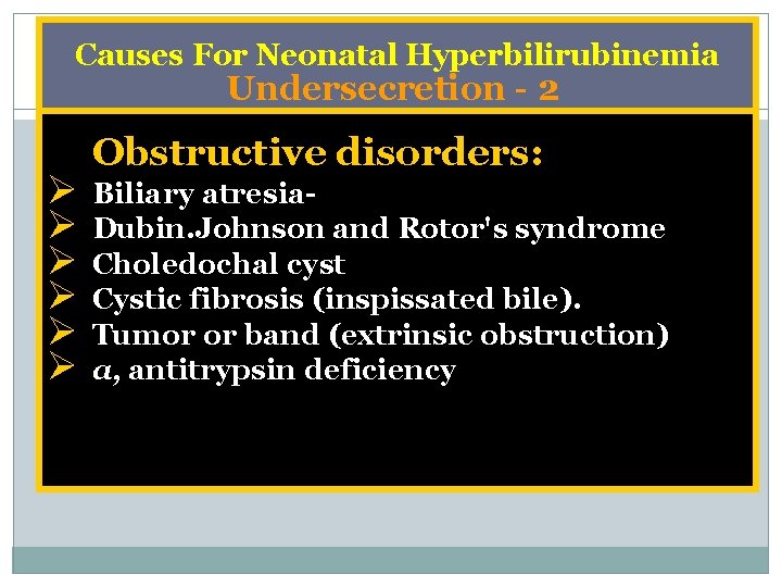
Causes For Neonatal Hyperbilirubinemia Undersecretion 2 Ø Ø Ø Obstructive disorders: Biliary atresia Dubin. Johnson and Rotor's syndrome Choledochal cyst Cystic fibrosis (inspissated bile). Tumor or band (extrinsic obstruction) a, antitrypsin deficiency
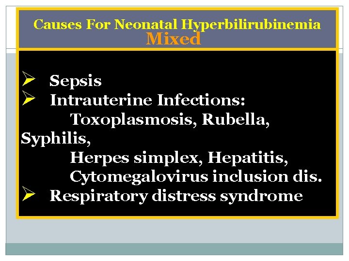
Causes For Neonatal Hyperbilirubinemia Mixed Ø Ø Sepsis Intrauterine Infections: Toxoplasmosis, Rubella, Syphilis, Herpes simplex, Hepatitis, Cytomegalovirus inclusion dis. Ø Respiratory distress syndrome
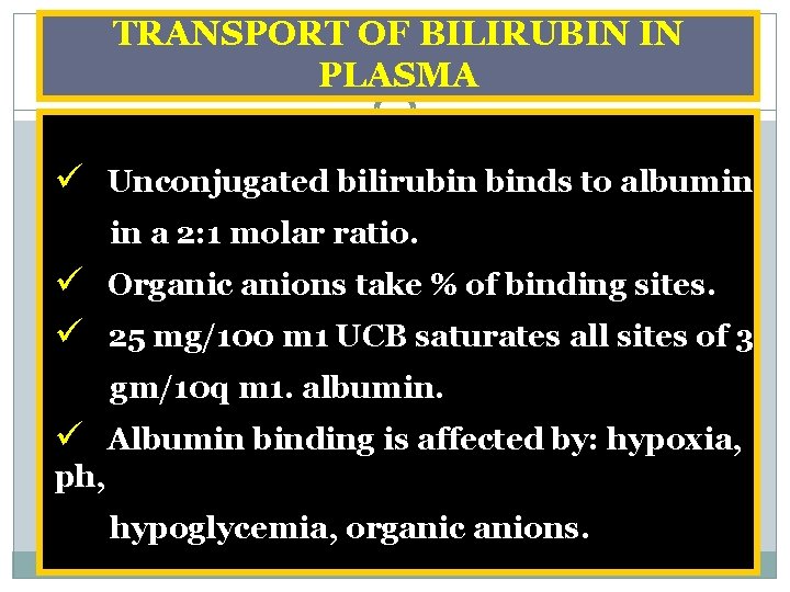
TRANSPORT OF BILIRUBIN IN PLASMA ü Unconjugated bilirubin binds to albumin in a 2: 1 molar ratio. ü Organic anions take % of binding sites. ü 25 mg/100 m 1 UCB saturates all sites of 3 gm/10 q m 1. albumin. ü Albumin binding is affected by: hypoxia, ph, hypoglycemia, organic anions.
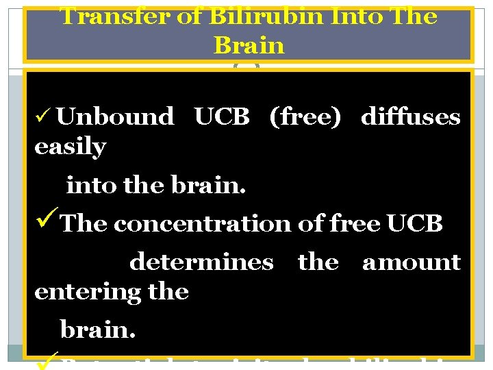
Transfer of Bilirubin Into The Brain ü Unbound UCB (free) diffuses easily into the brain. üThe concentration of free UCB determines the amount entering the brain.
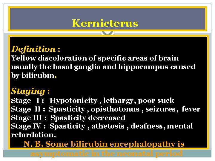
Kernicterus Definition : Yellow discoloration of specific areas of brain usually the basal ganglia and hippocampus caused by bilirubin. Staging : Stage I : Hypotonicity , lethargy, poor suck Stage II : Spasticity , opisthotonus , seizures, fever Stage III : Spasticity decreased Stage IV : Spasticity , athetosis , deafness, mental retardation. N. B. Some bilirubin encephalopathy is asymptomatic in the neonatal period
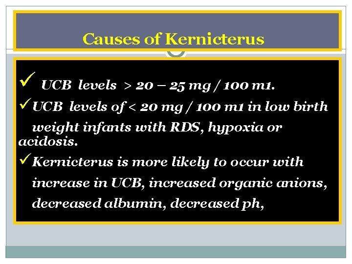
Causes of Kernicterus ü UCB levels > 20 – 25 mg / 100 m 1. üUCB levels of < 20 mg / 100 m 1 in low birth weight infants with RDS, hypoxia or acidosis. üKernicterus is more likely to occur with increase in UCB, increased organic anions, decreased albumin, decreased ph,
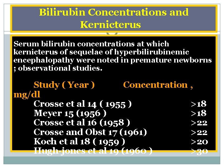
Bilirubin Concentrations and Kernicterus Serum bilirubin concentrations at which kernicterus of sequelae of hyperbilirubinemic encephalopathy were noted in premature newborns ; observational studies. Study ( Year ) Concentration , mg/dl Crosse et al 14 ( 1955 ) >18 Meyer 15 (1956 ) >18 Crosse et al 16 (1958 ) >22 Crosse and Obst 17 (1961) >22 Koch et al 18 ( 1959 ) >20 Hugh jones et al 19 (1960 ) >30
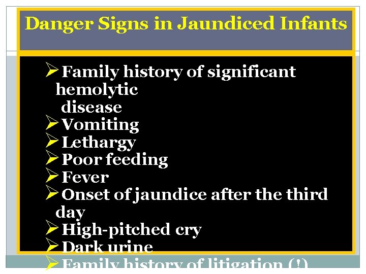
Danger Signs in Jaundiced Infants ØFamily history of significant hemolytic disease ØVomiting ØLethargy ØPoor feeding ØFever ØOnset of jaundice after the third day ØHigh pitched cry ØDark urine ØFamily history of litigation (!)
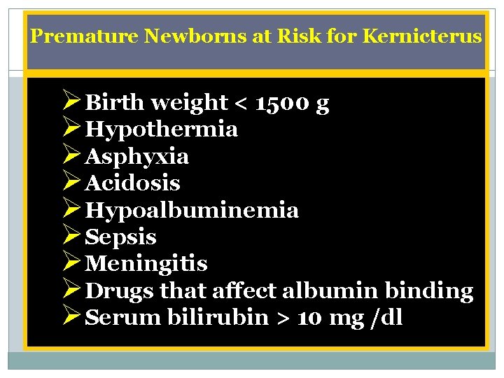
Premature Newborns at Risk for Kernicterus ØBirth weight < 1500 g ØHypothermia ØAsphyxia ØAcidosis ØHypoalbuminemia ØSepsis ØMeningitis ØDrugs that affect albumin binding ØSerum bilirubin > 10 mg /dl
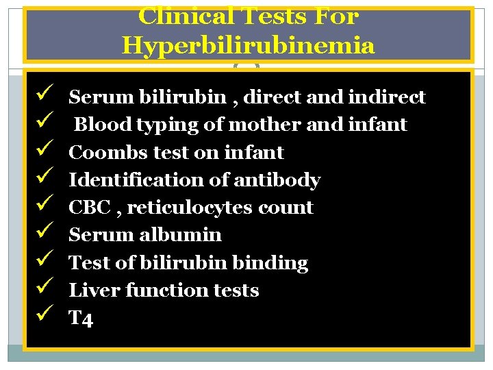
Clinical Tests For Hyperbilirubinemia ü ü ü ü ü Serum bilirubin , direct and indirect Blood typing of mother and infant Coombs test on infant Identification of antibody CBC , reticulocytes count Serum albumin Test of bilirubin binding Liver function tests T 4
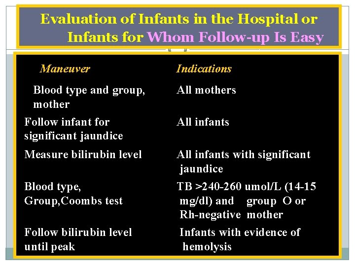
Evaluation of Infants in the Hospital or Infants for Whom Follow up Is Easy Maneuver Blood type and group, mother Indications All mothers Follow infant for significant jaundice All infants Measure bilirubin level All infants with significant jaundice Blood type, Group, Coombs test TB >240 -260 umol/L (14 -15 mg/dl) and group O or Rh-negative mother Follow bilirubin level until peak Infants with evidence of hemolysis
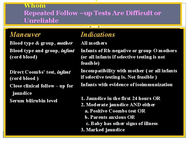
Evaluation and Follow up of Jaundiced Infants in Whom Repeated Follow –up Tests Are Difficult or Unreliable Maneuver Indications Blood type & group, mother Blood type and group, infant (cord blood) All mothers Infants of Rh-negative or group O mothers (or all infants if selective testing is not feasible) Incompatibility with mother ( or all infants if selective testing is. Not feasible ) Infants with evidence of isoimmunization Direct Coombs’ test, infant (cord blood ) Close clinical follow – up for jaundice Serum bilirubin level 1. Jaundice in the first 24 hours OR 2. Moderate jaundice AND either a. Positive Coombs test OR b. Parents anxious OR c. Baby has other signs of illness 3. Marked jaundice
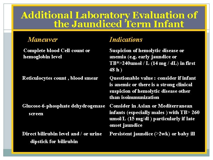
Additional Laboratory Evaluation of the Jaundiced Term Infant Maneuver Indications Complete blood Cell count or hemoglobin level Suspicion of hemolytic disease or anemia (e. g. early jaundice or TB*>240 umol / L (14 mg / d. L) in first 48 h ) Reticulocytes count , blood smear Questionable value : consider if infant is anemic or there is a strong clinical suspicion of hemolytic disease other than isoimmunization Glucose-6 -phosphate dehydrogenase Consider in Asian or Mediterranean infants (especially males ) with TB> 260 screen umol/L (15 mg/dl ) particularly if late onset jaundice Direct bilirubin level and / or urine dipstick for bilirubin Persistent jaundice (>2 wk) or baby ill
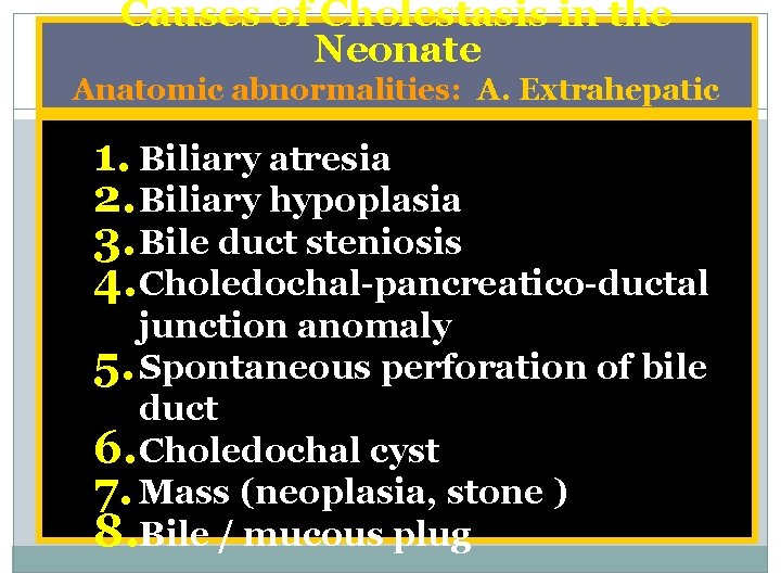
Causes of Cholestasis in the Neonate Anatomic abnormalities: A. Extrahepatic 1. Biliary atresia 2. Biliary hypoplasia 3. Bile duct steniosis 4. Choledochal pancreatico ductal junction anomaly 5. Spontaneous perforation of bile duct 6. Choledochal cyst 7. Mass (neoplasia, stone ) 8. Bile / mucous plug
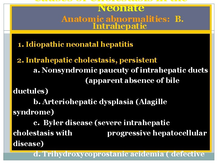
Causes of Cholestasis in the Neonate Anatomic abnormalities: B. Intrahepatic 1. Idiopathic neonatal hepatitis 2. Intrahepatic cholestasis, persistent a. Nonsyndromic paucuty of intrahepatic ducts (apparent absence of bile ductules) b. Arteriohepatic dysplasia (Alagille syndrome) c. Byler disease (severe intrahepatic cholestasis with progressive hepatocellular disease) d. Trihydroxycoprostanic acidemia ( defective
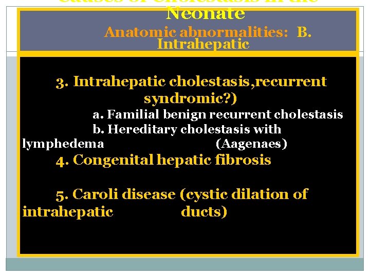
Causes of Cholestasis in the Neonate Anatomic abnormalities: B. Intrahepatic 3. Intrahepatic cholestasis, recurrent syndromic? ) a. Familial benign recurrent cholestasis b. Hereditary cholestasis with lymphedema (Aagenaes) 4. Congenital hepatic fibrosis 5. Caroli disease (cystic dilation of intrahepatic ducts)
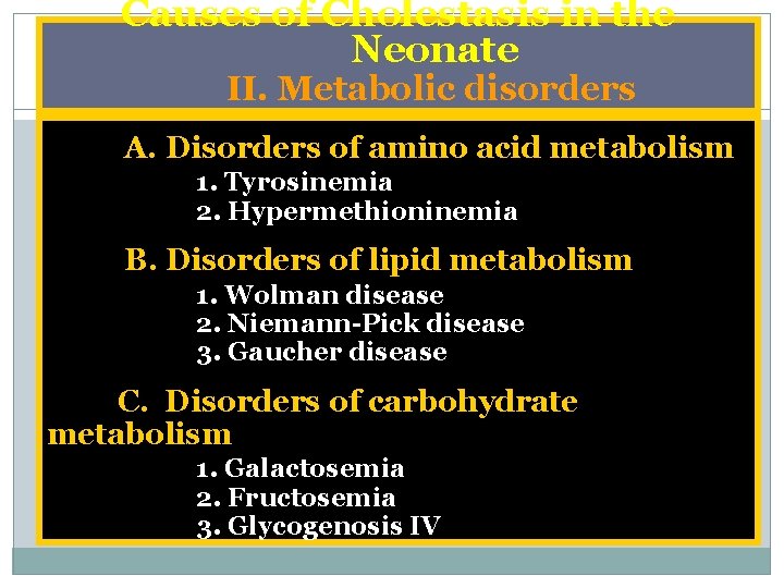
Causes of Cholestasis in the Neonate II. Metabolic disorders A. Disorders of amino acid metabolism 1. Tyrosinemia 2. Hypermethioninemia B. Disorders of lipid metabolism 1. Wolman disease 2. Niemann Pick disease 3. Gaucher disease C. Disorders of carbohydrate metabolism 1. Galactosemia 2. Fructosemia 3. Glycogenosis IV
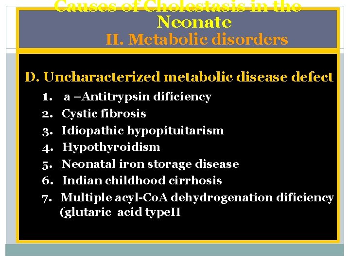
Causes of Cholestasis in the Neonate II. Metabolic disorders D. Uncharacterized metabolic disease defect 1. a –Antitrypsin dificiency 2. 3. 4. 5. 6. 7. Cystic fibrosis Idiopathic hypopituitarism Hypothyroidism Neonatal iron storage disease Indian childhood cirrhosis Multiple acyl Co. A dehydrogenation dificiency (glutaric acid type. II
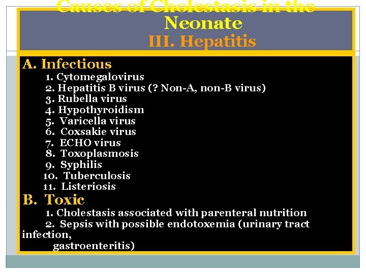
Causes of Cholestasis in the Neonate III. Hepatitis A. Infectious 1. Cytomegalovirus 2. Hepatitis B virus (? Non A, non B virus) 3. Rubella virus 4. Hypothyroidism 5. Varicella virus 6. Coxsakie virus 7. ECHO virus 8. Toxoplasmosis 9. Syphilis 10. Tuberculosis 11. Listeriosis B. Toxic 1. Cholestasis associated with parenteral nutrition 2. Sepsis with possible endotoxemia (urinary tract infection, gastroenteritis)

Causes of Cholestasis in the Neonate IV. Genetic / chromosomal A. Trisomy E B. Down syndrome C. Donahue syndrome ( Liprechaunism) V. Miscellaneous A. Histiocytosis X B. Shock C. Intestinal obstruction infection, gastroenteritis
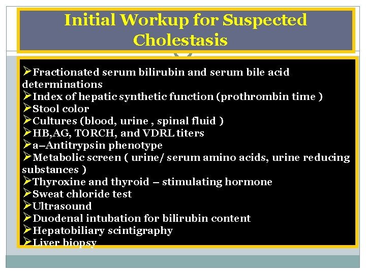
Initial Workup for Suspected Cholestasis ØFractionated serum bilirubin and serum bile acid determinations ØIndex of hepatic synthetic function (prothrombin time ) ØStool color ØCultures (blood, urine , spinal fluid ) ØHB, AG, TORCH, and VDRL titers Øa–Antitrypsin phenotype ØMetabolic screen ( urine/ serum amino acids, urine reducing substances ) ØThyroxine and thyroid – stimulating hormone ØSweat chloride test ØUltrasound ØDuodenal intubation for bilirubin content ØHepatobiliary scintigraphy ØLiver biopsy
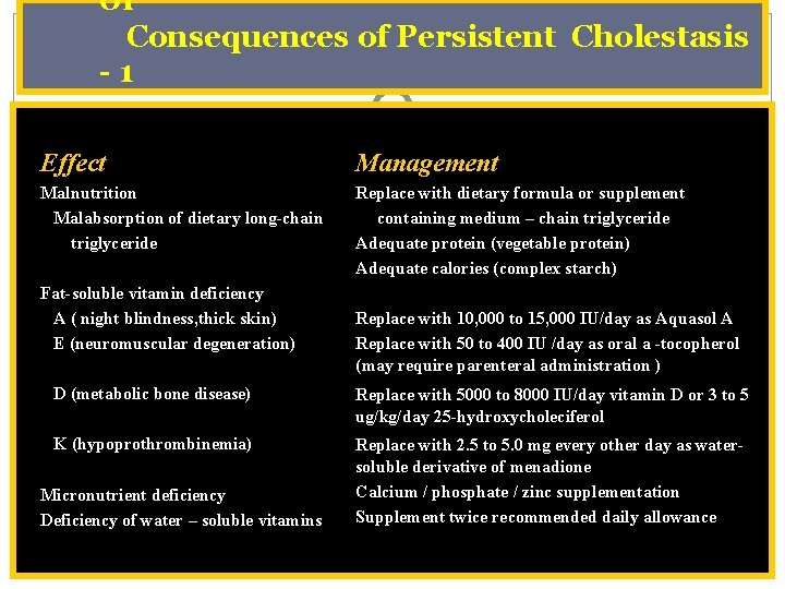
of Consequences of Persistent Cholestasis 1 Effect Management Malnutrition Malabsorption of dietary long-chain triglyceride Replace with dietary formula or supplement containing medium – chain triglyceride Adequate protein (vegetable protein) Adequate calories (complex starch) Fat-soluble vitamin deficiency A ( night blindness, thick skin) E (neuromuscular degeneration) Replace with 10, 000 to 15, 000 IU/day as Aquasol A Replace with 50 to 400 IU /day as oral a -tocopherol (may require parenteral administration ) D (metabolic bone disease) Replace with 5000 to 8000 IU/day vitamin D or 3 to 5 ug/kg/day 25 -hydroxycholeciferol K (hypoprothrombinemia) Replace with 2. 5 to 5. 0 mg every other day as watersoluble derivative of menadione Calcium / phosphate / zinc supplementation Supplement twice recommended daily allowance Micronutrient deficiency Deficiency of water – soluble vitamins
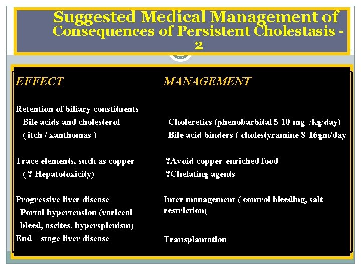
Suggested Medical Management of Consequences of Persistent Cholestasis 2 EFFECT MANAGEMENT Retention of biliary constituents Bile acids and cholesterol ( itch / xanthomas ) Choleretics (phenobarbital 5 -10 mg /kg/day) Bile acid binders ( cholestyramine 8 -16 gm/day Trace elements, such as copper ( ? Hepatotoxicity) ? Avoid copper-enriched food ? Chelating agents Progressive liver disease Portal hypertension (variceal bleed, ascites, hypersplenism) End – stage liver disease Inter management ( control bleeding, salt restriction( Transplantation
- Slides: 31