Mammary glands The mammary gland is a modified


 and sheep (ewe) each have two glands, each")


- Slides: 16
Mammary glands The mammary gland is a modified sweat gland that nourishes the young. It consists of the mamma and the teat. The number of glands and teats are not the same for the cow, the sow or the horse. However, the microscopic anatomy is very similar among species. In humans, the mammary glands are situated in the breasts , while in ruminants, the mammary glands are contained in the udders. The mammary glands of mammals other than primates, such as dogs and cats, are sometimes called dugs.
Development of the Mammary Gland • An ectodermal thickening developes along the ventral body wall extending from the thoracic to inguinal region - this is the mammary ridge. Cells aggregate, multiply and differentiate to form a chain of condensed mammary buds. Most mammary buds regress but those that remain each develops into a mammary gland. Proliferation of the mesenchyme surrounding the bud raises a teat (papilla) on the surface of the body. One or more epidermal sprouts grow from the mammary bud into the connective tissue of the teat and begin to canalize at about the time of birth.
• Each sprout is destined to form a separate duct system with associated glandular tissue. When there is only one sprout, the mammary gland arising from it has a single duct system leading to a single orifice on the tip of the teat When there are more, for example, two or four as in , the number of separate duct systems , each with an associated glandular mass and separate orifice be more. The growth of the ducts and gland tissue is continued after puberty and especially during the first pregnancy, forming the swelling that pushes the teat away from the body wall. The process is controlled by hormones from the hypophysis, ovaries, and other endocrine glands.
Anatomical structures • In the mare they are two in number, and are placed on either side of the median plane in the inguinal region. Each gland has the form of a short cone, much compressed transversely, and having a flat inner surface. It consists of the glandular mass or body of the gland the teat or nipple. The base is related to the abdominal wall, to which it is attached by areolar tissue, which contains a venous plexus, the superficial inguinal lymph glands, and a variable amount of fat. The apex is constituted by the teat, which is also flattened transversely and varies in length from one to two inches. • Between the bases of the teats is the intermammary groove. On the apex of each teat two or three small orifices are placed close together; these are the openings of the lactiferous ducts. •
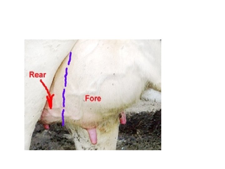
• The mammary gland of the dairy cow , consists of four separate glands each with a teat. Milk which is synthesized in one gland cannot pass over to any of the other glands. The right and left side of the udder are also separated by a median ligament, while the front and the hind quarters are more diffusely separated. •
• The lactating goat (nanny) and sheep (ewe) each have two glands, each drained by a single teat with a single streak canal. They are located in the inguinal region. The teats vary considerably in size. Goat teats and udder are generally larger than the sheep's. • The mammary glands in bitch are usually ten or twelve in number, and are arranged in two rows, as in the bitch. Each teat has commonly two excretory ducts.
Cow ewe goat Mare and sow Bitch and cat
Suspensory System • Suspensory System - A strong udder suspensory system is required to maintain proper attachments of the gland to the body. • There are seven tissues that provide support for the udder: • Skin covering the gland is only of very minor support. • Superficial fascia or Areolar subcutaneous tissue - This attaches the skin to underlying the tissue. • Coarse areolar or cordlike tissue - This tissue forms a loose bond between the dorsal surface of the front quarters and abdominal wall. Weakening of these causes the udder to break away from abdominal wall. • Subpelvic tendon - This tendon not actually part of the suspensory apparatus, but gives rise to the superficial and the deep lateral suspensory ligaments. It is attached to the pelvis at several points.
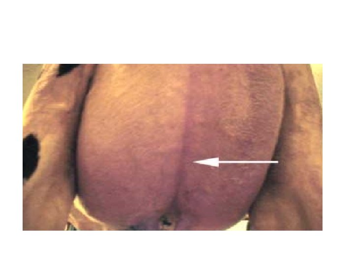
• Superficial layers of lateral suspensory ligament - These are mostly composed of fibrous tissue (with some elastic tissue), arising from the subpelvic tendon. They extend downward and forward from the pubic. When it reaches the udder it spreads out, continuing downward over the external udder surface beneath the skin and attaching to the areolar tissue. • Deep lateral suspensory ligament - The inner part of the lateral suspensory ligament also arises from the subpelvic tendon, but is thicker than the superficial layer, mostly fibrous tissue. It extends down over the udder and almost enveloping it. The lateral suspensory ligaments provide substantial support for the udder. • Median Suspensory Ligament - This is the most important part of the suspensory system in cattle. It is composed of two adjacent heavy yellow elastic sheets of tissue that arise from the abdominal wall and that attach to the medial flat surfaces of the two udder halves.
• The median suspensory ligament partially separates the left and right halves of the udder. Front and rear quarters are separated by a thin membrane and is not recognizable to the eye.
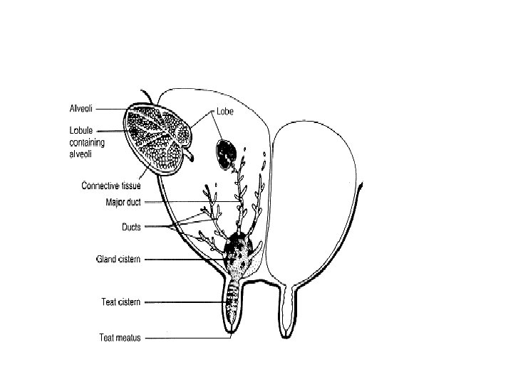
• Internal structures The lobes are the internal compartments of the mamma, separated by adipose tissue. The lobes are divided into lobules, consisting of connective tissue containing alveoli, which are clusters of milk secreting cells. The lactiferous ducts are large ducts conveying milk from the alveoli to the lactiferous sinus. The openings of the lactiferous ducts convey milk formed in the alveolus to the gland sinus. • The lactiferous sinus (milk sinus) is the milk storage cavity within the teat and glandular body. The gland sinus is part of the milk sinus within the glandular body and the teat sinus is part of the milk sinus within the teat.
• The teat is the projecting part of the mammary gland containing part of the milk sinus. The papillary duct (teat canal) is the canal leading from the teat sinus to the teat opening and may be single or multiple. The ostium (teat opening) is the opening of the papillary duct and the exit point for milk or entrance point for bacteria. The sphincter consists of muscular fibres surrounding the teat opening that prevent milk flow except during suckling or milking.
• Blood Supply • Arteries • The main blood supply to the inguinal mammary glands is from the external pudendal artery. • Veins : In most species thoracic and cranial abdominal mammary glands drain via cranial superficial epigastric veins.