Diseases of exocrine pancreas The exocrine pancreas constitutes


 - cystic fibrosis is the most common lethal genetic disease that")


- Slides: 38
Diseases of exocrine pancreas
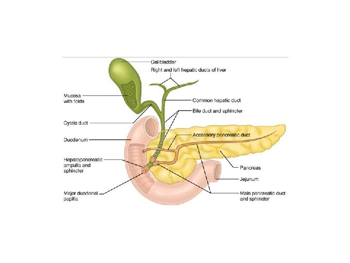
• The exocrine pancreas constitutes 80% to 85% of the organ and is composed of acinar cells that secrete enzymes needed for digestion.
• the accessory duct of Santorini, • the main pancreatic duct (the duct of Wirsung)
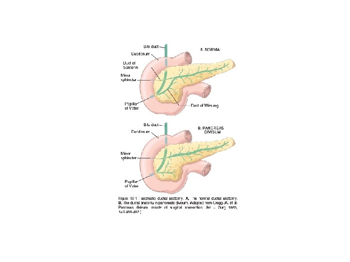
Congenital Anomalies • Pancreas Divisum: - is the most common congenital anomaly of the pancreas. - Pancreas divisum is caused by a failure of fusion of the fetal duct systems of the dorsal and ventral pancreatic primordia. As a result, the bulk of the pancreas (formed by the dorsal pancreatic primordium) drains into the duodenum through the small-caliber minor papilla. The duct of Wirsung in persons with divisum drains only a small portion of the head of the gland through the papilla of Vater. - inadequate drainage of the pancreatic secretions through the minor papilla increases susceptibility to pancreatitis.
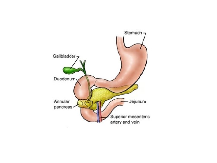
• Annular Pancreas: Annular pancreas is a band-like ring of normal pancreatic tissue that completely encircles the second portion of the duodenum. Annular pancreas can produce duodenal obstruction. • Ectopic Pancreas • Agenesis
Pancreatitis • Pancreatitis is divided into two forms, acute and chronic. • Both are initiated by injuries that lead to autodigestion of the pancreas by its own enzymes.
• Under normal circumstances, the following mechanisms protect the pancreas from self-digestion by its secreted enzymes: - Most digestive enzymes are synthesized as inactive proenzymes (zymogens), which are packaged within secretory granules. - Most proenzymes are activated by trypsin, which itself is activated by duodenal enteropeptidase (enterokinase) in the small bowel; thus, intrapancreatic activation of proenzymes is normally minimal. - Acinar and ductal cells secrete trypsin inhibitors, including serine protease inhibitor Kazal type l (SPINK 1), which further limit intrapancreatic trypsin activity.
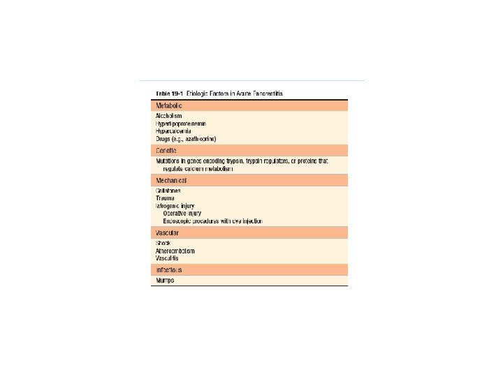
Acute Pancreatitis • Acute pancreatitis is characterized by reversible pancreatic parenchymal injury associated with inflammation and has diverse etiologies, including: toxic exposures (e. g. , alcohol), pancreatic duct obstruction (e. g. , biliary calculi), inherited genetic defects, vascular injury, infections.
• Biliary tract disease and alcoholism account for approximately 80% of cases of acute pancreatitis in Western countries. • The male-to female ratio is 1 : 3 in the group with biliary tract disease and 6 : 1 in those with alcoholism.
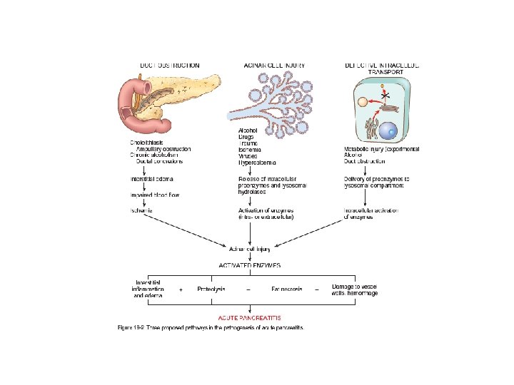
• Pathogenesis: Acute pancreatitis results from inappropriate release and activation of pancreatic enzymes, which destroy pancreatic tissue and elicit an acute inflammatory reaction. Inappropriate intrapancreatic activation of trypsin can in turn cause the activation of other proenzymes such as prophospholipase and proelastase, which then degrade fat cells and damage the elastic fibers of blood vessels, respectively. Trypsin also converts prekallikrein to its activated form, thus bringing into play the kinin system and, by activation of coagulation factor XII, the clotting and complement systems as well. The resulting inflammation and small-vessel thromboses damage acinar cells, further amplifying intrapancreatic activation of digestive enzymes.
• Alcohol consumption may cause pancreatitis through all of these mechanisms: - increases contraction of the sphincter of Oddi (the muscle at the Papilla of Vater), - secretion of protein-rich pancreatic fluid that leads to the deposition of inspissated protein plugs and obstruction of small pancreatic ducts. - direct toxic effects on acinar cells---- oxidative stress---generate free radicals in acinar cells, leading to membrane lipid oxidation and free radical production.
• Other proven or suspected triggers of acute pancreatitis: - hypertriglyceridemia - hypercalcemic states - Medications - Traumatic injury of acinar cells - Ischemic injury of acinar cells, caused by shock, vascular thrombosis, embolism, and vasculitis - Infections, including mumps - Hereditary factors (defect that increases or sustains the activity of trypsin)----- recurrent attacks of severe acute pancreatitis often beginning in childhood and ultimately leading to chronic pancreatitis.
• Clinical Features: Abdominal pain is the cardinal manifestation of acute pancreatitis. the pain is constant and intense and is referred to the upper back and occasionally to the left shoulder. Anorexia, nausea, and vomiting frequently accompany the pain. Elevated plasma levels of amylase and lipase support the diagnosis of acute pancreatitis.
• Full-blown acute pancreatitis is a medical emergency/acute abdomen. • Many of the systemic features of severe acute pancreatitis can be attributed to release of toxic enzymes, cytokines, and other mediators into the circulation and explosive activation of a systemic inflammatory response, resulting in: leukocytosis disseminated intravascular coagulation Edema acute respiratory distress syndrome. Shock acute renal tubular necrosis
• Laboratory findings include: - marked elevation of serum amylase levels during the first 24 hours - Rising serum lipase level by 72 to 96 hours -Glycosuria occurs in 10% of cases. - Hypocalcemia may result from precipitation of calcium soaps in necrotic fat.
• The key to the management of acute pancreatitis is “resting” the pancreas by total restriction of oral intake and by supportive therapy with intravenous fluids and analgesia. • most individuals with acute pancreatitis recover fully, about 5% with severe acute pancreatitis die in the first week of illness. • Sequelae can include a sterile pancreatic abscess and a pancreatic pseudocyst.
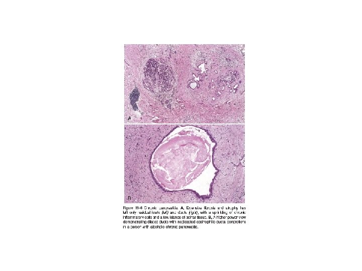
Chronic Pancreatitis • Chronic pancreatitis is defined as prolonged inflammation of the pancreas associated with irreversible destruction of exocrine parenchyma, fibrosis, and, in the late stages, the destruction of endocrine parenchyma.
• The most common cause of chronic pancreatitis by far is long term alcohol abuse. In addition to alcohol, chronic pancreatitis has been associated with the following conditions: - Long-standing obstruction of the pancreatic duct by calculi or neoplasms. - Autoimmune injury to the gland - Hereditary pancreatitis
• Pathogenesis: Chronic pancreatitis most often follows repeated episodes of acute pancreatitis. leads to local production of inflammatory mediators that promote fibrosis and acinar cell loss. While the cytokines produced during chronic and acute pancreatitis are similar, fibrogenic factors tend to predominate in chronic pancreatitis. These fibrogenic cytokines include transforming growth factor β (TGF-β) and platelet-derived growth factor, which induce the activation and proliferation of periacinar myofibroblasts (pancreatic stellate cells), resulting in the deposition of collagen and fibrosis
• - Clinical Features: may follow repeated bouts of acute pancreatitis. persistent abdominal and back pain. Attacks may be precipitated by alcohol abuse, overeating (which increases demand on the pancreas), or the use of opiates and other drugs that increase the tone of the sphincter of Oddi. - pancreatic insufficiency (malabsorption/weight loss) and diabetes mellitus develop due to destruction of the exocrine and endocrine pancreas. Pancreatic pseudocysts develop in about 10% of patients hereditary pancreatitis have a 40% lifetime risk of developing pancreatic cancer - A very helpful finding is visualization of calcifications within the pancreas by computed tomography and ultrasonography.
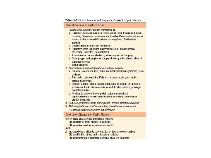
Cystic Fibrosis (Mucoviscidosis) - cystic fibrosis is the most common lethal genetic disease that affects Caucasian populations. It follows an autosomal recessive transmission pattern.
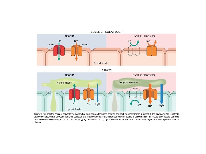
• In normal duct epithelia, chloride is transported by plasma membrane channels (chloride channels). • The primary defect in cystic fibrosis results from abnormal function of an epithelial chloride channel protein encoded by the cystic fibrosis transmembrane conductance regulator (CFTR) gene on chromosome 7 q 31. 2.
• it is now recognized that CFTR can regulate multiple ion channels and cellular processes. • the interaction of CFTR with the epithelial sodium channel (ENa. C) has possibly the most pathophysiologic relevance in cystic fibrosis. The ENa. C is situated on the apical surface of exocrine epithelial cells and is responsible for sodium uptake from the luminal fluid, rendering it (the luminal fluid) hypotonic. hence, in cystic fibrosis, ENa. C activity increases, markedly augmenting sodium uptake across the apical membrane.
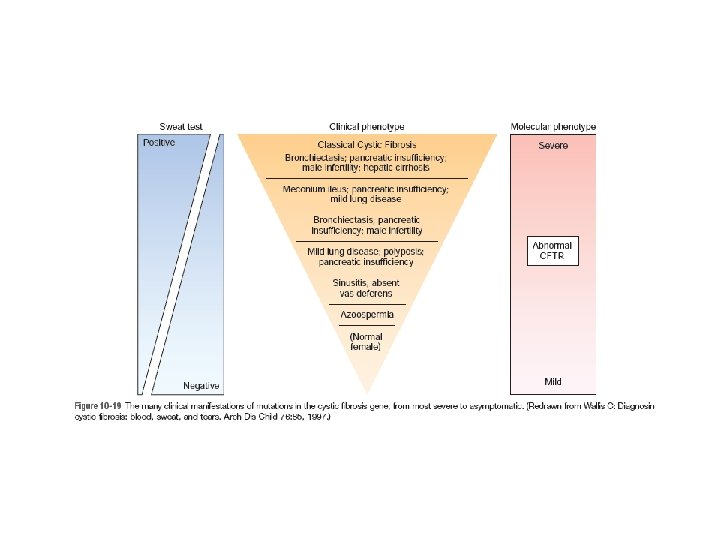
• The one exception to this rule happens to be the human sweat ducts, where ENa. C activity decreases as a result of CFTR mutations; therefore, a hypertonic luminal fluid containing high sweat sodium chloride (the sine qua non of classic cystic fibrosis) is formed. This is the basis for the “salty” sweat that mothers can often detect in their affected infants.
• functions of CFTR are tissue-specific: The major function of CFTR in the sweat gland ducts is to reabsorb luminal chloride ions and augment sodium reabsorption via the ENa. C. Therefore, in the sweat ducts, loss of CFTR function leads to decreased reabsorption of sodium chloride and production of hypertonic sweat. in the respiratory and intestinal epithelium, the CFTR is one of the most important avenues for active luminal secretion of chloride. At these sites, CFTR mutations result in loss or reduction of chloride secretion into the lumen. Active luminal sodium absorption is increased (due to loss of inhibition of ENa. C activity), and both of these ion changes increase passive water reabsorption from the lumen, lowering the water content of the surface fluid layer coating mucosal cells. the pathogenesis of respiratory and intestinal complications in cystic fibrosis seems to stem from an isotonic but low-volume surface fluid layer
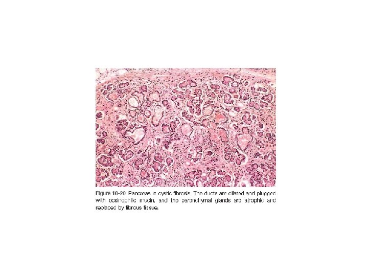
• CFTR regulates transport of bicarbonate ions. Epithelia harboring these mutant CFTR alleles secret acidic fluid (due to absence of bicarbonate ions). The acidity of secretions results in decreased luminal p. H that can lead to a variety of adverse effects such as increased mucin precipitation and plugging of ducts, and increased binding of bacteria to plugged mucins. Pancreatic insufficiency, a feature of classic cystic fibrosis, is virtually always present when there are CFTR mutations with abnormal bicarbonate conductance.
• exocrine pancreatic insufficiency in more than 80% of patients. The result is failure of the intraluminal phase of nutrient absorption.