Spondylolisthesis and spondylolysis spondylolysis Spondylolysis is defined as


")


 is")


 – commonest Degenerative ( 25%)")


.")


 may occur due to a decrease in")
 : - occurs due to congenitally defective superior sacral")
 : painless , unduly protruding")


- Slides: 43
Spondylolisthesis and spondylolysis
spondylolysis
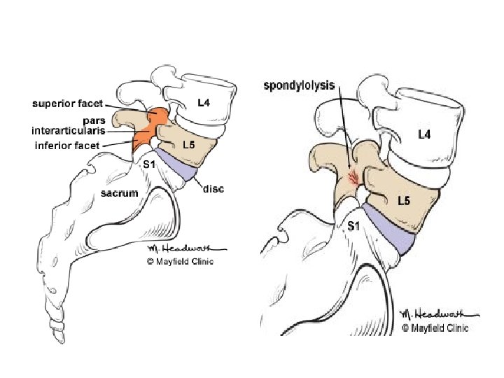
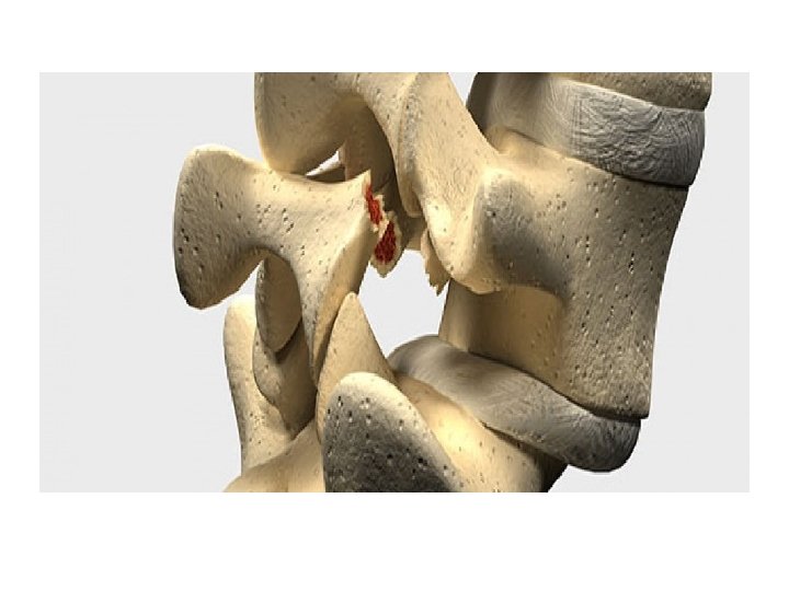
• Spondylolysis is defined as a defect or non-displaced stress fracture ( uni-bilateral) in the pars interarticularis of the vertebral arch without forward displacement. • Occurs in the pediatric age group. • The pars interarticularis , or isthmus , is the portion of the neural arch that connects the lamina with the pedicle, facet joints, and transverse process. • The vast majority of cases (85%) occur in the lower lumbar vertebrae (L 5) , but spondylolysis may also occur in the cervical vertebrae. • A pars defect is twice as common in boys than in girls,
• Etiology : - mechanical factors play the major role especially when congenital abnormalities are present. - Repeated extension strains of lumbar spine makes P. I more susceptible for shear stresses. - The pars interarticularis becomes impinged from the inferior facet of the cephalad vertebrae, which results in microfractures and attempts at repair.
• Clinical presentation : - Symptomatic only in 10% of patients. - Usually come with low back pain aggravated by extension movements. - some discomfort can be elicited with deep percussion over the midline of the lumbar area. No tenderness found. - Full range of movements with normal N. V exam. - The 1 -leg hyperextension test elicits pain on the involved side.
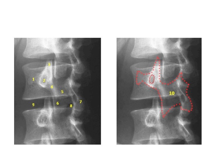
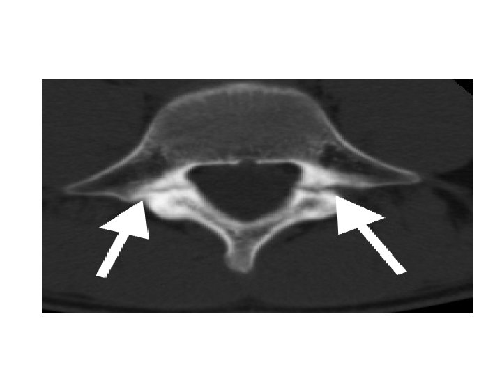
• Imaging studies : - X-ray : AP radiographic findings are usually normal in spondylolysis. The oblique views ( Rt. And Lt ) are particularly screened for the "Scotty dog" lesion in the pars interarticularis. The pars defect is represented by the collar on the Scotty dog. - ( 99 m Tc) / SPECT bone scan : superior to CT/MRI : these scans can aid in establishing the acuity of the lesion or in identifying the site of the problem in an athlete with negative plain radiography results but whose clinical course is suggestive of a pars interarticularis fracture.
• Treatment : - Acute phase : Bracing and rest are the cornerstones of treatment for this type of lesion. Pain control and avoiding sports are also part of the acute phase or rehabilitation. - Recovery phase : Flexibility and strengthening of the paraspinal, iliopsoas, and abdominal muscles along with endurance training of the back are all especially important.
• Return to play of an athlete with lumbosacral spondylolysis (lumbar spondylolysis) is first begun with low-level sport activities after the follow-up visit at 4 -6 weeks, after which gradual increase in intensity as tolerated is allowed under supervision. Return to full activity is permitted only when patients are totally asymptomatic with full range of motion. Patients must also have normal flexibility and normal strength and balance.
Spondylolisthesis
Spondyloisthesis • ‘Spondylolisthesis’ means forward translation of one segment of the spine upon another. • Location : nearly always between L 4 and L 5, or between L 5 and the sacrum. • Normal discs, laminae and facets constitute a locking mechanism that prevents each vertebra from moving forwards on the one below. Forward shift (or slip) occurs only when this mechanism has failed.
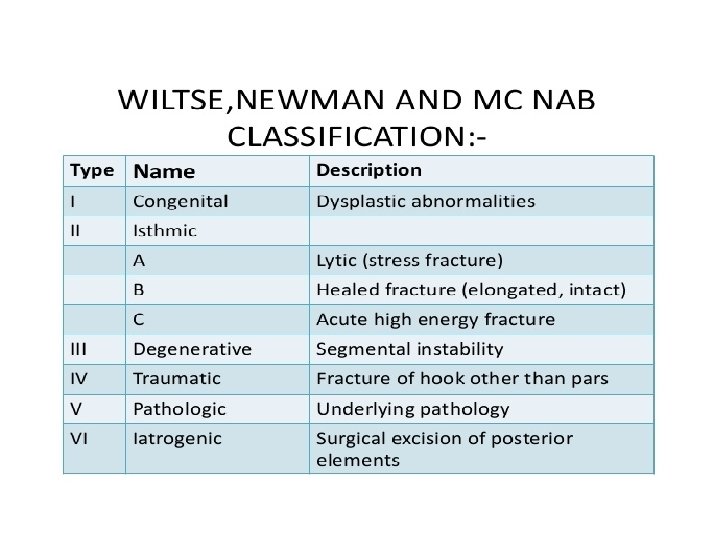
Classification • - 6 types : Isthmic ( 50%) – commonest Degenerative ( 25%) Dysplastic (20%) Pathological ( TB , neoplasm ) Post-traumatic Post-operative ( excessive bone removal in decompression ).
• Isthmic : 50 % - Typically presents in the teenage or early adulthood years even the presentation may be in the late middle age. - Location : mostly (82 %) at L 5/S 1 and may be L 4/L 5. - Etiology : defect in pars interarticularis – on both sides( acquired by microtrauma or congenital ) or elongation of P. I due to repetitive breaking and healing. • The condition is more common than usual in those whose spines are subjected to extraordinary stresses (e. g. competitive gymnasts and weight-lifters). • Genetic factors may play a role.
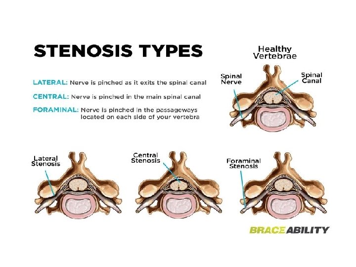
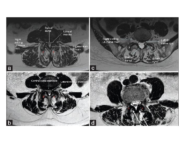
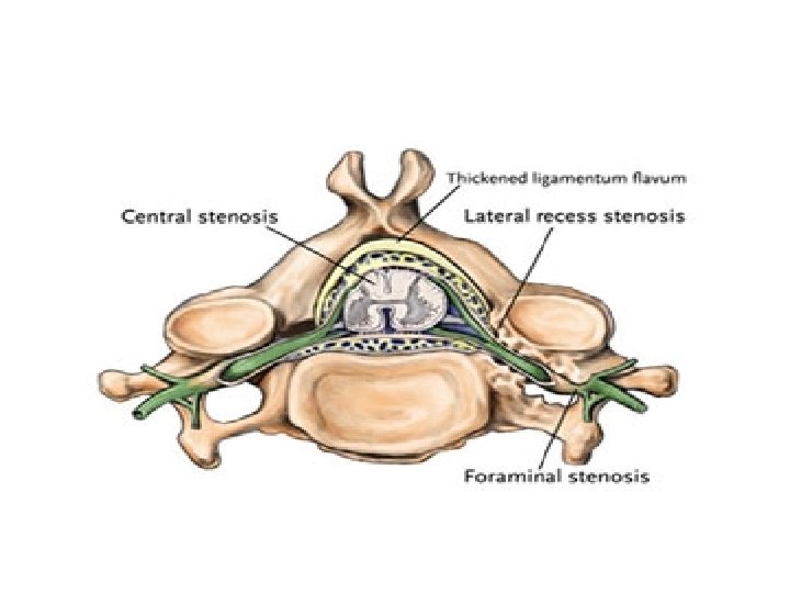
• Pathophysiology : Compression can lead to either foraminal stenosis mainly , lateral recess stenosis or rarely central stenosis. § foraminal stenosis : mainly : – Adult isthmic spondylolisthesis at L 5/S 1 often leads to radicular symptoms caused by compression of the exiting L 5 nerve root in the L 5 -S 1 foramen. compression can be caused by : • hypertrophic fibrous repair tissue of the pars defect • uncinate spur formation of the posterior L 5 body • bulging of the L 5/S 1 disc § lateral recess stenosis • caused by facet arthrosis mainly or hypertrophic ligamentum flavum. § central stenosis • rare due to fact that these slips are usually only Grade I or II
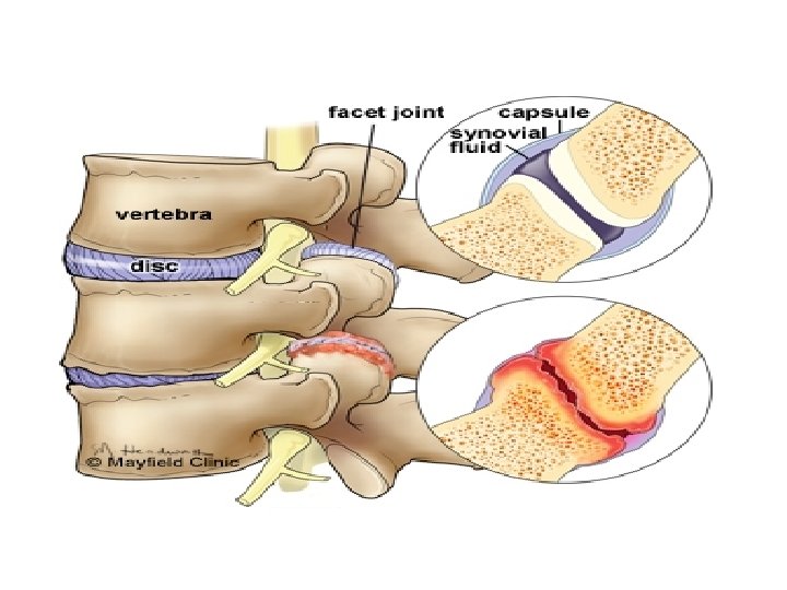
• Degenerative : 25% - Typically in middle aged women ( H. factors). - Location : mostly at L 4/L 5. - Etiology : degeneration ( with OA , crystal arthropathy) in the facets and disc with intact lamina … here there is no pars interarticularis defect. - This can be worsened by the presence of congenitally sagittal facet joint orientation.
• Pathophysiology : - The forward slip of the vertebral body here is mainly allowed by I. D degeneration and facet joint degeneration. - disc degeneration leads to facet capsule degeneration and microinstability … with further degeneration >> macroinstability.
- neurologic symptoms caused mainly by central and lateral recess stenosis and to a less degree foraminal stenosis. § Central and lateral recess stenosis : • degenerative slip at L 4/5 will affect the descending L 5 nerve root in the lateral recess. - caused by slippage, hypertrophy of ligamentum flavum, and encroachment into the spinal canal of osteophytes from facet arthrosis.
§ Foraminal stenosis ( L 4 compression) may occur due to a decrease in the space vertically ( by loss of disc height and osteophytes that push the nerve root superiorly against the inferior surface of the pedicle ) or ant. posteriorly.
• Dysplastic ( 20%) : - occurs due to congenitally defective superior sacral facets. - The displacement is usually severe and occurs slowly. Prognosis is usually bad. - usually associated with spina bifida occulta.
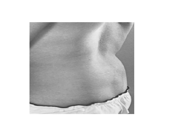
• Clinical presentation : - In children (dysplastic) : painless , unduly protruding abdomen and peculiar spastic gait. - In adolescents and adults (isthmic) backache is the usual presenting symptom; it is often intermittent, coming on after exercise or strain. L 5 radiculopathy occurs in one or both legs. - Degenerative spondylolisthesis : usually women aged over 50 years. They always have backache, L 4 and L 5 compression occur , claudication due to spinal stenosis.
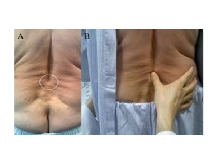
• PE : - Lumbar spine may be flat or hyperlordotic. - Flat buttocks with more vertical sacrum ( it may extend to the waist) Sometimes scoliosis. - Transverse loin creases. - Spastic gait due to hamstring tightness. - Movement of spine is stiff if degenerative. - palpable Step-off deformity.
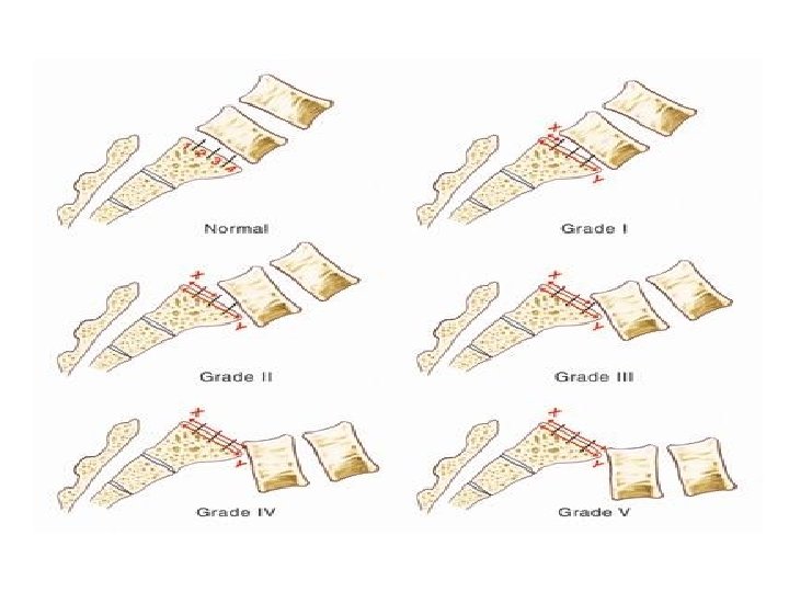
• Grading :
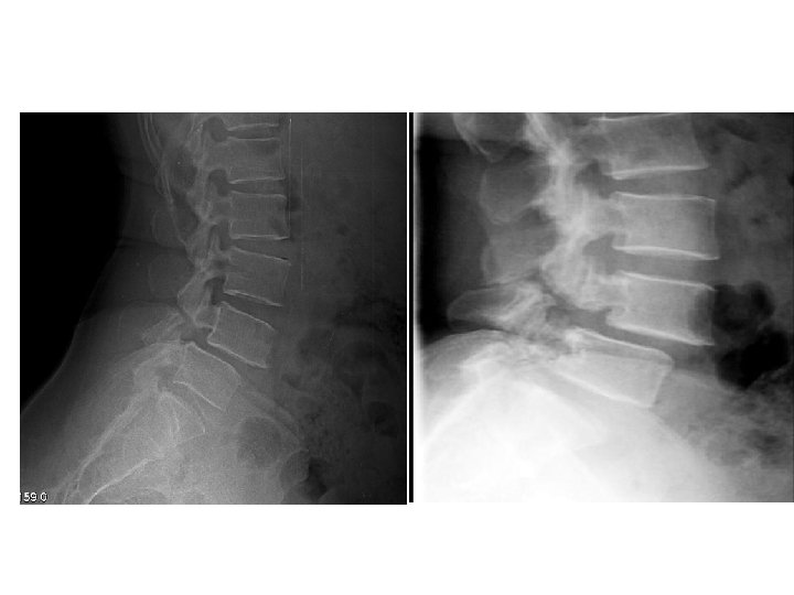
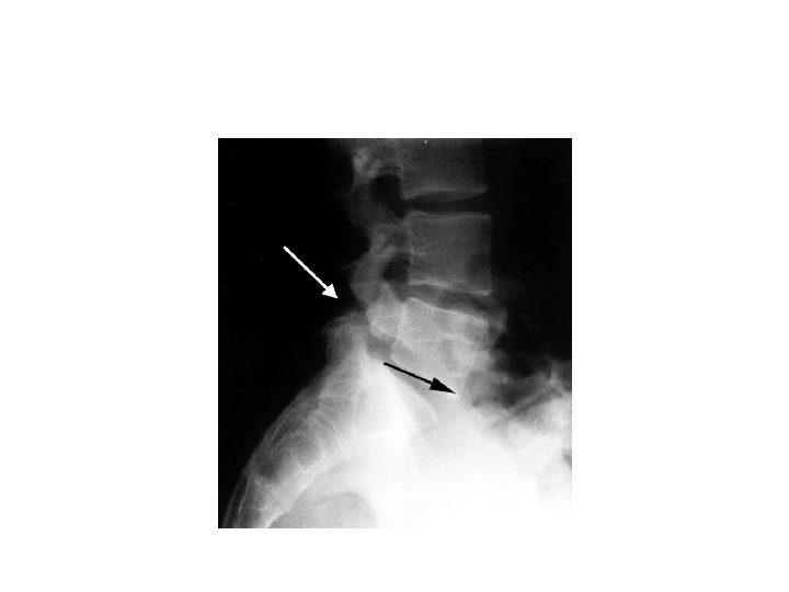
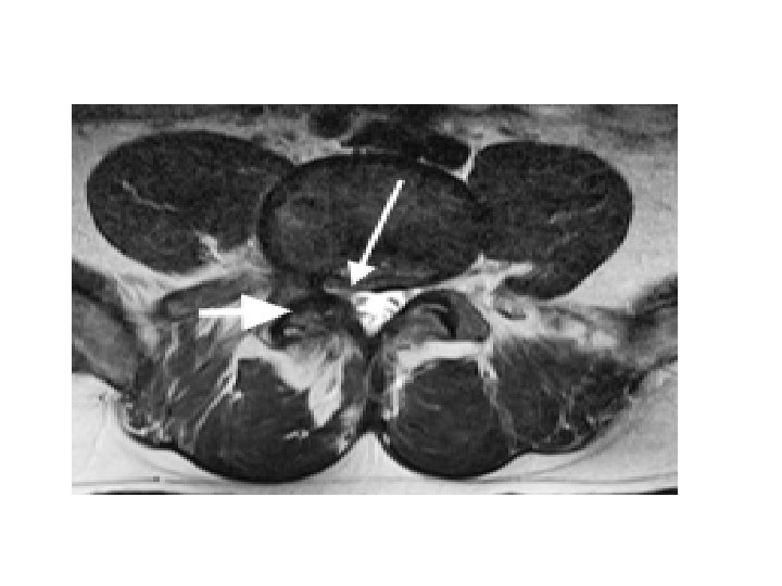
• Imaging : - X-ray : AP , lateral , oblique * on lateral view : forward slip is evident. * on oblique view : gap in pars interarticularis. - MRI : to evaluate impingement of neural elements. - CT : for bony pathology. - CT myelogram : when MRI contraindicated.
• Treatment : - Conservative treatment is suitable for most patients. This includes : * activity restriction * NSAIDs * bracing in the acute phase. * physiotherapy
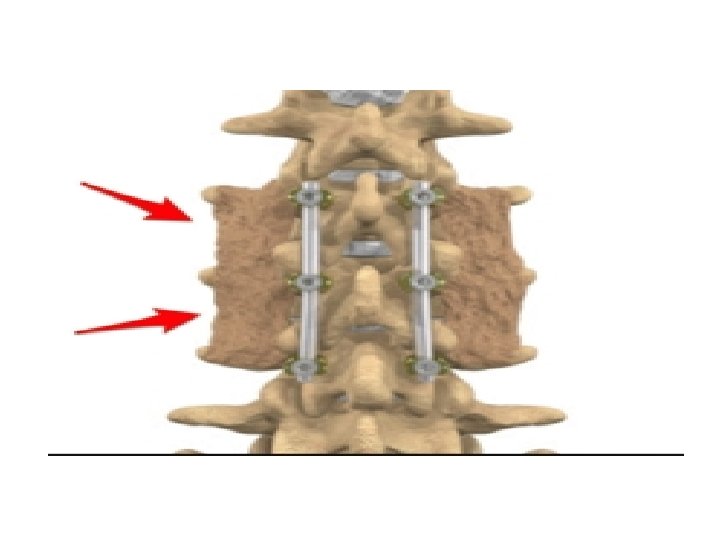
• Operative treatment : - The goal of surgery is to stabilize the segment with listhesis and decompress any of the neural elements under pressure. Restoration of normal sagittal alignment must also be achieved - Indications : (1) if the symptoms are disabling and interfere significantly with work and recreational activities; (2) if the slip is more than 50 per cent and progressing; (3) if neurological compression is significant.
• For children, posterior intertransverse fusion in situ is almost always successful; if neurological signs appear, decompression can be carried out later. • For adults, either posterior or anterior fusion is suitable. • For ‘degenerative’ group, where neurological symptoms predominate, decompression without fusion may suffice.
THANK YOU