
Lumbar degenerative spondylolisthesis indications for fusion Presenters name
Lumbar degenerative spondylolisthesis indications for fusion Presenter‘s name Arial 24 pt Meeting Arial 24 pt Presenter‘s title Arial 20 pt City, Month, Year Arial 20 pt
Learning outcomes • Identify the clinical and radiological features of instability in patients presenting for the surgical treatment of lumbar spinal canal stenosis • Discuss the evidence in relation to this treatment • Describe the surgical techniques appropriate to manage this condition • Identify and manage common complications relating to the management of this condition
Spinal canal stenosis: considerations • Anatomy / Pathology • Stability: pre and post decompression • Age of patient • Functional requirements • Extent degenerative disease • Severity of back versus leg pain
Spinal canal stenosis: considerations • Anatomy & Pathology • Orientation of facet joints • Facet-joint fluid or gas • Disc height • Foraminal compromise • Stability of segment • Presence of synovial cyst • Degenerative scoliosis
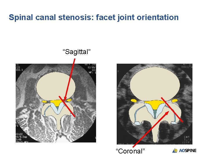
Spinal canal stenosis: facet joint orientation “Sagittal” “Coronal”
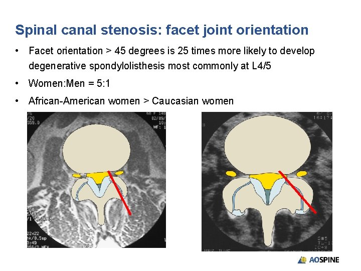
Spinal canal stenosis: facet joint orientation • Facet orientation > 45 degrees is 25 times more likely to develop degenerative spondylolisthesis most commonly at L 4/5 • Women: Men = 5: 1 • African-American women > Caucasian women
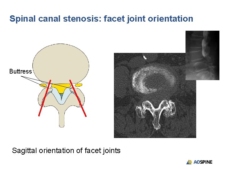
Spinal canal stenosis: facet joint orientation Buttress Sagittal orientation of facet joints
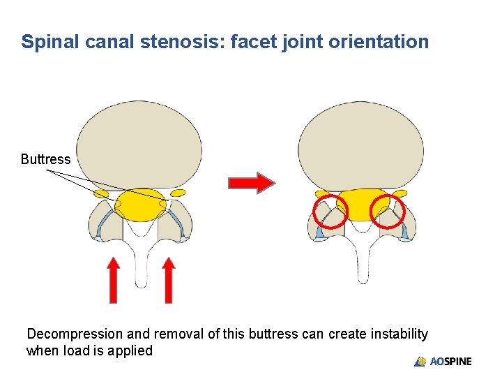
Spinal canal stenosis: facet joint orientation Buttress Decompression and removal of this buttress can create instability when load is applied
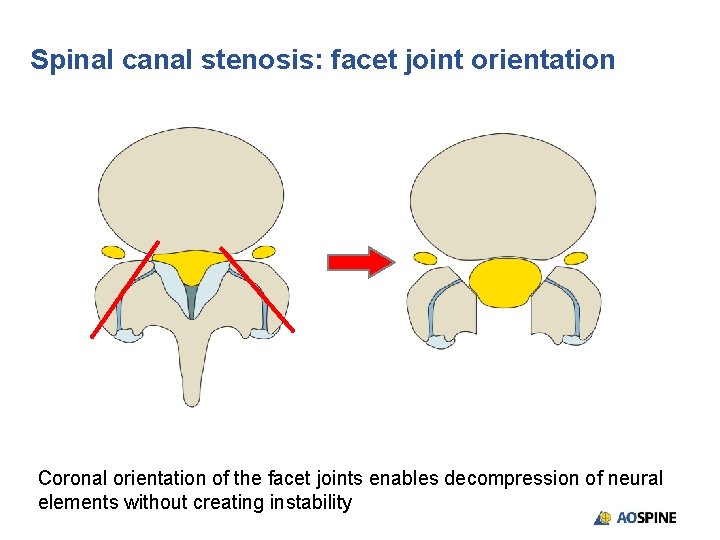
Spinal canal stenosis: facet joint orientation Coronal orientation of the facet joints enables decompression of neural elements without creating instability
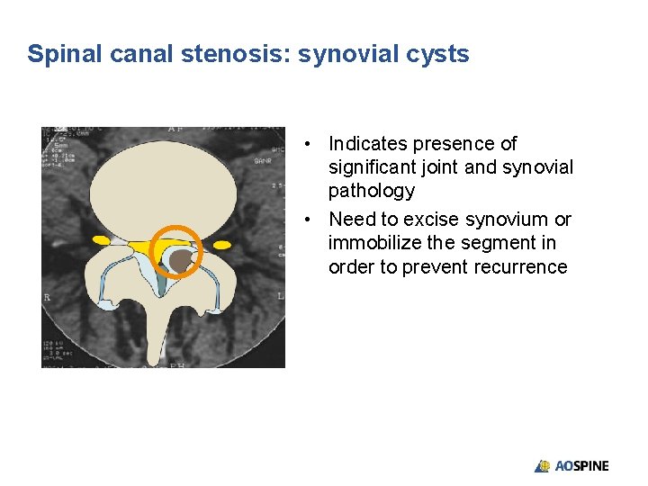
Spinal canal stenosis: synovial cysts • Indicates presence of significant joint and synovial pathology • Need to excise synovium or immobilize the segment in order to prevent recurrence
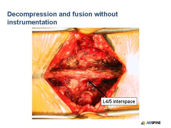
Decompression and fusion without instrumentation L 4/5 interspace
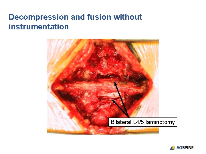
Decompression and fusion without instrumentation Bilateral L 4/5 laminotomy
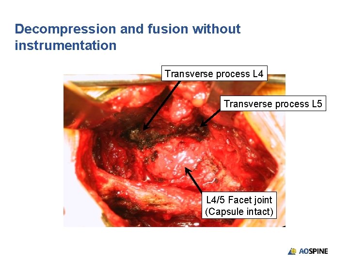
Decompression and fusion without instrumentation Transverse process L 4 Transverse process L 5 L 4/5 Facet joint (Capsule intact)
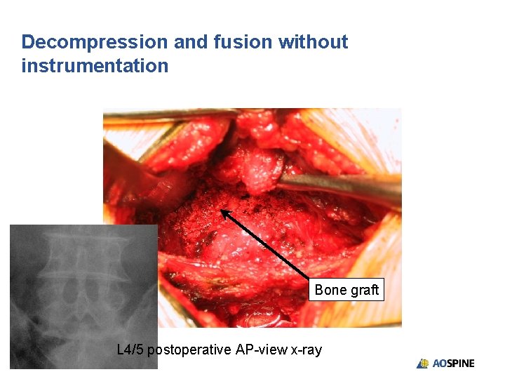
Decompression and fusion without instrumentation Bone graft L 4/5 postoperative AP-view x-ray
Decompression and fusion without instrumentation • “Microdiscectomy and uninstrumented single-segment lumbar fusion for spinal canal stenosis with degenerative spondylolisthesis was associated with an 86% satisfaction rate. ” (Mc. Culloch JA. SPINE. 1998. 23(20): 2243– 2252)
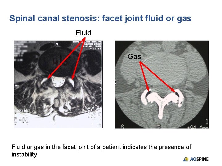
Spinal canal stenosis: facet joint fluid or gas Fluid Gas Fluid or gas in the facet joint of a patient indicates the presence of instability
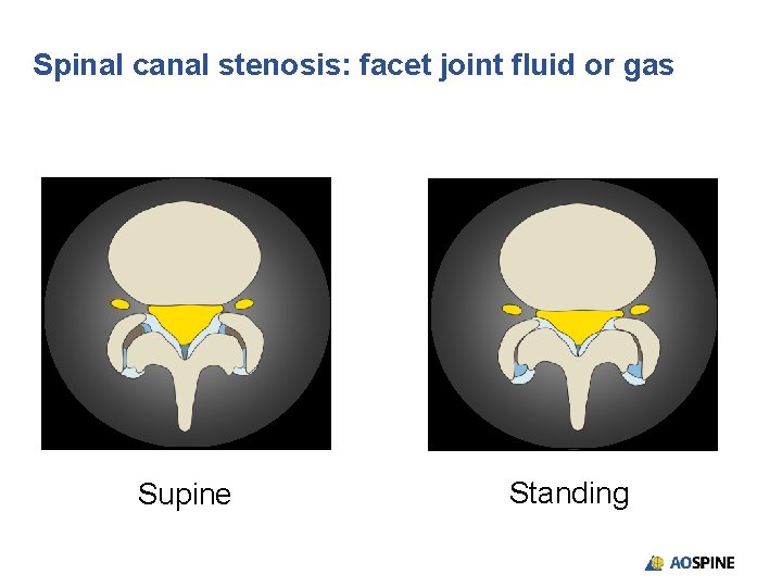
Spinal canal stenosis: facet joint fluid or gas Supine Standing
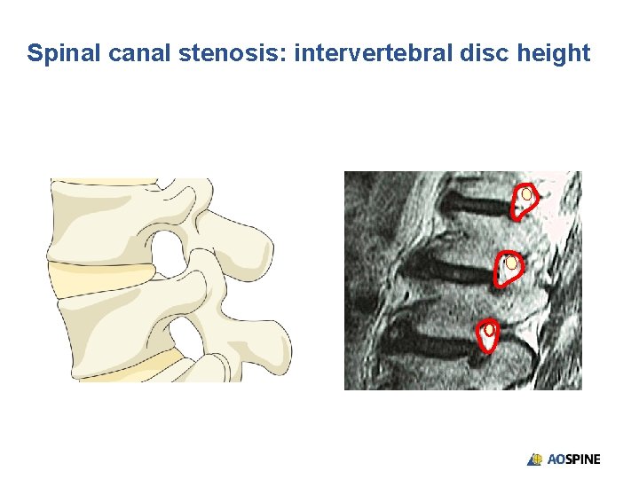
Spinal canal stenosis: intervertebral disc height
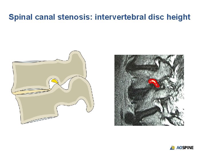
Spinal canal stenosis: intervertebral disc height
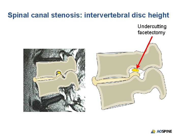
Spinal canal stenosis: intervertebral disc height Undercutting facetectomy
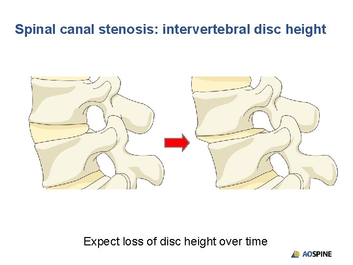
Spinal canal stenosis: intervertebral disc height Expect loss of disc height over time
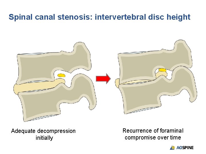
Spinal canal stenosis: intervertebral disc height Adequate decompression initially Recurrence of foraminal compromise over time
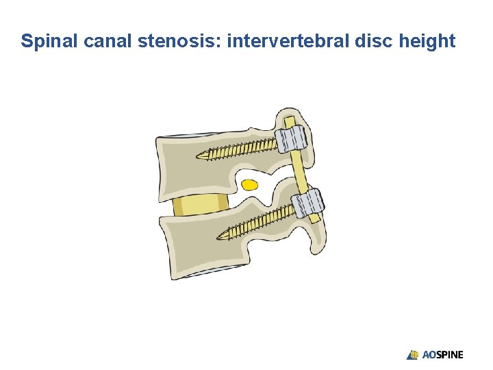
Spinal canal stenosis: intervertebral disc height
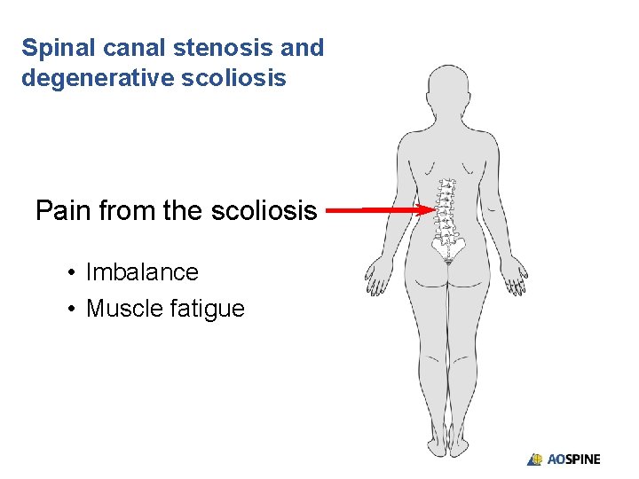
Spinal canal stenosis and degenerative scoliosis Pain from the scoliosis • Imbalance • Muscle fatigue
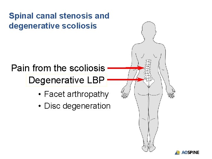
Spinal canal stenosis and degenerative scoliosis Pain from the scoliosis Degenerative LBP • Facet arthropathy • Disc degeneration
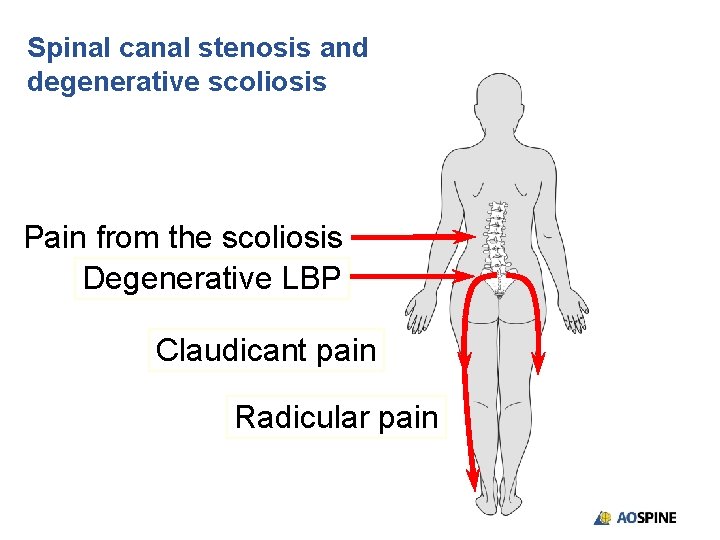
Spinal canal stenosis and degenerative scoliosis Pain from the scoliosis Degenerative LBP Claudicant pain Radicular pain
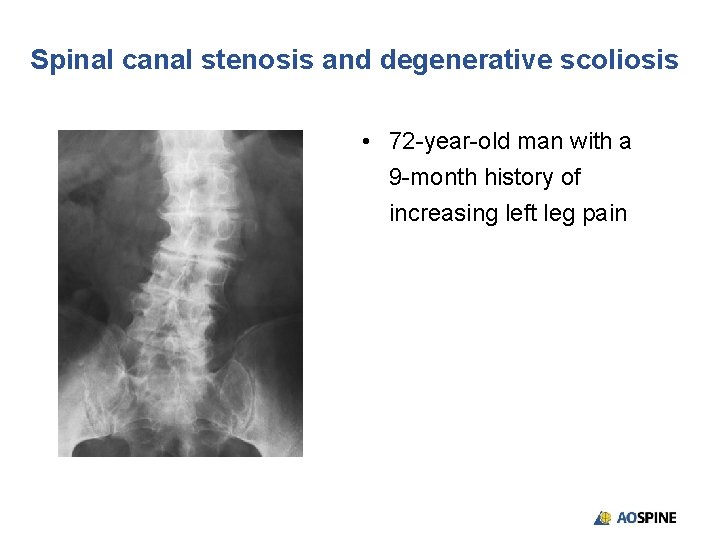
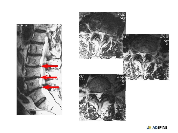
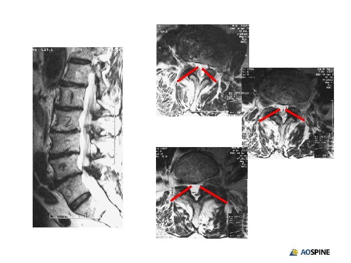
Spinal canal stenosis and degenerative scoliosis • 72 -year-old man with a 9 -month history of increasing left leg pain
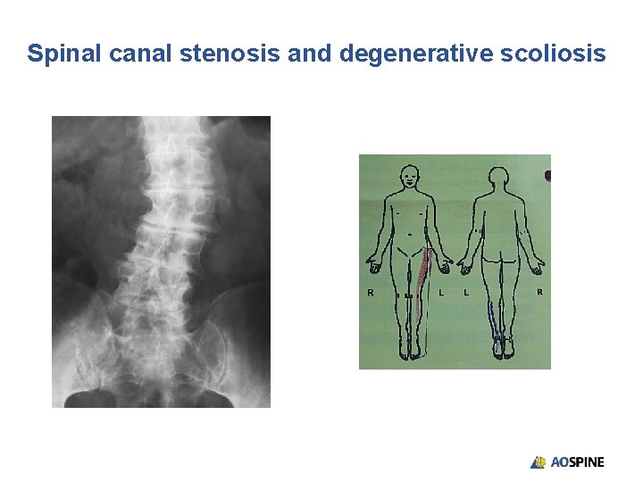
Spinal canal stenosis and degenerative scoliosis
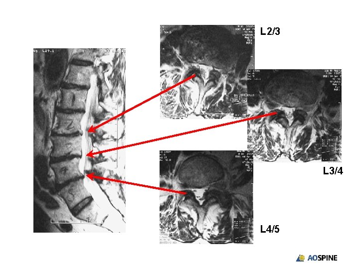
L 2/3 L 3/4 L 4/5
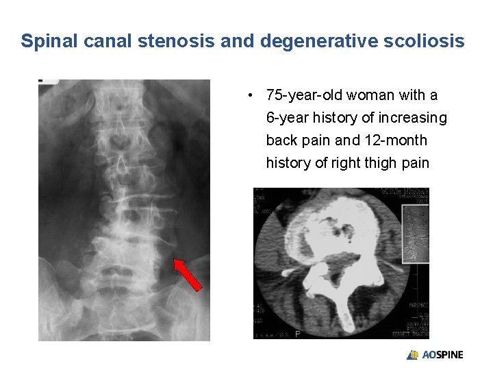
Spinal canal stenosis and degenerative scoliosis • 75 -year-old woman with a 6 -year history of increasing back pain and 12 -month history of right thigh pain
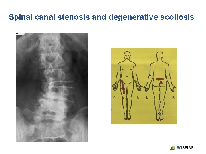
Spinal canal stenosis and degenerative scoliosis
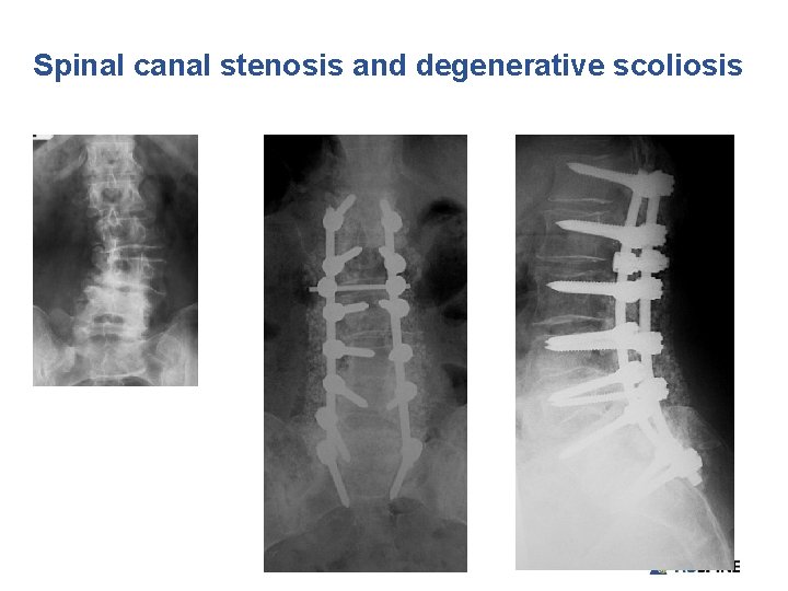
Spinal canal stenosis and degenerative scoliosis
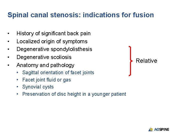
Spinal canal stenosis: indications for fusion • • • History of significant back pain Localized origin of symptoms Degenerative spondylolisthesis Degenerative scoliosis Anatomy and pathology • • Sagittal orientation of facet joints Facet joint fluid or gas Synovial cysts Preservation of disc height in a younger patient Relative
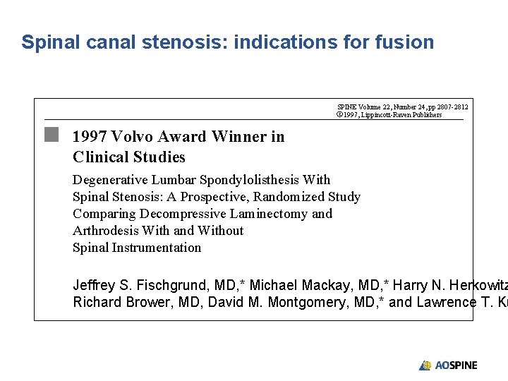
Spinal canal stenosis: indications for fusion SPINE Volume 22, Number 24, pp 2807 -2812 C 1997, Lippincott-Raven Publishers 1997 Volvo Award Winner in Clinical Studies Degenerative Lumbar Spondylolisthesis With Spinal Stenosis: A Prospective, Randomized Study Comparing Decompressive Laminectomy and Arthrodesis With and Without Spinal Instrumentation Jeffrey S. Fischgrund, MD, * Michael Mackay, MD, * Harry N. Herkowitz Richard Brower, MD, David M. Montgomery, MD, * and Lawrence T. Ku
Spinal canal stenosis: indications for fusion • “Clinical outcome was excellent or good in 76% of the patients in whom instrumentation was placed, and in 85% of those in whom no instrumentation was placed (p = 0. 45)” (Fischgrund JS, Mackay M, Herkowitz HN, et al. SPINE. 1997. 22(24): 2807– 2812)
Spinal canal stenosis: indications for fusion • “In patients undergoing single level postero-lateral fusion for degenerative spondylolisthesis with spinal stenosis, the use of pedicle screws may lead to a higher fusion rate, but there is no improvement in clinical outcome” (Fischgrund JS, Mackay M, Herkowitz HN, et al. SPINE. 1997. 22(24): 2807– 2812)
Take-home messages • The only clear indication for fusion when performing a lumbar decompression is the presence of significant instability • The presence of a degenerative spondylolisthesis in isolation does not indicate instability • Factors such as the facet orientation, disc and foraminal height, and the presence of synovial cysts, degenerative spondylolisthesis, or degenerative scoliosis as well as the age of the patient and functional requirements should all be considered when deciding if fusion is indicated and whether or not instrumentation should be added
Excellence in Spine
- Slides: 40