Somatic and Special Senses Sensation Sensory Receptors detect


, and auditory ossicle (3 small bones) ◦ auditory")


- Slides: 51
Somatic and Special Senses
Sensation Sensory Receptors detect changes in our body and surroundings ◦ Trigger nerve impulses Travels to CNS for interpretation Perceive a sensation
Types of Receptors Chemoreceptors: changes in chemical concentration of substances Pain Receptors: tissue damage
Types of Receptors Thermoreceptors: changes in temperature Mechanoreceptors: changes in pressure or movement Photoreceptors: light energy
Sensations �A feeling that occurs when the brain interprets sensory impulses �Differences in impulse is due to which region of the brain receives the impulse ◦ Sound vs. touch �Projection: cerebral cortex causes the sensation to seem to come from the stimulated receptor ◦ Eyes see and ears hear
Sensory Adaptation sensory receptors are continuously stimulated Impulses leave at a decreasing rate until the signal stops After adaptation, impulse can be triggered if the stimulus strength changes ◦ Strong scent in a room
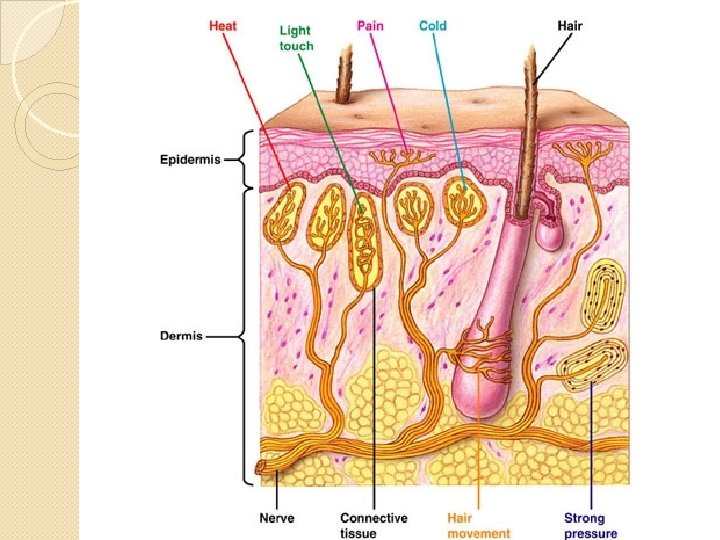
Somatic Senses Receptors in the skin, muscles, joints, and viscera Touch and Pressure: 3 types of receptors ◦ Sensory nerve fibers: epithelial tissue
Touch and Pressure Meissner’s corpuscles: small, oval masses of flattened connective tissue cells ◦ Hairless portions of the skin: lips, fingertips, palms, soles, nipples, and external genital organs ◦ Respond to the motion of objects that barely contact the skin, light touch
Touch and Pressure Pacinian Corpuscles: relatively large structures composed of connective tissue fibers and cells ◦ Subcutaneous tissues, muscle tendons, and joint ligaments ◦ Respond to heavy pressure ◦ Sensation of deep pressure
Temperature Heat receptors: respond to warmer temp. 45ºC-25ºC, 113ºF-77ºF ◦ Above this range, pain receptors are stimulated burning sensation Cold receptors: respond to colder temp. ◦ 10ºC -20ºC, 50ºF-68ºF ◦ Below this, pain receptors simulate freezing sensation
Pain Free nerve endings Widely distributed throughout the skin and internal tissues ◦ Excepts in nervous tissue of the brain Protection Unpleasant stimulus signals a person to remove the stimulus
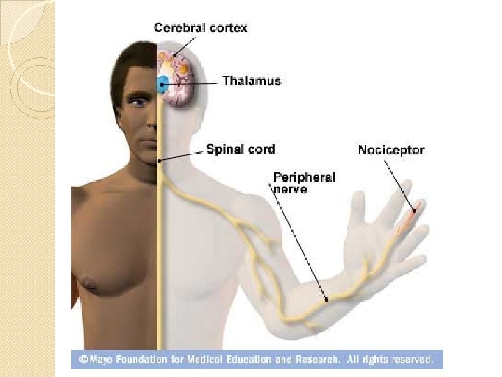
Visceral Pain receptors are the only receptors in viscera whose stimulation produces sensation Intestinal tissues are stretches or smooth muscles in intestinal walls undergo spasms Referred Pain: the origin of the pain is different than where it is perceived ◦ Heart pain appears in left shoulder or left upper arm
Pain Nerve Fibers Acute Pain Fibers: thin, myelinated fibers ◦ Conduct nerve impulses rapidly ◦ sharp pain from the skin Chronic Pain Fibers: thin, unmyelinated fiber ◦ Conduct impulses more slowly ◦ Dull, aching pain from deeper tissues ◦ Difficult to pinpoint
Regulation of Pain Impulses �Awareness of pain arises when impulses reach the thalamus �Cerebral cortex: determine pain intensity, locates pain source, and mediates emotional and motor responses �Nerve fibers release biochemicals that block pain signals ◦ Enkephalins: suppress acute and chronic pain, relieve sever pain ◦ Similar to morphine and other opiates ◦ Serotonin: stimulates other neurons to release enkephalines ◦ Endorphins: extreme pain and natural pain control
Smell, Taste, and Hearing
Smell Olfactory organs smell ◦ Upper region of the nasal cavity ◦ Chemoreceptors Smell and taste function closely together ◦ Aid in food selection
Olfactory Organs Olfactory receptors: yellowish brown masses ◦ Covers upper parts of the nasal cavity, the superior nasal conchae, and portion of the nasal septum ◦ Bipolar neurons Covered by hair like cilia at the distal ends Sensitive parts of the receptors Gases must dissolve into the watery fluids surrounding the cilia before receptors detect them
Olfactory Nerve Pathways Olfactory bulbs analyze the impulse ◦ Travels to limbic system ◦ Interpreted in the temporal lobes and at the base of frontal lobes
Olfactory Stimulation Olfactory impulses may result when gaseous molecules combine with specific sites on cilia of receptor cells Olfactory receptor cells adapt rapidly
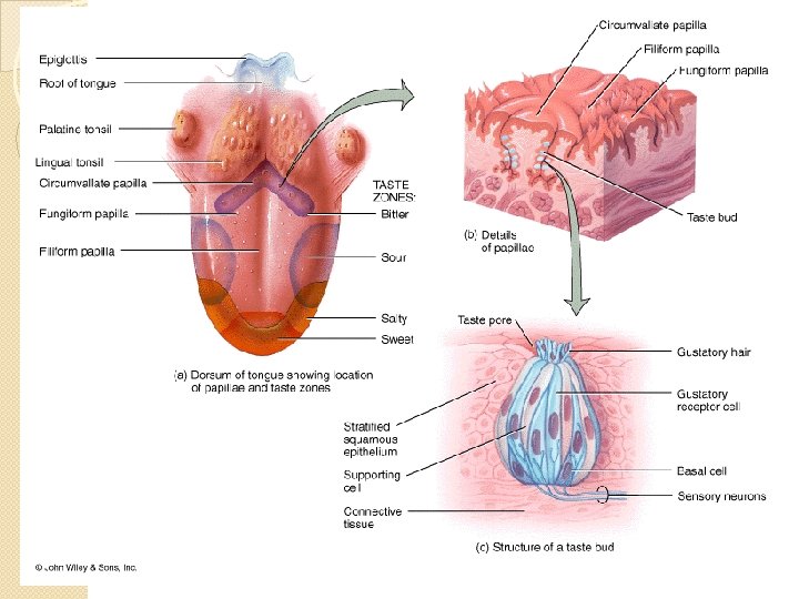
Sense of Taste Buds: specialized organ of taste ◦ Surface of the tongue, roof of the mouth, walls of pharynx Taste Cell: gustatory cells, receptors ◦ Taste Port: opening ◦ Taste hair: protrude from taste cell Sensitive part of receptor cell
Taste Before a chemical can be tasted, it must be dissolved in water ◦ Provided by salivary glands ◦ Generates a sensory impulse on a neighboring neuron
Taste Sensations Each receptor type is concentrated in certain regions of the tongue ◦ Sweet: sugar ◦ Salty: table salt quinine ◦ Alkaline Flavor: Sour: lemon Bitter: caffeine or Metallic taste, odor, texture, and temperature Burning: chili peppers or ginger
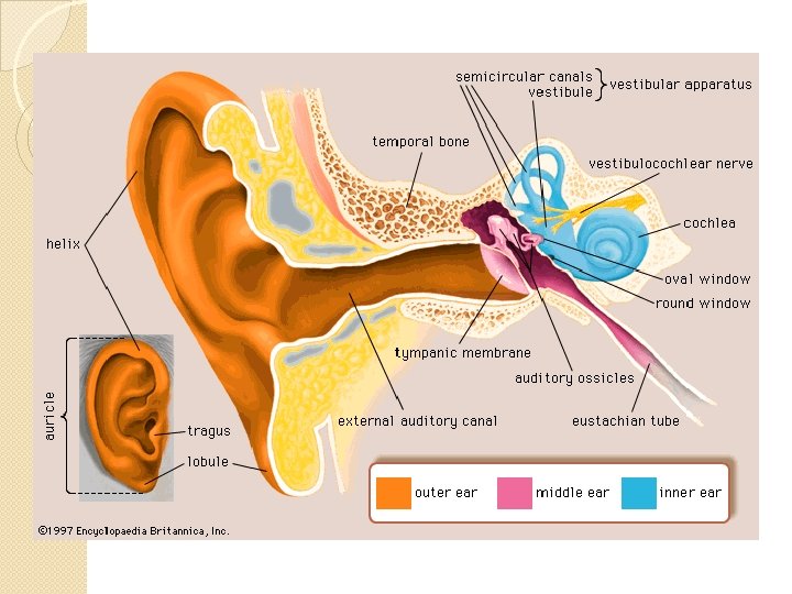
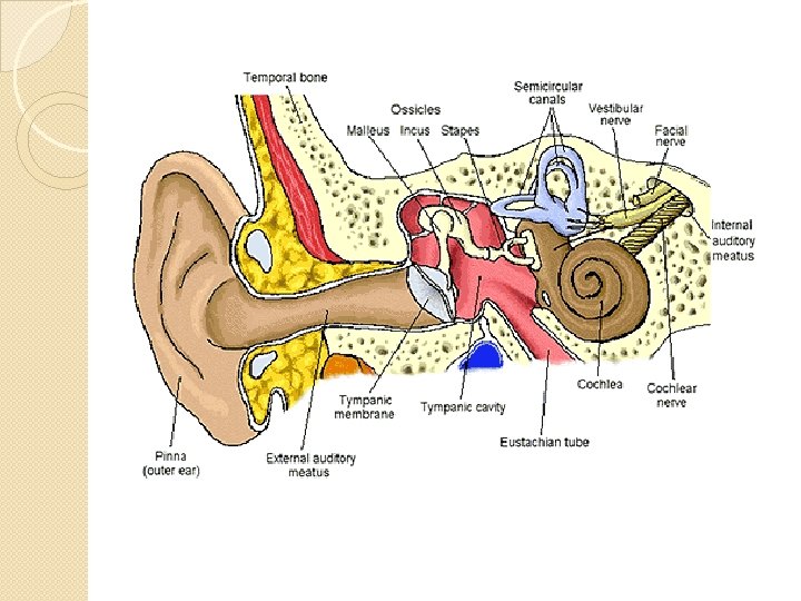
Sense of Hearing Vibrating objects produce sound ◦ Sound waves ◦ Musical instruments: vibrating string or reed ◦ Voice: vibrating vocal cords External Ear: ◦ Auricle or pinna: outer, funnel-like structure Collects sound waves and directs them ◦ External Auditory meatus: S-shaped tube Leads inward through temporal bone
Middle Ear tympanic cavity, eardrum(tympanic membrane), and auditory ossicle (3 small bones) ◦ auditory ossicles of the middle ear conduct sound waves form the eardrum to the oval window on the inner ear
Auditory Tube Connects the middle ear to the throat Maintain equal air pressure on both sides of the ear drum ◦ Detect problems when you have a sudden change in altitude ◦ Equalizing the air pressure causes a popping sound hearing returns
Inner Ear Complex system of connected tubes and chambers ◦ Osseous and membranous labyrinths Corti contains the hearing receptors that vibrations in the fluid of the inner ear stimulate Different frequencies of vibrations stimulate different sets of receptor cells
Equilibrium and Sight
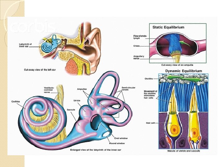
Equilibrium Static Equilibrium ◦ Maintains the stability of the head and body when they are motionless ◦ Hair cells project upward in mass of gelatinous material Moving the head bends the hair cells in response to gravity Nerve impulse travels to CNS
Equilibrium Dynamic Equilibrium ◦ Balances the head and body when they are moved or rotated suddenly ◦ Hair cells extending upward in a gelatin mass ◦ Movement of hair cells stimulate a nerve signal Other Structures assisting in equilibrium ◦ Eyes and mechanorecpetors of joints ◦ In the neck
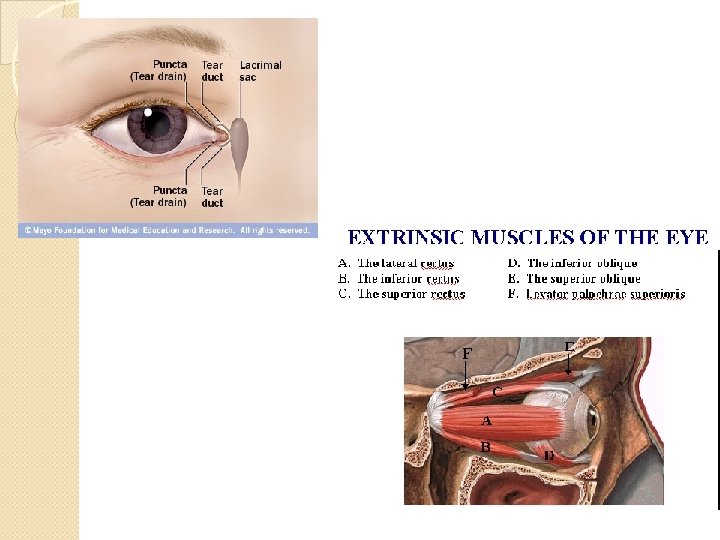
Sight Visual accessory organs ◦ Eyelids 4 layers: skin, muscle, connective tissue, and conjunctiva (mucus membrane) ◦ Lacrimal apparatus: secretes tears Moistens and lubricates eye’s surface Contain antibacterial agents ◦ Extrinsic muscles: 6 muscles that move the eye arise from the orbit bone
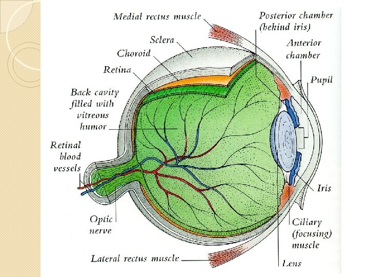
Structure of the Eye Hollow spherical structure about 2. 5 cm in diameter 3 Layers ◦ Outer tunic: fibrous ◦ Middle tunic: vascular ◦ Inner tunic: nervous Fluid filled ◦ Supports walls and shape
Parts of the Eye Outer Tunic ◦ Cornea: focus entering light rays Transparent connective tissue Loss of cornea transparency is leading cause of blindness ◦ Sclera: white portion of the eye 5/6 of outer tunic Protection & muscle attachment ◦ Optic Nerve: back of eye
Parts of the Eye Middle Tunic ◦ Choroid Coat: posterior 5/6 of the globe Contains melanocytes that absorb light Maintain darkness inside the eye
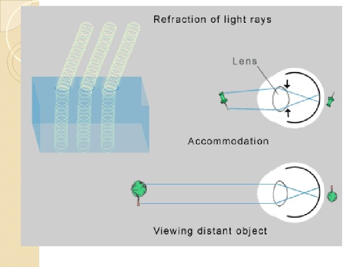
◦ Ciliary Body: extends from choriod coat Internal ring around the front of the eye ◦ Lens: clear, membrane like structure Lies directly behind the iris and pupil Accommodation: lens can adjust shape to focus on an image
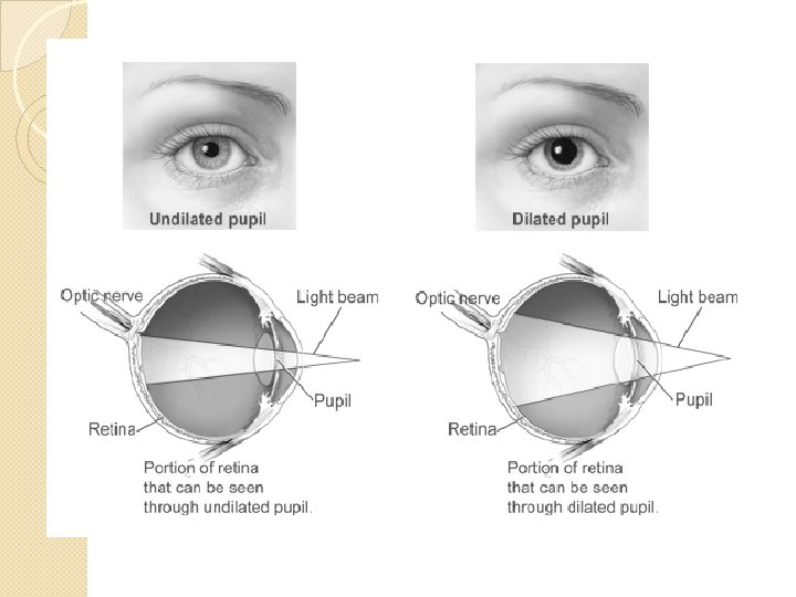
Middle Tunic Iris: thin diaphragm composed of connective tissue and smooth muscle fibers ◦ Colored portions of the eye ◦ Muscle control the size of the pupil Pupil: circular opening in the center of the iris ◦ Opening that light passes into the eye
Inner Tunic Retina: photorecptors ◦ Continuous with optic nerve Fovea centralis: region producing sharpest vision Optic disk: nerve fibers leave the eye and join optic nerve ◦ Blind spot: no receptor here Vitreous Humor: transparent, jelly-like fluid ◦ Supports internal parts of the eye ◦ Maintains shape
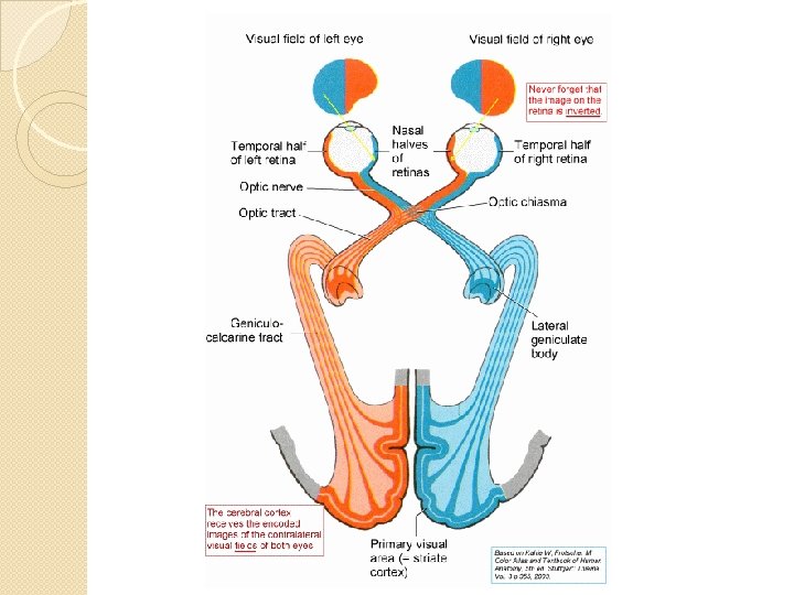
Light Refraction Bending of light waves to focus an image on the retina Convex surface of lens causes the light waves to converge ◦ Image focuses on retina like a project shows a movie Image is upside down and reversed left-to-right Visual cortex corrects the image
Visual Receptors Rods: long, thin projections ◦ 100 x more sensitive dim light ◦ Black and white ◦ General outlines of objects Cones: short, blunt projections ◦ Colored images ◦ Sharp images
Visual Pigments Decompose in the presence of light and triggers a complex series of reaction that initiate nerve impulses ◦ Rods: rhodopsin ◦ Cones: erythrolable (red), chlorolable (green), cyanolable (blue)
Visual Pigments