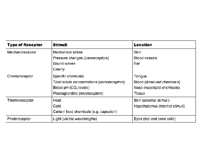
Types of Senses SENSES Receptors distributed over Receptors

Types of Senses SENSES Receptors distributed over Receptors localized within a large part of the body specific organs Special senses General senses Located in skin, muscles, joints. Located in internal organs Somatic Visceral Smell Touch Pressure Pain Balance Taste Temperature Hearing Sight Proprioception Pain Pressure

Special Senses • Olfaction • Taste • Visual system • Hearing and balance

Olfaction • The olfactory system gives humans their sense of smell by collecting odorants from the environment and transducing them into neural signals. • The receptors are chemoreceptors. • They detect odorants dissolved in solution.


15. 2 Taste • Papillae with taste buds – Taste bud: supporting cells surrounding taste (gustatory) cells. – Taste cells have microvilli (gustatory hairs) extending into taste pores – Replaced about every 10 days – Receptor cells are chemoreceptors

Taste Types • Sour. Most sensitive receptors on lateral aspects of the tongue. • Salty. Most sensitive receptors on tip of tongue. Shares lowest sensitivity with sweet. • Bitter. Most sensitive receptors on posterior aspect. Highest sensitivity. Sensation produced by alkaloids, which are toxic. • Sweet. Most sensitive receptors on tip of tongue. Shares lowest sensitivity with salty. • Umami (Glutamate). Scattered sensitivity. Caused by amino acids.

Neuronal Pathways for Taste • CN VII: sensations from anterior one-third of tongue • Cranial nerve IX and X carry information from posterior one-third tongue
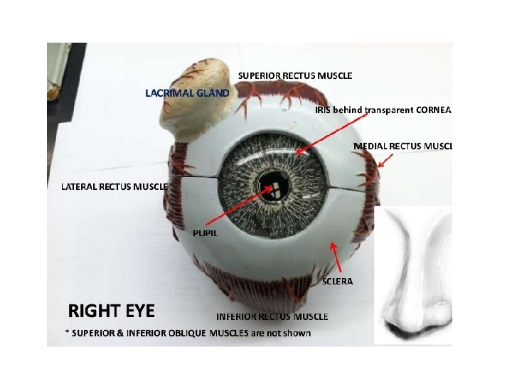
 Pupil Eyebrow Iris Sclera Caruncle Medial canthus Lateral canthus")
Visual System Superior palpebra (eyelid) Pupil Eyebrow Iris Sclera Caruncle Medial canthus Lateral canthus Inferior palpebra (eyelid)

Lacrimal Apparatus Puncta Lacrimal gland 1 1 Tears are produced in the lacrimal gland exit the gland rough several lacrimal ducts. Lacrimal canaliculi 2 The tears pass over the surface of the eye. 3 Tears enter the lacrimal canaliculi. 2 Lacrimal sac 3 4 Tears are carried through the lacrimal sac to the nasolacrimal duct. 5 Tears enter the nasal cavity from the nasolacrimal duct. 4 Nasolacrimal duct 5 Tears and saliva controlled by CN VII Lacrimal ducts
 Lateral rectus Medial")
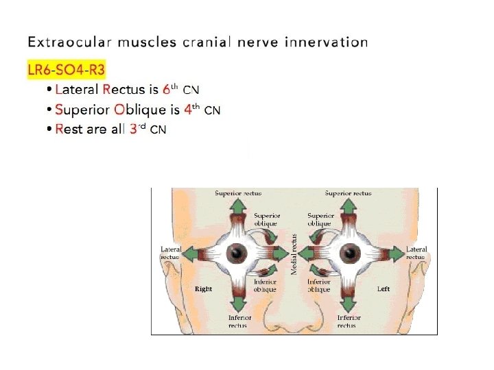
Extrinsic Eye Muscles Posterior View Optic nerve Levator palpebrae superioris (cut) Lateral rectus Medial rectus Superior oblique Trochlea (a) Superior view Anterior View Levator palpebrae superioris (cut) Superior Trochlea Superior oblique Optic nerve Superior rectus Lateral rectus Inferior rectus (b) Lateral view Inferior oblique Inferior

Wall of the Eyeball The wall of the eyeball is made up of three layers – fibrous (outer), vascular/muscular (middle) and sensorineural (inner) layers.
 • Sclera: white outer layer. Maintains")
Anatomy of the Eye: Fibrous Layer (outer layer) • Sclera: white outer layer. Maintains shape, protects internal structures. • Cornea: Layer of stratified squamous epithelium on the outer surface. Avascular, transparent, allows light to enter eye; bends and refracts light.

Anatomy of the Eye: Vascular Layer • Middle layer. Iris: colored part of the eye. Controls light entering the pupil. Smooth muscle determines size of pupil. – Dilating and constricting pupil, ciliary body: CN III occulomotor – Ciliary body: produces aqueous humor that fills anterior chamber • Ciliary muscles: control lens shape; smooth muscle. Ciliary processes attached to suspensory ligaments of lens – Choroid: associated with sclera. Very thin, pigmented.

Anatomy of the Eye: Sensoryneural area: Retina

Opthalmoscopic View of Retina • Lens focuses light on macula lutea and fovea centralis Macula – Macula lutea: small yellow spot Fovea centralis – Fovea centralis: area of greatest visual acuity; photoreceptor cells tightly packed • Optic disc: blind spot. Area through which blood vessels enter eye, where nerve processes from sensory retina meet and exit from eye Optic disc Retinal vessels (a) (b)

Chambers of the Eye • Anterior compartment: anterior to lens; filled with aqueous humor • Vitreous chamber: posterior to lens. Filled with jelly-like vitreous humor. Helps maintain intraocular pressure, holds lens and retina in place, refracts light.

Lens • Held by suspensory ligaments attached to ciliary muscles. Changes shape as ciliary muscles contract and relax.

Structure and Function of the Retina • Sensory retina: three layers of neurons: photoreceptor, bipolar, and ganglionic • Pigmented retina: single layer of cells; filled with melanin. With choroid, enhances visual acuity by isolating individual photoreceptors, reducing light scattering

15 -22

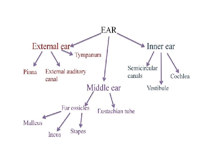
The ear: an organ of hearing and equilibrium that detects and analyzes noises by transduction (or the conversion of sound waves into electrochemical impulses) and maintains the sense of balance (equilibrium). The ear contains sense organs that serve two different functions: hearing and postural equilibrium Anatomically the ear has three distinguishable parts: the outer, middle, and inner ear.
. Includes auricle and external auditory canal")
External ear: hearing. Terminates at eardrum (tympanic membrane). Includes auricle and external auditory canal Middle ear: hearing. Air-filled space containing auditory ossicles Inner ear: hearing and balance. Interconnecting fluid-filled tunnels and chambers within the temporal bone

External Ear • External ear – Auricle or pinna: elastic cartilage covered with skin – External auditory canal: lined with hairs and ceruminous glands. Produce cerumen – Tympanic membrane • Thin membrane of two layers of epithelium with connective tissue between • Sound waves cause it to vibrate • Border between external and middle ear

The shapes of the outer ear and the external auditory canal help amplify and direct the sound. Sound waves entering the external auditory canal eventually hit the tympanic membrane, or eardrum (tympanum = drum), the boundary between the outer and middle ears. The tympanic membrane marks the border between the outer ear and middle ear. The tympanic membrane serves as a transmitter of sound by vibrating in response to sounds traveling down the external auditory canal, and beginning sound conduction in the middle ear.

Middle Ear • Middle ear – Separated from the inner ear by the oval and round windows – Auditory or eustachian tube: opens into pharynx, equalizes pressure – Ossicles: malleus, incus, stapes: transmit vibrations from eardrum to oval window – Oval window: connection between middle and inner ear. Foot of the stapes rests here.

The oval window Transmits sound vibrations to the inner ear. The round window Dampens the sound vibrations of the inner ear.
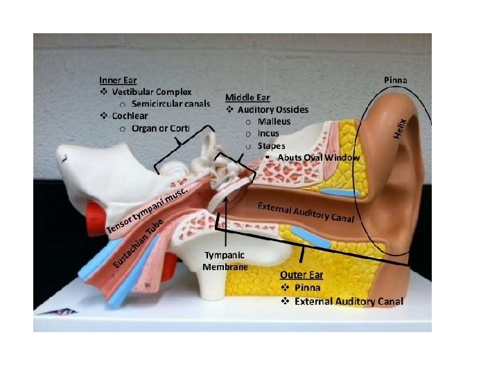
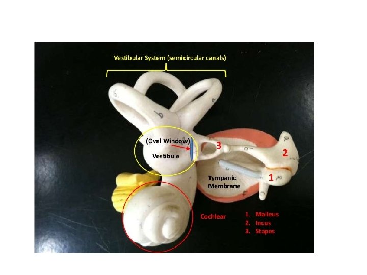

The middle ear is an air-filled chamber on the other side of the tympanic membrane and inside the cranial bone. The auditory (or eustachian tube), emerges from the middle ear and opens into the nasopharynx. The middle cavity contains three small pieces of bone (the auditory ossicles)- malleus (hammer), incus (anvil) and stapes (stirrup). The handle of malleus is attached to the central part of tympanic membrane. Malleus is attached to the incus, and incus is attached to stapes. The foot plate of stapes is placed on the oval window of the cochlea. They transmits the sound vibrations of the tympanic membrane to the fluid in the inner ear.

The inner ear has two main organs. The vestibular apparatus and cochlea. Receptors for hearing and balance are mechanoreceptors

The vestibular apparatus is the organ of equilibrium. It has three semicircular canals

The cochlea is the organ of hearing 15 -36

15 -37

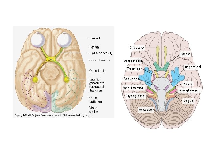
CN VIII Vestibulocochlear Nerve

http: //academic. pgcc. edu/~aimholtz/A and. P/Prac/2050_Lab 20/Brainmode l. html http: //academic. pgcc. edu/~aimholtz/A and. P/Prac/2050_Lab 17/neuron. ht ml 15 -39

15 -40
- Slides: 40