RESPIRATORY SYSTEM PHYSICAL EXAMINATION Prof Dr Reha Cengizlier


 – General")
 – Asymmetry (the abnormality")


 sec; generally bradicardia and")


 • Sucrepitan")
 Orta raller (sub krepitan,")
: – Probably represent opening of small airways and")
: – Musical sound heard on expiration. In severe cases they")

: A creaking sound caused by stiff pleural membranes such")


- Slides: 49
RESPIRATORY SYSTEM – PHYSICAL EXAMINATION Prof. Dr. Reha Cengizlier 8. 9. 2014
Solunum sistemi muayenesi Amaç: • Öğrencilerin solunumun; – Özelliklerini – Normal ve anormal bulguların ayırımını – Muayene bulguları ile tanıya yaklaşmasını – Patolojik solunum seslerinin oluşma mekanizmasını ve – İsimlendirmeyi öğrenmesi
Solunum sistemi muayenesi Hedef • Bilgi ve beceri: – Solunum sisteminin bölümlerini sayabilmeli – Muayene yöntemlerini sayabilmeli – Patolojik solunum seslerinin oluş mekanizmasını anlatabilmeli – Patolojik solunum seslerini sayabilmeli – Belli hastalıklara tanı koyduran tipik solunum seslerini söyleyebilmeli – Solunum sistemi muayenesinin nasıl yapıldığını düzenli olarak anlatabilmeli
Solunum sistemi muayenesi • Tutum: – Doğru tanıya ve tedaviye giden yolun en önemli aşamalarından biri olarak muayenenin önemini kavramalı
Inspection General inspection: – Evidence of respiratory distress at rest or when walking, e. g. obvious breathlessness, talking in short phrases rather than full sentences, use of accessory muscles, exhalation with pursed lips – Evidence of other respiratory symptoms, e. g. cough, audible wheeze – Does the patient appear to be pyrexial (check their temperature)
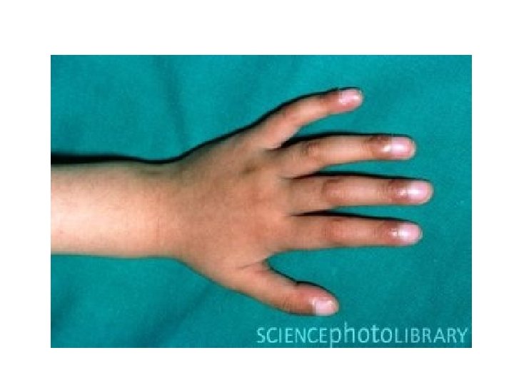
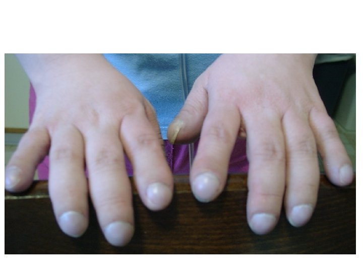
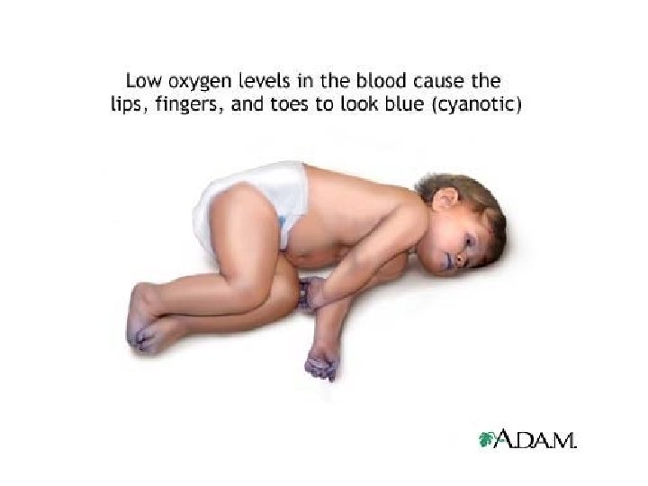
Inspection Hands, Face and Neck – Finger clubbing – Cyanosis-(perypheral or central) – General appearance, e. g. Cushingoid as a result of long-term use of steroids – Anemia (conjunctiva) – Goitre (any possible tracheal obstruction) – Lymphadenopathy
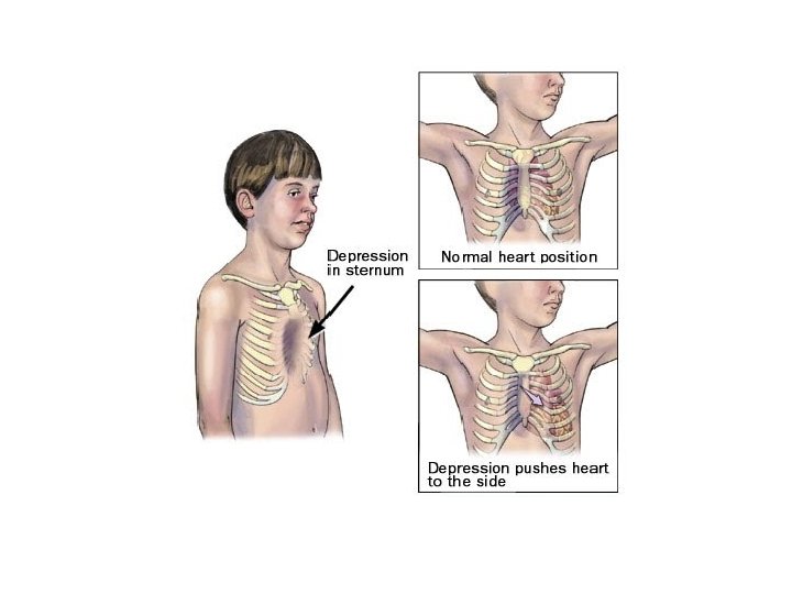
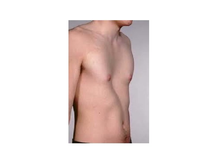
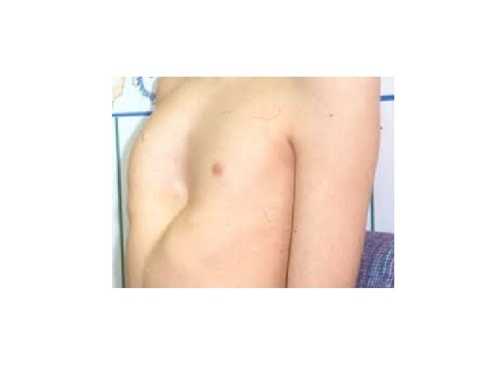
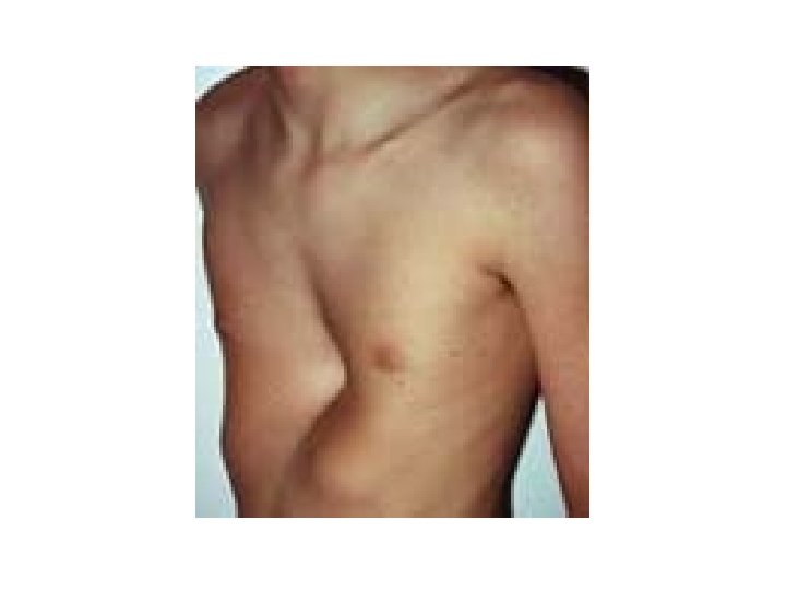
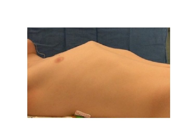
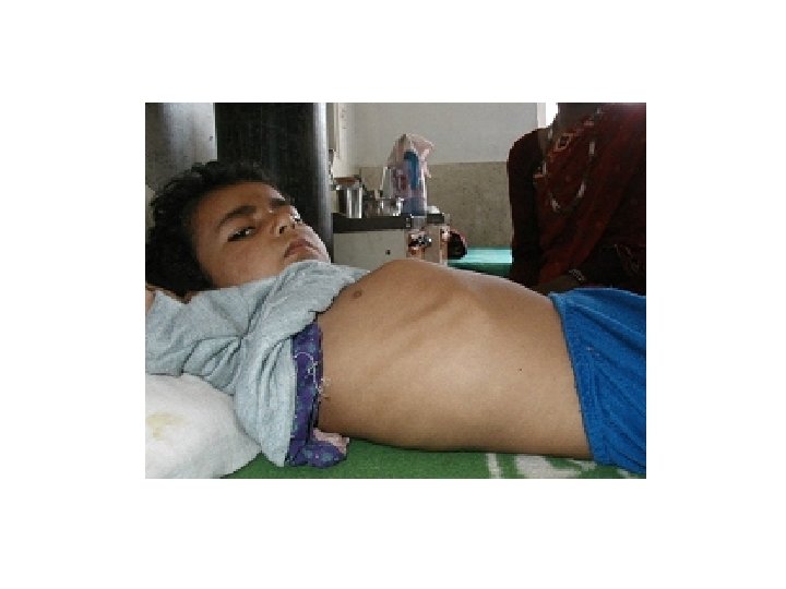
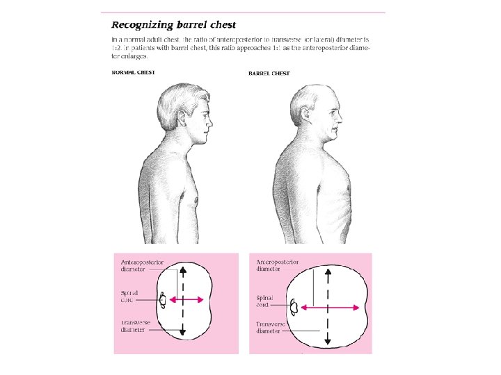
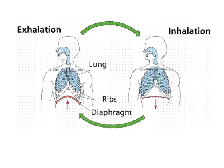
Inspection Chest • Chest shape: – Overinflated (severe acute asthma) – Asymmetry (the abnormality is on the side that moves less, e. g. pneumothorax, collapse, consolidation or effusion) – Other abnormalities include pigeon chest (pectus carinatum), funnel chest (pectus excavatum), kyphosis and/or scoliosis – Respiratory rate • Operation scars • Paradoxical chest movement may indicate a fractured rib
Respiratory rate Newborn 1 mo-1 yr 1 -5 yrs 5 -12 yrs >12 yrs 40 (30 -60) 25 -35 20 -30 20 -25 16 -20
Dyspnea • Exaggerated use of accessory muscles • Intercostal, supraclavicular, and subcostal retractions • Flaring of nostrils • Tachypnea
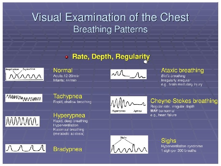
Breathing patterns • Apnea: Respiratory arrest longer than 15 (20) sec; generally bradicardia and cyanosis accompanies • Periodic respiration: In preterm babies; in the first days of life, repeating respiratory arrest attacks, shorter than 20 sec.
Breathing patterns • Cheyne-Stokes: Progressively deeper breathing followed by temporary apnoea, which may occur with heart failure, cerebrovascular disease, head injury, carbon monoxide poisoning or brain tumors, or be a normal variant during sleep or at high altitude • Ortopnea: Breathing only in upright position (heart failure ) • Bradypnea: Low frequence breathing (metabolic alkalosis and CNS depression) • Hyperpnea: Rapid deep breathing
Breathing patterns • Hypopnea: Slow and shallow breathing • Kussmaul: Deep and rapid breathing, (often associated with severe metabolic acidosis) • Paradoxical: Inspiratory depression of thorax (intercostal muscle paralysis, URT obstruction, preterm baby)
Auscultation • • Infants; in arms Children; upright position Hands over head for axillar area ( big children) Ask the children to take deep breaths in and out with their mouth open • Infants are better audible while crying • Place the stethoscope over each of the 5 lobes of the lungs in turn, on the front and back of the chest
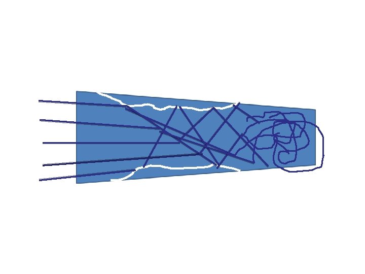
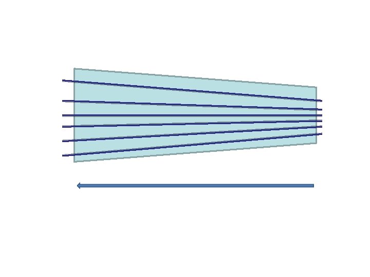
Auscultation • Normal breath sounds are called vesicular • They are described as quiet and gentle • There is usually no gap between the inspiratory and expiratory phase sounds • If; – Inspirium is longer (normal) – Expirium is longer (obstructive problem)
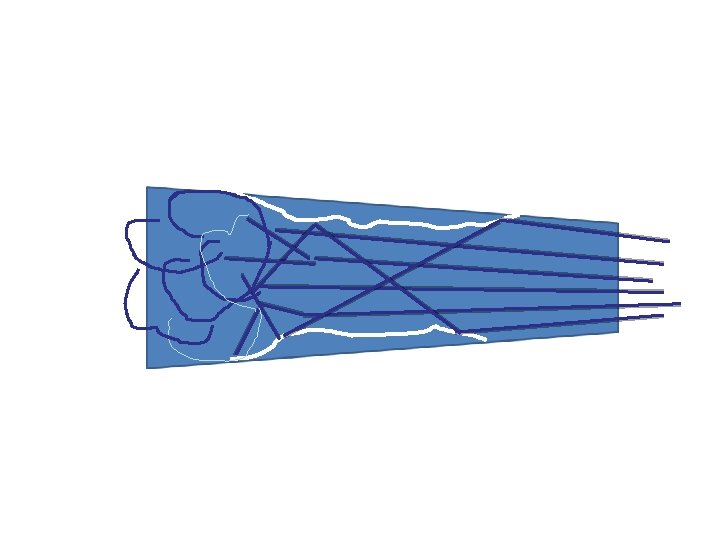
Pay attention to; • Inspirium-expirium relationship • Adititonal sounds • Dispersion of breathing sounds
Auscultation • Pathologic sounds – Rales: Generally inspiratory • Coarse (Trcheitis, bronchitis) • Sucrepitan (Bronchitis, bronchiolitis, bronchectasis, tbc) • Crepitan (Pneumonia, pulmonary congestion, edema) – Rhonchi: Generally expiratory • Sonor (ronflan rale) (Foreign body aspiration, Asthma, bronchitis) • Sibilan (wheezing, sibilan rale)(Asthma, bronchiolitis, cystic fybrosis)
Patolojik solunum sesleri Raller Kaba raller Ronküsler İnce raller (Krepitan) Orta raller (sub krepitan, sukrepitan) Sonor ronküs Sibilan ronküs (Sibilan ral) (Wheezing)
Auscultation • Rales (sometimes called crackles): – Probably represent opening of small airways and alveoli – They may be normal at the lung bases if they clear on coughing or after taking a few deep breaths – Typical finding of bronchopneumonia – Basal rales are a classical feature of pulmonary congestion with left ventricular failure – May be more diffuse in pulmonary fibrosis
Auscultation • Rhonchi (wheezes): – Musical sound heard on expiration. In severe cases they may be both inspiratory and expiratory. Imply narrowing of the airways – The loudness of rhonchi gives no indication of the severity of the condition – Typical in; Bronchial asthma and bronchiolitis
Auscultation • Tuber sufl: – Generated by turbulent air flow in large airways (similar sounds can be heard in healthy patients by listening over the trachea) – Sounds are harsh and poor in nature. Unlike normal vesicular breath sounds, there is a gap between the inspiratory and expiratory phase sounds – Bronchial breathing suggests consolidation or fibrosis, which permits the sound to be conducted more effectively to the chest wall
Auscultation • Pleural rub (=frotman): A creaking sound caused by stiff pleural membranes such as with pleurisy • Stridor: harsh inspiratory sound caused by partial obstruction of a large airway
Auscultation • Vocal resonance: – Place the stethoscope at various levels over the back and ask the patient to whisper “ 41 -42" each time. Note how well the sound is transmitted – The sound is muffled over a normal lung, increased if there is consolidation, and decreased or absent if there is effusion or collapse • Whispering pectoriloquy: – Is elicited as for vocal fremitus but ask the patient to whisper "one, two, three" – Whispering pectoriloquy is the increased quality and loudness of whispers that are heard with a stethoscope over an area of lung consolidation
Palpation • Use the index finger to feel the trachea and to determine whether the trachea feels central or is deviated – The trachea is deviated away from pneumothorax and effusion and towards collapse and consolidation – The trachea may also be deviated by a mass, e. g. enlarged lymph nodes
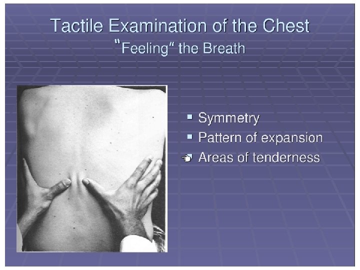
Palpation • Chest expansion: – Usual chest expansion in an adult is 4 -5 cm and should be symmetrical – Symmetrical reduction: overinflated lungs (e. g. bronchial asthma, emphysema), stiff lungs (e. g. pulmonary fibrosis), ankylosing spondylitis. – Asymmetrical reduction of chest wall expansion: absent expansion (e. g. empyema and pleural effusion) or reduced expansion (e. g. pulmonary consolidation and collapse)
Palpation • Tactile vocal fremitus: – To assess tactile vocal fremitus, use the ulnar side of the hand, by the hypothenar eminence with the palms facing upwards. Place it at various levels over the back, each time asking the patient to say "ninety-nine". Note how the sound is transmitted to the hand – Tactile vocal fremitus is increased over areas of consolidation and decreased or absent over areas of effusion or collapse • Feel for the apex beat of the heart; it will be displaced if the mediastinum is displaced or distorted
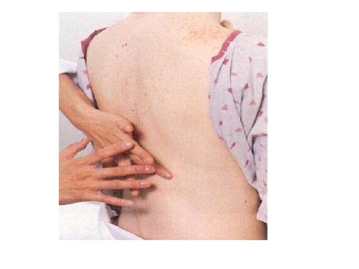
Percussion • For percussion of the chest, it is usual to use the middle finger of the dominant hand to do this • The chest is percussed by placing the nondominant hand on the chest and using the dominant middle finger to tap the other middle finger over the middle phalanx • A hyper-resonant sound suggests hyperinflation or a pneumothorax • A dull sound is easier to distinguish from normal. It may suggest collapse or consolidation, or a pleural effusion
Percussion • Dullness – – – Plural fluid Consolidation Wide atelectasis Tumor Pleural thickening • Hypersonor – Pneumothorax) – Obstructive disease • Asthma • Bronchiolitis • Emphysema
RESULT • • • Quiet environment Child’s at least upper body naked With a trusted parent Be patient EXPERIENCE IS VERY IMPORTANT AUSCULT, AUSCULT