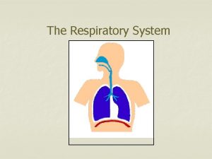
Respiratory System cells need oxygen to release energy


- Slides: 26

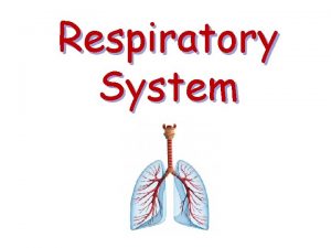
Respiratory System

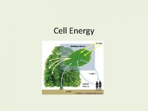
�cells need oxygen to release energy from food �without energy, cells cannot carry out their main functions �respiration ensures that the body gets the energy needed to function

Respiration involves: �External respiration (breathing) � inhaling oxygen from the air and exhaling to expel carbon dioxide from the lungs � O 2 is taken up by capillaries of lung alveoli and CO 2 is released from blood �Internal respiration � metabolic process O 2 is released to tissues /cells and CO 2 is transferred to the blood �Cellular respiration � O 2 is used by cells for producing energy ATP (adenosine triphosphate)

Image from: http: //web. carteret. edu/keoughp/LFreshwater/CPAP/Circulatory/circulatory_class_notes_files/image 012. jpg

Respiratory System Structures

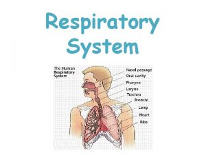
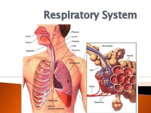
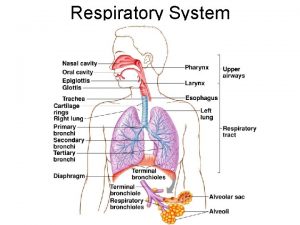
nasal cavity pharynx epiglottis larynx trachea lung bronchus bronchiole diaphragm

Nasal Cavity �separated from the mouth cavity by a bony platform called the hard palate �nasal cavity warms, moistens, and cleans the air before it hits the lungs �many blood vessels help warm the air (surface area is important!) �epithelial cells provide moisture and mucus Image from: http: //www. medart. plus. com/Artwork_GIFs/nasalcav. GIF

Pharynx �forms a tube common to respiratory and digestive systems �extends from back of nasal cavity to the larynx (voice box) �nasopharynx (top part) � have ciliated epithelial cells that trap fine particles � contains the tonsils and adenoids (lymphoid tissue) �oropharynx and laryngopharynx (bottom part) � behind the mouth – passage for food and air � epithelial cells here are tough because of the wear and tear from food passage �divides into two tubes: esophagus (food) and larynx (air)

Image from: http: //www. cedars-sinai. edu/Patients/Programs-and-Services/Head-and-Neck-Cancer-Center/Treatment/Images/pharynx_openmouth_web-80182. jpg

uvula : What is it for? __________________ Image from: http: //mentalfloss. com/article/30806/what-does-dangly-thing-back-your-throat-do

Larynx �box-like structure at the opening of the airway �formed by cartilage structures to prevent collapse �thyroid cartilage is largest – makes the Adam’s apple �leaf-like flap is called the epiglottis �prevents food from entering airway Images from: http: //www. cedars-sinai. edu/Patients/Programs-and-Services/Head-and-Neck-Cancer-Center/Treatment/Images/larynx_model_web-80443. jpg http: //0. tqn. com/d/np/singing/41 -1. jpg

vocal cords – air passage causes them to vibrate and produce sounds (VIDEO: http: //www. youtube. com/watch? v=-XGds 2 GAv. GQ ) �muscles tighten or loosen the flaps to control pitch �thickness and length of the vocal cords also affect sound �we shape the mouth and position the tongue to modify the sounds (some languages also use uvula) Images from: http: //www. cancer. gov/publications/patient-education/wyntk-larynx/All. Pages

Trachea �tube, about 12 cm, made of smooth muscle with “C” shaped cartilage rings to support it �gap in rings is at back against esophagus rings support the trachea while allowing esophagus to expand during swallowing Image from: http: //bastianmedicalmedia. com/wp-content/gallery/trachea/6 -trachea. jpg

Bronchi, Bronchioles, and Alveoli �trachea splits into two bronchi (also have rings) for the left and right lungs �bronchi branch into more tubes called bronchioles , and these continue to branch into subsequently smaller tubes (smaller ones lack rings) �alveolar ducts on the ends of the branches lead into chambers (air sacs) called alveoli Image from: http: //www. abpischools. org. uk/res/coresourceimport/resources 04/asthma/images/5 c 2 Alv. Brjpg. gif

Lungs �each bronchus plus all of its bronchioles and alveoli make up a lung �lungs have no muscles �are elastic and can respond passively to the action of the rib muscles and diaphragm Image from: http: //guardianlv. com/wp-content/uploads/2014/02/Human-Lungs-Being-Grown-in-Fish-Tanks-State-Texas-Scientists. jpg

�we have two lungs – the right has three lobes and the left has two (why is the left smaller? ) �well-protected by the ribs and supported by the diaphragm GIF from: http: //stream 1. gifsoup. com/view 6/4645075/lungs-breathing-o. gif

�many of the epithelial cells of the entire respiratory tract secrete mucus and have cilia to trap and remove particles �cilia beat upwards so that the mucus can be swallowed (yum!) or discharged by coughs and sneezes Image from: https: //elcaminogmi. dnadirect. com/img/content/tests/pcd/trachea. jpg

�this gets rid of potentially dangerous material, including bacteria �not always successful – people get diseases like emphysema and lung cancer – especially smokers (nicotine paralyses the cilia ) Image from: http: //media-cache-ak 0. pinimg. com/736 x/ed/00/44/ed 004490 d 4 fbfb 3 b 27 ebec 88 c 261 ab 6 b. jpg

Pleura �two membrane sacs that contain the lungs �outer sac is stuck to the inner thoracic cavity �inner sac is stuck to the lungs �are close together with a thin film of fluid between them Image from: http: //mesothelioma-navy. com/wp-content/uploads/2012/10/Pleural-mesothelioma-diagram-of-lung-Converted-300 x 300. png

The fluid is important! �isolates each lung �lubricates lungs for less friction, and the fluid tension adheres the lungs to the thoracic cavity �the fluid cannot be compressed, so the lungs follow the fluid when breathing in and out �disrupt the pleura and you can get a collapsed lung Image from: http: //www. primehealthchannel. com/wp-content/uploads/2013/03/Collapsed-lung-Picture. gif

Mechanics of Breathing

How breathing works… �we must force air in and out of the system to get air fast enough to live (diffusion is not enough) �ordinary breathing is involuntary – we do not think about it but we do have some voluntary control �the diaphragm is a dome-shaped muscle that bulges upward (located right under the lungs) �it flattens somewhat when contracted �works in unison with the intercostal muscles of the 12 pairs of ribs

Image from: https: //www. xtremepapers. com/images/gcse/biology/the_respiratory_system/inhalation_exhalation. png

Inhalation/inspiration (active) �the diaphragm and external intercostal muscles contract �diaphragm is dome-shaped when relaxed, but contracts and lowers during inhalation �intercostals pull the ribs up and outward �the volume inside lungs increases �air pressure inside the alveoli decreases, creating a partial vacuum (negative pressure) the difference in pressure between the alveoli and the atmosphere causes air to rush into the alveoli **air comes in because the lung opens up** **the lungs do not open up because air is forced in! ** �inspiration is active (contraction), but the flow of air is passive (negative pressure)

Exhalation/expiration �is usually passive no effort is required (relaxing) �the thoracic cavity is elastic – diaphragm relaxes and it recoils back into place �intercostals relax and the rib cage moves down and inward �air pushes out, lung volume drops �active expiration is possible – the inner layer of intercostal muscles can contract and force the rib cage downward and inward �also, the abdominal wall muscles can push on the viscera, and push up the diaphragm, expelling more air

OVERVIEW �diaphragm and intercostal muscles contract �ribs pull up and forward �volume of chest goes up due to negative pressure � inhale �diaphragm and intercostal muscles relax �ribs move down and inward �the volume drops because air is physically forced out � exhale
 Why do cells need oxygen?
 Respiratory zone
 Modified release dosage forms
 Ocusert definition
 Extended release vs sustained release
 How does a cell obtain energy
 Why does a cell need energy
 Circularory system
 Goblet cells of respiratory epithelium
 What compounds do cells need
 Why do we need cells
 What cells need isotonic solutions to be at homeostasis
 Onodi cells and haller cells
 Regulation of tubular reabsorption
 Thyroid parafollicular cells
 Haploid and diploid venn diagram
 Why dna is more stable than rna?
 Chlorocruorin
 Prokaryote vs eukaryote worksheet
 Plant vs animal cell venn diagram
 Prokaryotic cells vs eukaryotic cells
 Cell organelle jeopardy
 Masses of cells form and steal nutrients from healthy cells
 Younger cells cuboidal older cells flattened
 Cuál es la diferencia entre la célula animal y vegetal
 Is a red blood cell prokaryotic or eukaryotic
 Nondisjunction in meiosis
