Respiratory System Respiratory System Oxygen Delivery System The


. Respiratory portion of")


- Slides: 18
Respiratory System
Respiratory System: Oxygen Delivery System The respiratory system is the set of organs that allows a person to breathe and exchange oxygen and carbon dioxide throughout the body. The integrated system of organs involved in the intake and exchange of oxygen and carbon dioxide between the body and the environment and including the nasal passages, larynx, trachea, bronchial tubes, and lungs. The respiratory system performs two major tasks: Exchanging air between the body and the outside environment known as external respiration. Bringing oxygen to the cells and removing carbon dioxide from them referred to as internal respiration.
1. 2. 3. 4. 5. 6. Functions of Respiratory system: Supplies the body with oxygen and disposes of carbon dioxide Filters inspired air Produces sound Contains receptors for smell Removal of some excess water and heat Helps regulate blood p. H Breathing (pulmonary ventilation). consists of two cyclic phases: Inhalation, also called inspiration - draws gases into the lungs. Exhalation, also called expiration - forces gases out of the lungs.
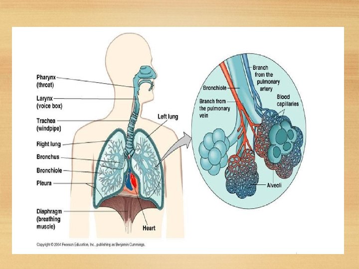
: External Respiration: Upper respiratory tract Air from the outside environment enters the nose or mouth during inspiration (inhalation). Composed of the nose and nasal cavity, paranasal sinuses, pharynx (throat), larynx. All part of the conducting portion of the respiratory system.
Nose: Also called external nares. Divided into two halves by the nasal septum. Contains the paranasal sinuses where air is warmed. Contains cilia which is responsible for filtering out foreign bodies.
Internal nares - opening to exterior External nares - opening to pharynx Nasal conchae - folds in the mucous membrane that increase air turbulence and ensures that most air contacts the mucous membranes Provides an airway for respiration Moistens and warms entering air Filters and cleans inspired air Resonating chamber for speech Detects odors in the air stream
Paranasal Sinuses Four bones of the skull contain paired air spaces called the paranasal sinuses - frontal, ethmoidal, sphenoidal & maxillary. Decrease skull bone weight. Warm, moisten and filter incoming air. Add resonance to voice. Communicate with the nasal cavity by ducts. Lined by pseudostratified ciliated columnar epithelium.
Pharynx Common space used by both the respiratory and digestive systems. Commonly called the throat. Originates posterior to the nasal and oral cavities and extends inferiorly near the level of the bifurcation of the larynx and esophagus. Common pathway for both air and food. Walls are lined by a mucosa and contain skeletal muscles that are primarily used for swallowing. Flexible lateral walls are distensible in order to force swallowed food into the esophagus.
Three Sections of the Pharynx Nasopharynx Superior-most region of the pharynx. Covered with pseudostratified ciliated columnar epithelium Oropharynx Back portion of the mouth that Contains non-keratinized stratified squamous epithelium Laryngopharynx bottom section of the pharynx where the respiratory tract divides into the esophagus and the larynx.
Lower Respiratory Tract Conducting airways (trachea, bronchi, up to terminal bronchioles). Respiratory portion of the respiratory system (respiratory bronchioles, alveolar ducts, and alveoli).
Larynx: Voice box is a short, somewhat cylindrical airway ends in the trachea. It connects throat (pharynx) with the windpipe(trachea). At the top of the larynx is the epiglottis. This small flap prevents food & water from entering the voice box. Supported by a framework of nine pieces of cartilage (three individual pieces an three cartilage pairs) that are held in place by ligaments and muscles.
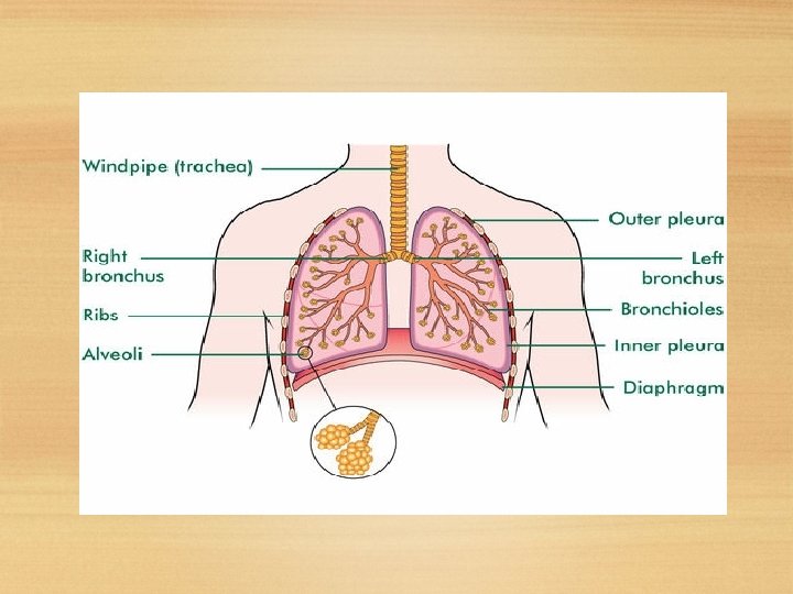
Trachea A flexible tube also called windpipe. Extends through the mediastinum and lies anterior to the esophagus and inferior to the larynx. Cartilage rings reinforce and provide rigidity to the tracheal wall to ensure that the trachea remains open at all times. At the level of the sternal angle, the trachea bifurcates into two smaller bronchi. tubes, called the right and left primary Each primary bronchus projects laterally toward each lung. .
Lung s Each lung has a conical shape. Its wide, concave base rests upon the muscular diaphragm. Both lungs are bordered by the thoracic wall anteriorly, laterally, and posteriorly, and supported by the rib cage. Toward the midline, the lungs are separated from each other by the mediastinum. Left lung divided into 2 lobes by oblique fissure smaller than the right lung cardiac notch accommodates the heart Right lung divided into 3 lobes by oblique and horizontal fissure located more superiorly in the body due to liver on right side
The outer surface of each lung and the adjacent internal thoracic wall are lined by a serous membrane called pleura. The outer surface of each lung is tightly covered by the visceral pleura. while the internal thoracic walls, the lateral surfaces of the mediastinum, and the superior surface of the diaphragm are lined by the parietal pleura (covering the rib cage). Pleural Cavities The potential space between the two pleura of the lungs is a pleural cavity. The pleural membranes produce a thin, serous pleural fluid that circulates in the pleural cavity and acts as a lubricant, ensuring minimal friction during breathing. Pleural effusion – pleuritis with too much fluid
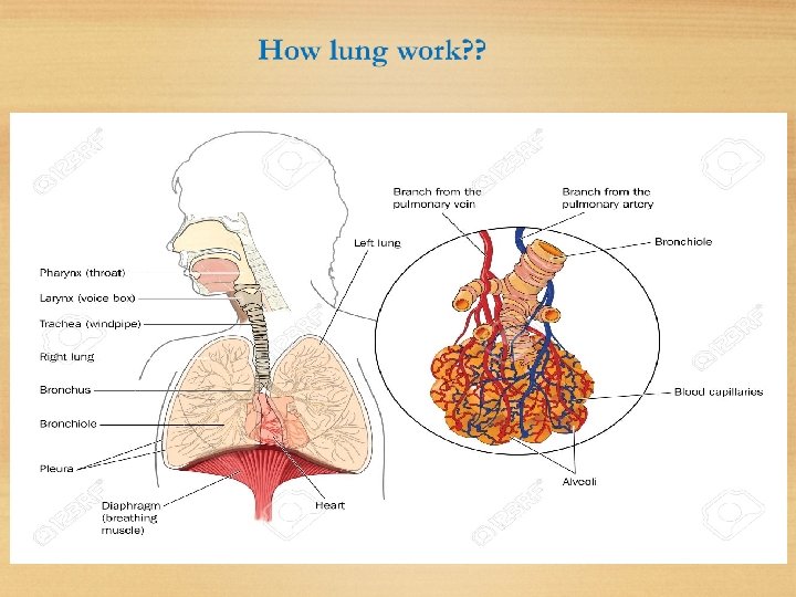
How lung work? ? Air enters your lungs through a system of pipes called the bronchi. The alveoli are where the important work of gas exchange takes place between the air and your blood. Covering each alveolus is a whole network of little blood vessel called capillaries, It is important that the air in the alveoli and the blood in the capillaries are very close together, so that oxygen and carbon dioxide can move (or diffuse) between them. When you breathe in, air comes down the trachea and through the bronchi into the alveoli. This fresh air has lots of oxygen in it, and some of this oxygen will travel across the walls of the alveoli into your blood stream. Travelling in the opposite direction is carbon dioxide, which crosses from the blood in the capillaries into the air in the alveoli and is then breathed out. In this way, you bring in to your body the oxygen that you need to live, and get rid of the waste product carbon dioxide.