PETCT Fusion Imaging and SUV Assessment Using MIMvista






- Slides: 38
PET-CT Fusion Imaging and SUV Assessment Using MIMvista (Resisting RECIST) Pete Anderson MD, Ph. D Professor, Pediatrics pmanders@mdanderson. org
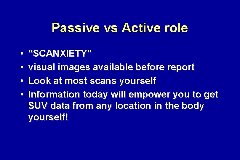
Passive vs Active role • • “SCANXIETY” visual images available before report Look at most scans yourself Information today will empower you to get SUV data from any location in the body yourself!
PET-CT Imaging • • • Standard Uptake Value (SUV) FDG is the prototype : “ I Have A DREAM” Sensitive means to document responses Numeric data from PET-CT using Mimvista Downloading of DICOM images from QC CD for central review (e. g. SARC studies)
Assessing Response of Sarcomas. NOT Always Easy • Clinical: pain improved, alkaline phosphatase and/or LDH better, exam better function • Imaging: shrinkage on CT (Choi) and MRI • Osteosarcoma: bone scan changes very slowly and can even increase if some calcification associated with healing occurs • Sometimes “more” or “worse” lung metastases are just increased calcification
RECIST (Response Evaluation Criteria in Solid Tumors) • 30% decrease in sum of longest diameter of target lesion(s): PR • PROG >20% • CR (complete disappearance)- unusual • Most: stable (but patient seems better!) • Ignores stromal or bone components
PET-CT Imaging: A Reality Check for the Both Clinician and Patient • Is the ongoing or proposed treatment worth the toxicity (chemo) or morbidity *Surgery/RT • Any disease elsewhere? • 3 -D imaging: “Seeing is believing” • Helps set priority of intervention(s) such as local control vs chemotherapy
MIMvista PET-CT Fusion Images • First-hand info about tumor location • FUSION images allow clinician to determine where highest FDG uptake is in-around-near a target lesion. YOU get the SUV! • VISUAL PICTURE and numeric data of where there is active tumor • LOCAL Control strategy (surgery RT, RFA), more chemotherapy , or just follow-up after therapy (esp RT) – “at a glance”
MIMvista Color Fusion Images • MIMvista for desktop PC (Cleveland OH (www. mimvista. com) • Right click on image (e. g. in MD Anderson Clinic. Station or other) • DICOM images are downloaded into the MIMvista program (~1 -2 min) • Displays fusion images for YOUR analysis
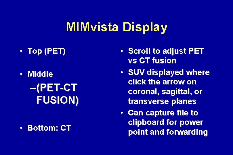
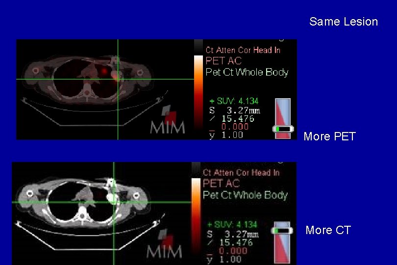
MIMvista Display • Top (PET) • Middle – (PET-CT FUSION) • Bottom: CT • Scroll to adjust PET vs CT fusion • SUV displayed where click the arrow on coronal, sagittal, or transverse planes • Can capture file to clipboard for power point and forwarding
Same Lesion More PET More CT
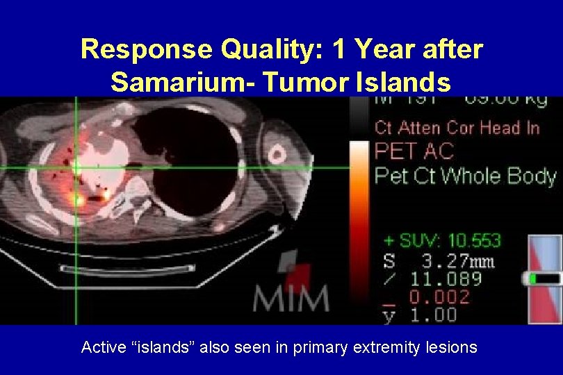
SUV on a Fusion Image - anywhere you point the mouse • All areas of tumor not alike: heterogeneity of edges of tumor vs middle of tumor • Areas near or in vital structures (e. g. spine, mediastinum, liver, chest wall) • Ossified lesions may have small “islands” of intense FDG uptake (incomplete response to chemotherapy, focal relapse)
Indicator (Target) Lesion • More PR than RECIST (Mahajan SIOP) –RT of OS 0/8 by RECIST – 6/8 improved SUV on PET-CT • PET-CT may allow better F/U after RT + chemo
MIMvista PET-CT Fusion Examples • Osteosarcoma • Ewings
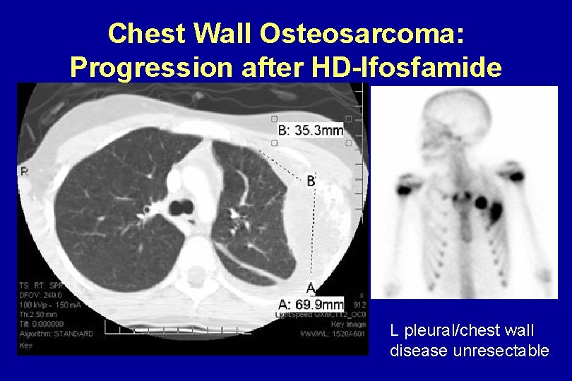
Chest Wall Osteosarcoma: Progression after HD-Ifosfamide L pleural/chest wall disease unresectable
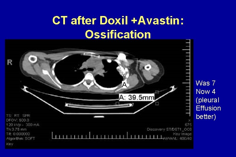
CT after Doxil +Avastin: Ossification Was 7 Now 4 (pleural Effusion better)
PET: Avid Hmmmm……
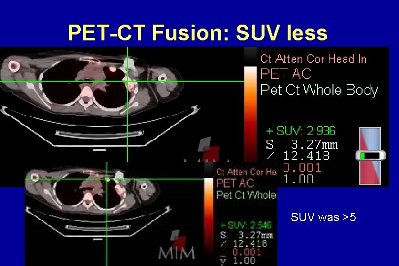
PET-CT Fusion: SUV less SUV was >5
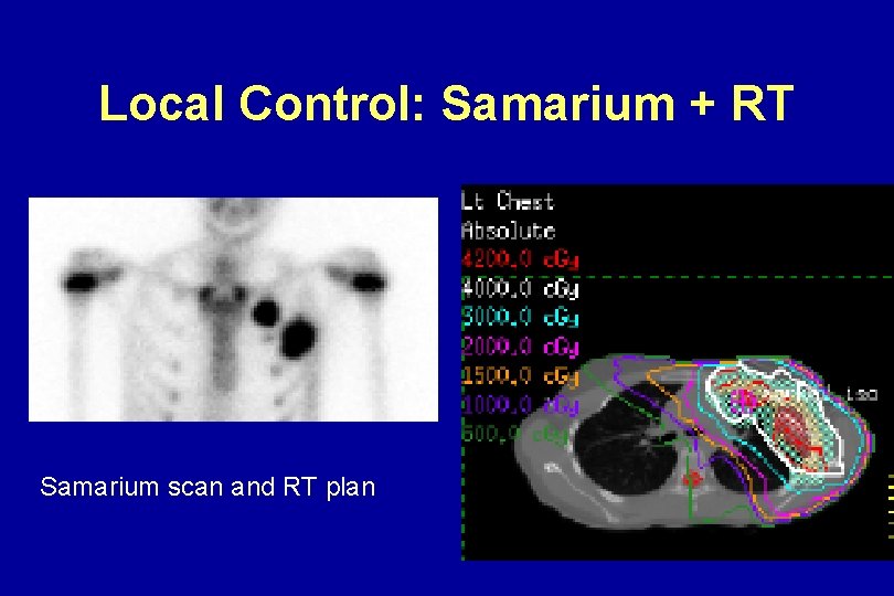
Local Control: Samarium + RT Samarium scan and RT plan
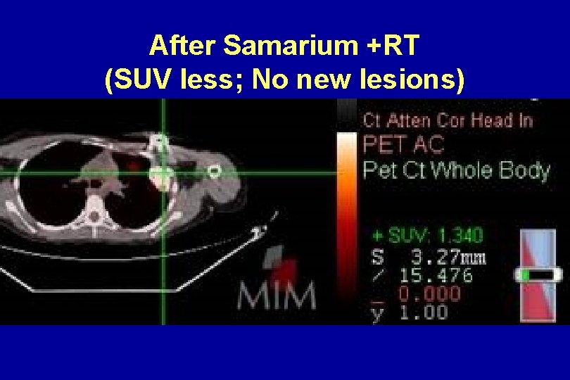
After Samarium +RT (SUV less; No new lesions)
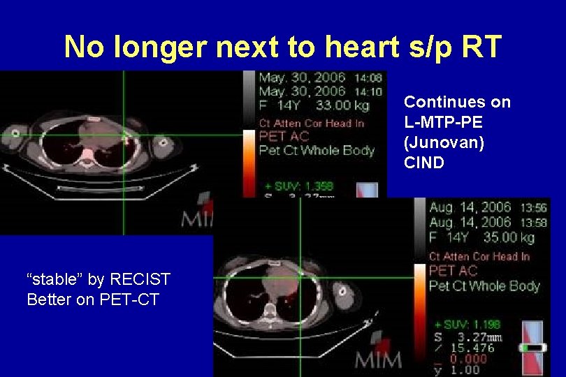
No longer next to heart s/p RT Continues on L-MTP-PE (Junovan) CIND “stable” by RECIST Better on PET-CT
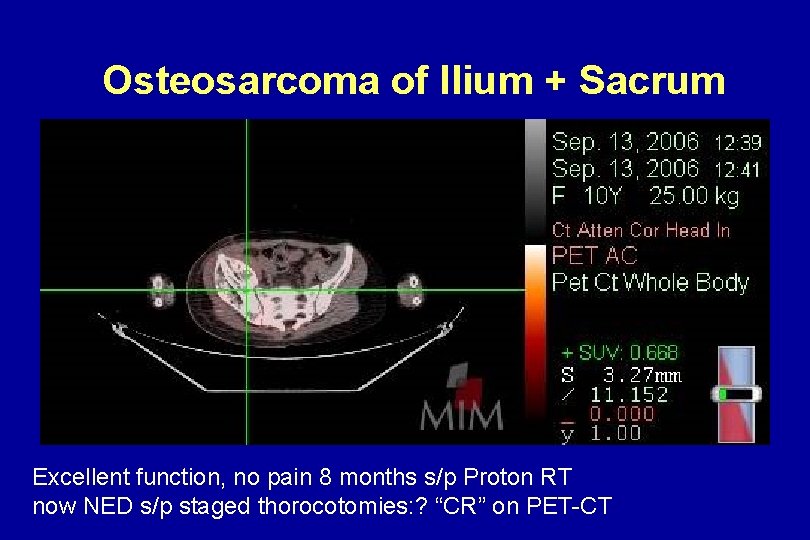
Osteosarcoma of Ilium + Sacrum Excellent function, no pain 8 months s/p Proton RT now NED s/p staged thorocotomies: ? “CR” on PET-CT
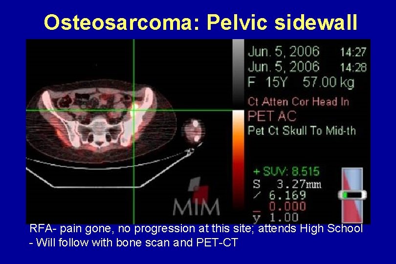
Osteosarcoma: Pelvic sidewall RFA- pain gone, no progression at this site; attends High School - Will follow with bone scan and PET-CT
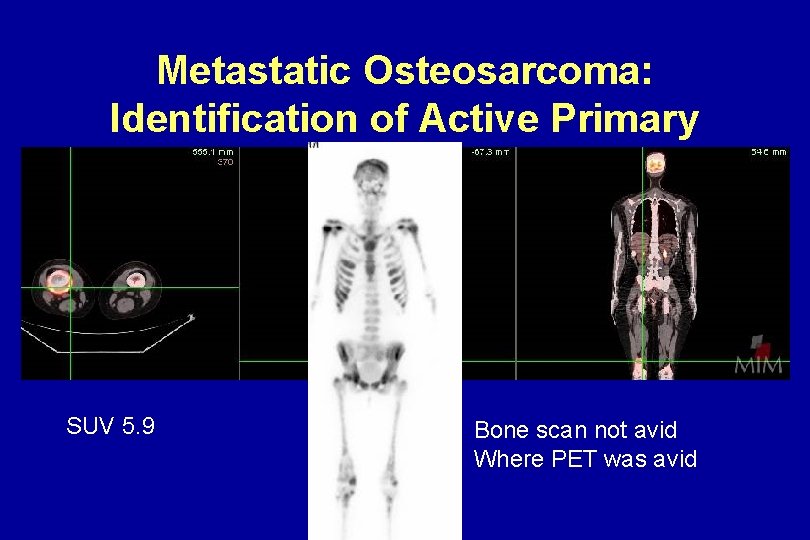
Metastatic Osteosarcoma: Identification of Active Primary SUV 5. 9 Bone scan not avid Where PET was avid
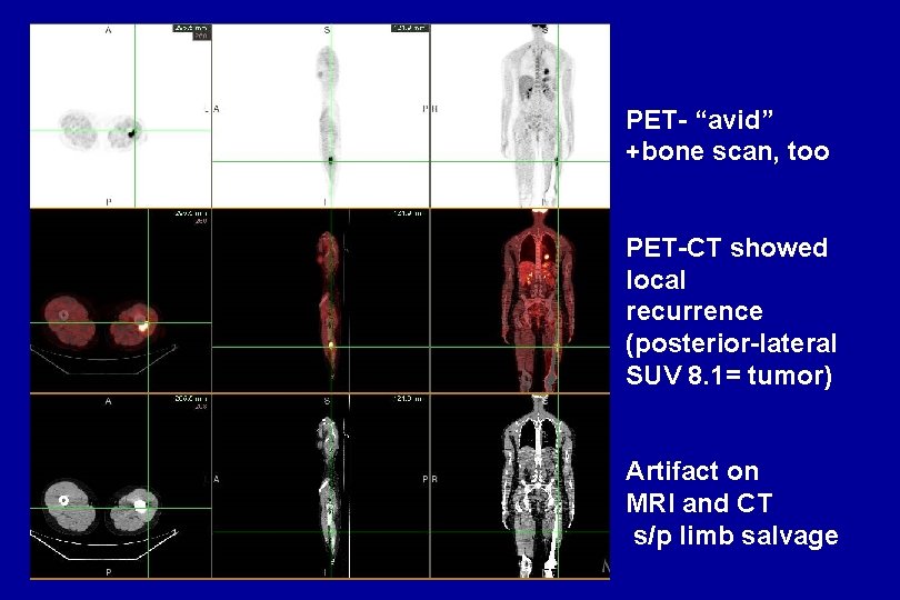
PET- “avid” +bone scan, too PET-CT showed local recurrence (posterior-lateral SUV 8. 1= tumor) Artifact on MRI and CT s/p limb salvage
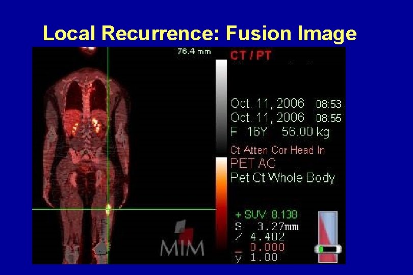
Local Recurrence: Fusion Image
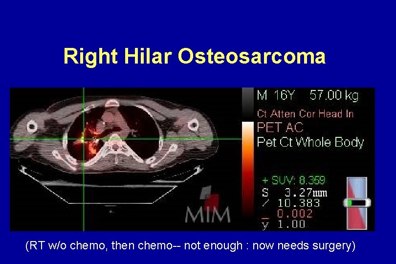
Right Hilar Osteosarcoma (RT w/o chemo, then chemo-- not enough : now needs surgery)
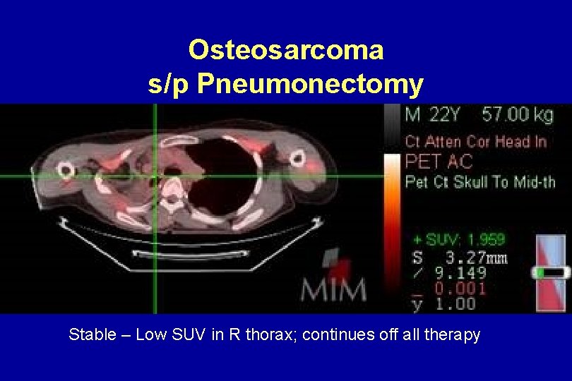
Osteosarcoma s/p Pneumonectomy Stable – Low SUV in R thorax; continues off all therapy
Response Quality: 1 Year after Samarium- Tumor Islands Active “islands” also seen in primary extremity lesions
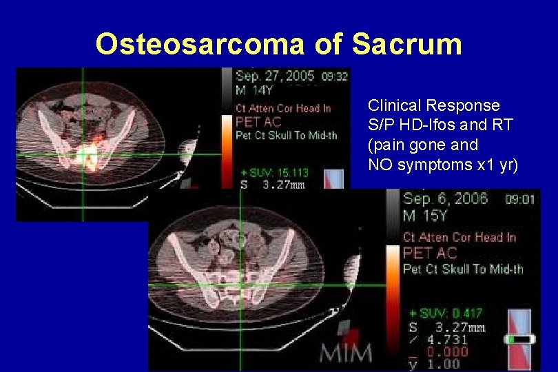
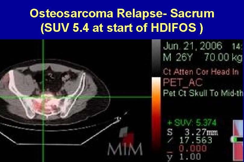
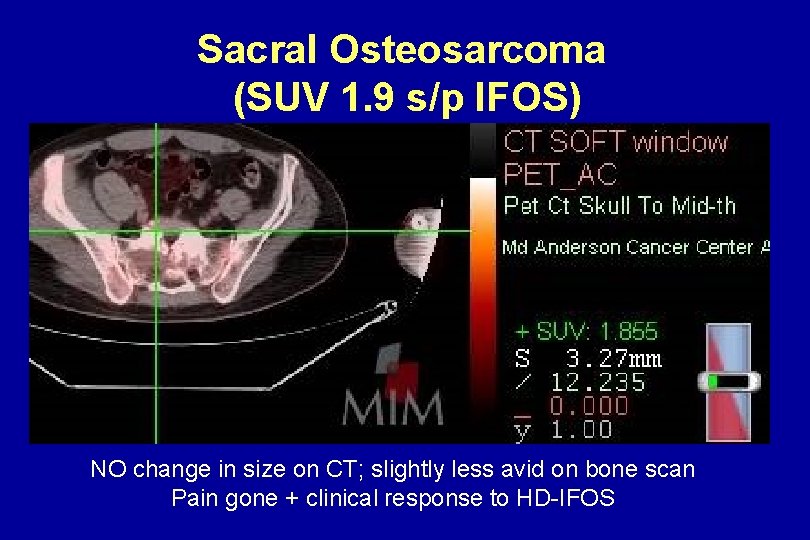
Osteosarcoma of Sacrum Clinical Response S/P HD-Ifos and RT (pain gone and NO symptoms x 1 yr)
Osteosarcoma Relapse- Sacrum (SUV 5. 4 at start of HDIFOS )
Sacral Osteosarcoma (SUV 1. 9 s/p IFOS) NO change in size on CT; slightly less avid on bone scan Pain gone + clinical response to HD-IFOS
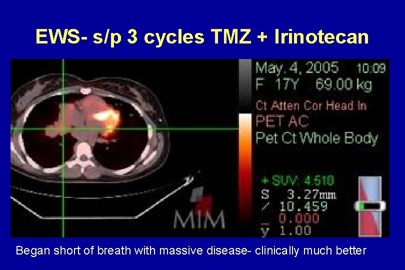
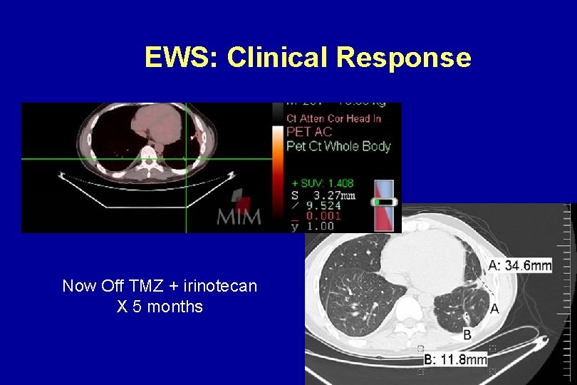
EWS- s/p 3 cycles TMZ + Irinotecan Began short of breath with massive disease- clinically much better
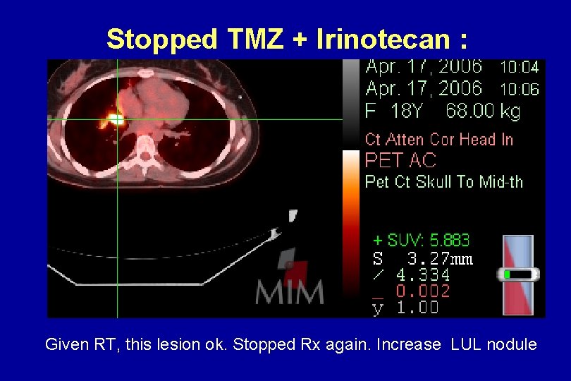
Stopped TMZ + Irinotecan : Relapse Given RT, this lesion ok. Stopped Rx again. Increase LUL nodule
EWS: Clinical Response Now Off TMZ + irinotecan X 5 months
Summary • MIMvista PET-CT fusion can provide same -day visual information to clinicians and patients about FDG metabolic activity • PET-CT can now be used in the clinic for to capture numeric response data (SUV) • SUV obtained-anywhere you click • Additional data possible (tumor volumes) • Image capture and sharing possible
Conclusion • MIMvista information can help YOU to communicate better -with patients, families, referring physicians, surgeons, radiotherapists • Response data? SARC to determine • COLOR fusion images with numeric data provides best of both worlds in the clinic – Response + numbers “at a glance”
THANKS! Questions? pmanders@mdanderson. org