Peptic ulcer disease Dr Tarek Mazzawi MD Ph

- Slides: 42
Peptic ulcer disease Dr. Tarek Mazzawi, MD, Ph. D Department of Internal Medicine Faculty od Medicine Al-Balqa’a Applied University 2020
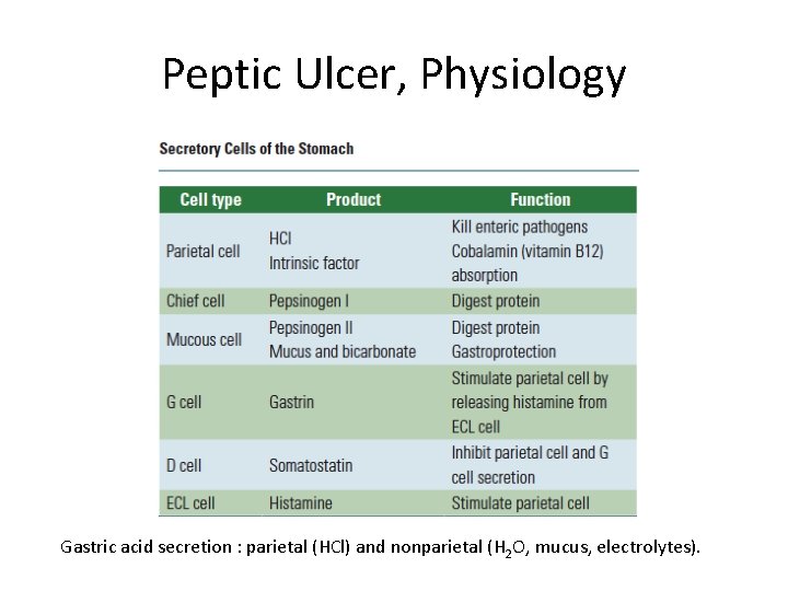
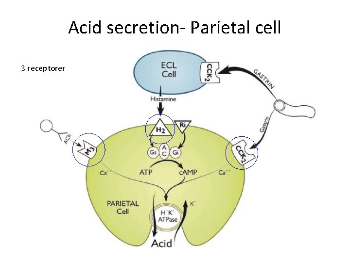
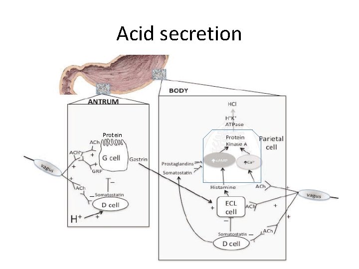
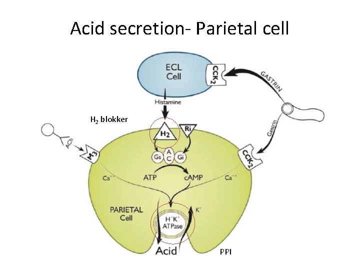
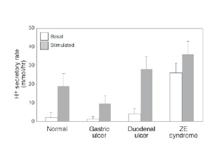
Peptic Ulcer, Physiology Gastric acid secretion : parietal (HCl) and nonparietal (H 2 O, mucus, electrolytes).
Acid secretion- Parietal cell 3 receptorer
Acid secretion Protein _ _
Acid secretion- Parietal cell H 2 blokker PPI
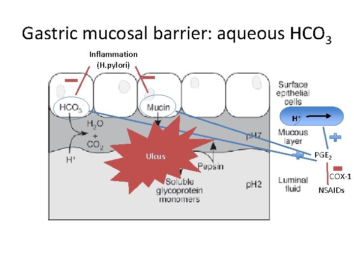
Gastric mucosal barrier: aqueous HCO 3 Inflammation (H. pylori) H+ Ulcus PGE 2 COX-1 NSAIDs
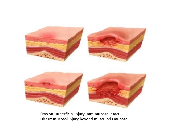
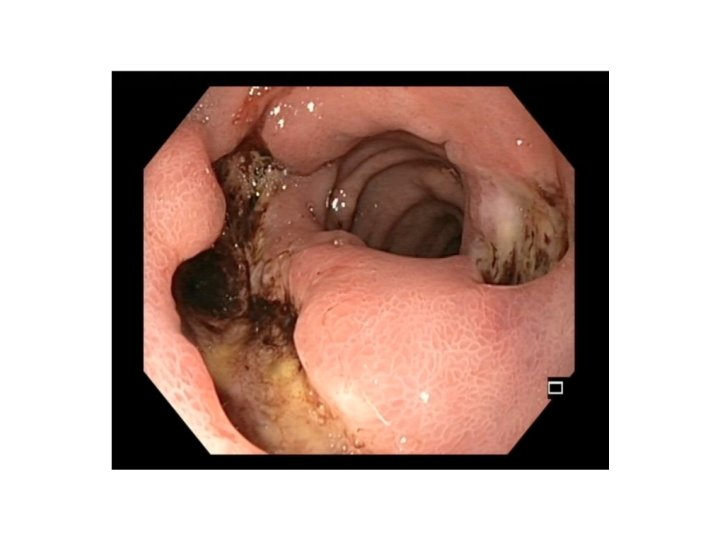
Erosion: superficial injury, mm. mucosa intact. Ulcerr: mucosal injury beyond muscularis mucosa.
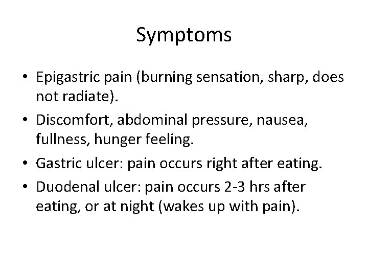
Symptoms • Epigastric pain (burning sensation, sharp, does not radiate). • Discomfort, abdominal pressure, nausea, fullness, hunger feeling. • Gastric ulcer: pain occurs right after eating. • Duodenal ulcer: pain occurs 2 -3 hrs after eating, or at night (wakes up with pain).
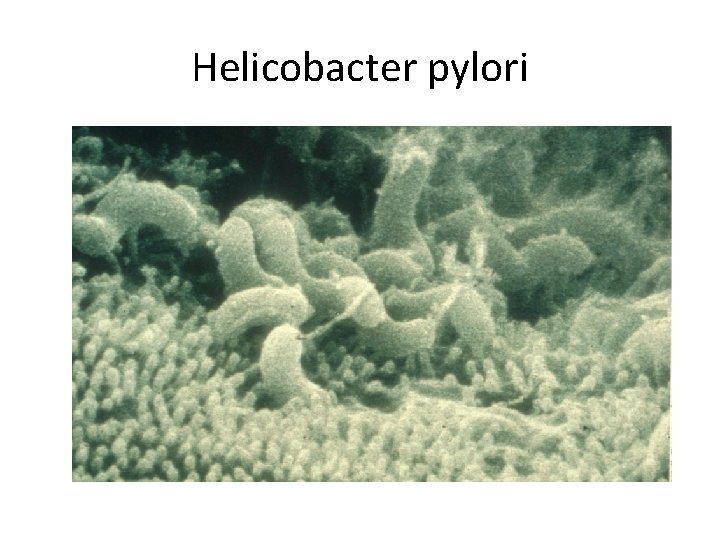
Helicobacter pylori
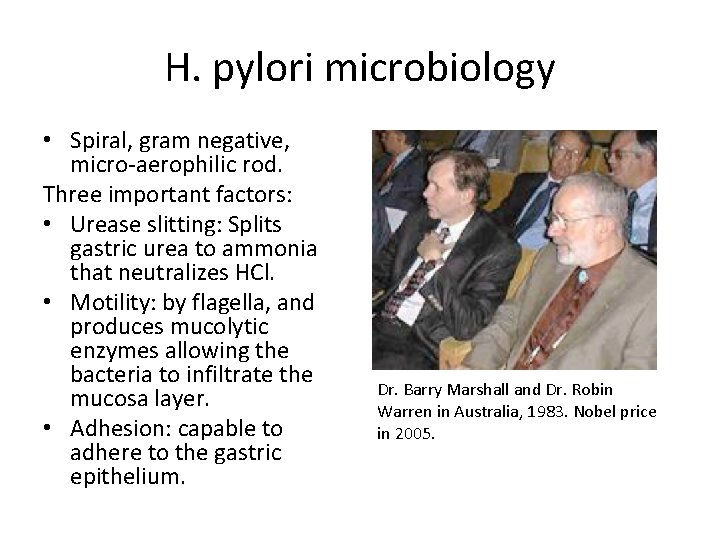
H. pylori microbiology • Spiral, gram negative, micro-aerophilic rod. Three important factors: • Urease slitting: Splits gastric urea to ammonia that neutralizes HCl. • Motility: by flagella, and produces mucolytic enzymes allowing the bacteria to infiltrate the mucosa layer. • Adhesion: capable to adhere to the gastric epithelium. Dr. Barry Marshall and Dr. Robin Warren in Australia, 1983. Nobel price in 2005.
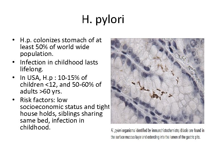
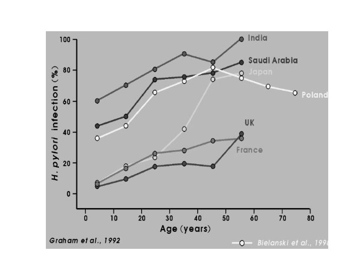
H. pylori • H. p. colonizes stomach of at least 50% of world wide population. • Infection in childhood lasts lifelong. • In USA, H. p : 10 -15% of children <12, and 50 -60% of adults >60 yrs. • Risk factors: low socioeconomic status and tight house holds, siblings sharing same bed, infection in childhood.
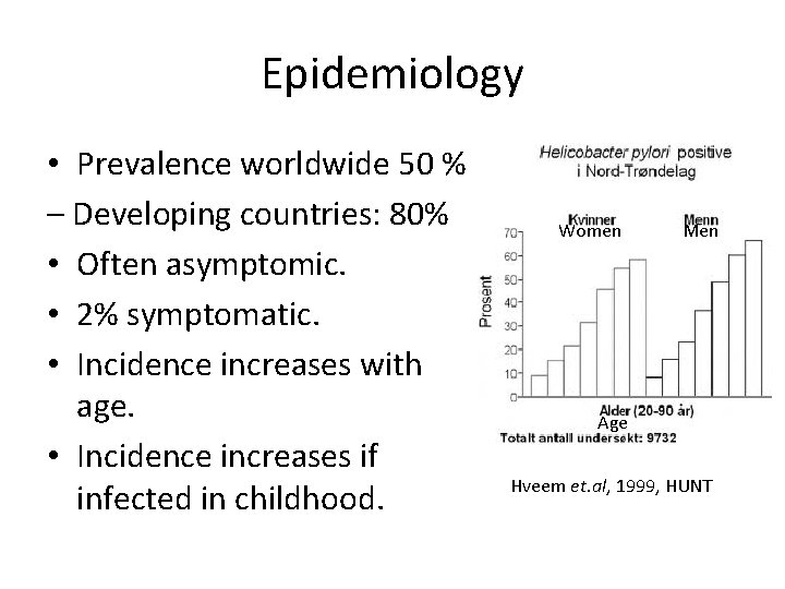
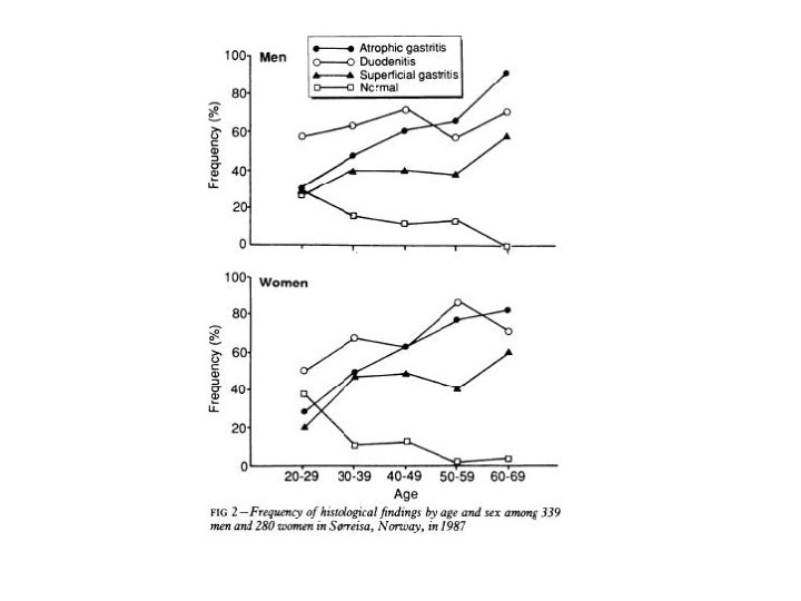
Epidemiology • Prevalence worldwide 50 % – Developing countries: 80% • Often asymptomic. • 2% symptomatic. • Incidence increases with age. • Incidence increases if infected in childhood. Women Men Age Hveem et. al, 1999, HUNT
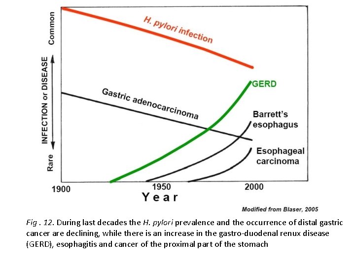
Fig. 12. During last decades the H. pylori prevalence and the occurrence of distal gastric cancer are declining, while there is an increase in the gastro-duodenal renux disease (GERD), esophagitis and cancer of the proximal part of the stomach
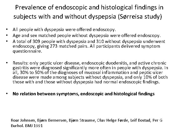
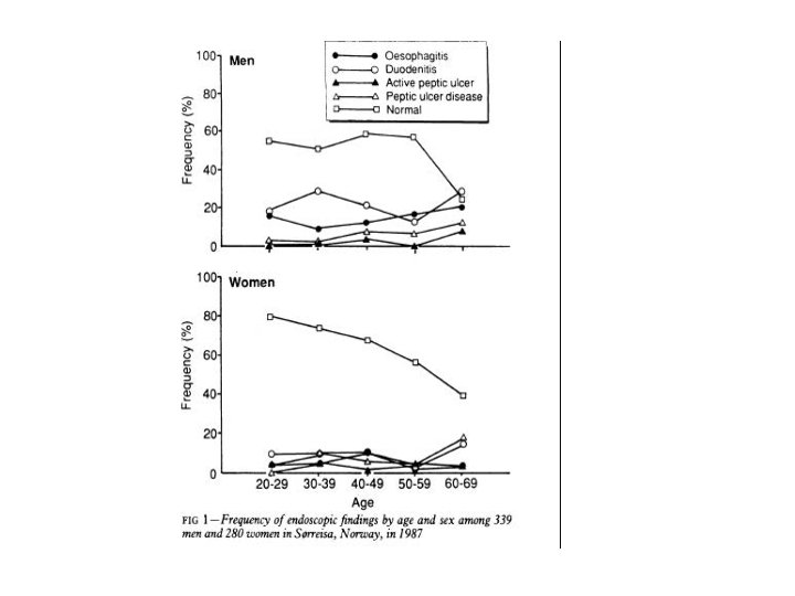
Prevalence of endoscopic and histological findings in subjects with and without dyspepsia (Sørreisa study) • All people with dyspepsia were offered endoscopy. • Age and sex matched people without dyspepsia were offered endoscopy. • A total of 309 people with dyspepsia and 310 without dyspepsia underwent endoscopy, giving 273 matched pairs. All participants delivered symptom questionnaire. • Results: only peptic ulcer disease, endoscopic duodenitis, and active chronic gastritis were diagnosed significantly more often in people with dyspepsia. In all, 30% to 50% of the diagnoses of mucosal inflammation and peptic ulcer disease were made among subjects without dyspepsia, and only 10% of both those with and those without dyspepsia had normal endoscopic findings. • No relation between symptoms, endoscopic and histological findings Roar Johnsen, Bjørn Bernersen, Bjørn Straume, Olav Helge Førde, Leif Bostad, Per G Burhol. BMJ 1991
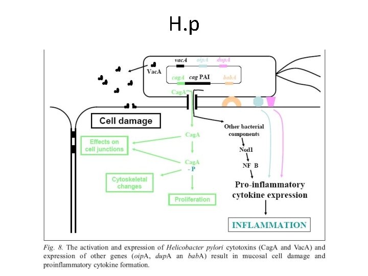
H. p
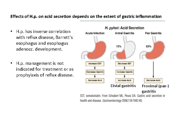
Effects of H. p. on acid secretion depends on the extent of gastric inflammation • H. p. has inverse correlation with reflux disease, Barrett’s esophagus and esophagus adenocc. development. • H. p. management is not indicated for treatment or as prophylaxis of reflux disease. Distal gastritis Proximal (pan-) gastritis
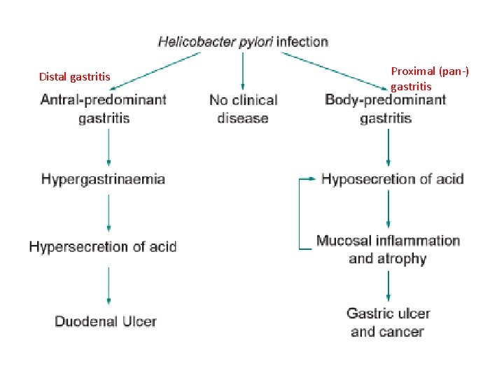
Distal gastritis Proximal (pan-) gastritis
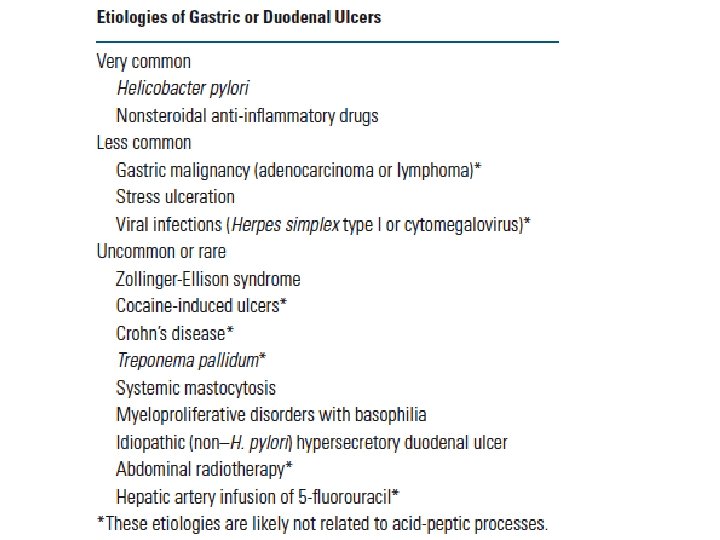
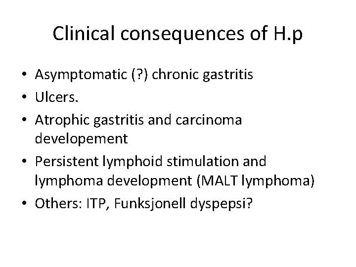
Clinical consequences of H. p • Asymptomatic (? ) chronic gastritis • Ulcers. • Atrophic gastritis and carcinoma developement • Persistent lymphoid stimulation and lymphoma development (MALT lymphoma) • Others: ITP, Funksjonell dyspepsi?
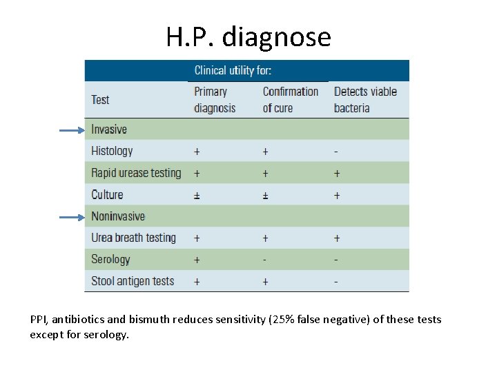
H. P. diagnose PPI, antibiotics and bismuth reduces sensitivity (25% false negative) of these tests except for serology.
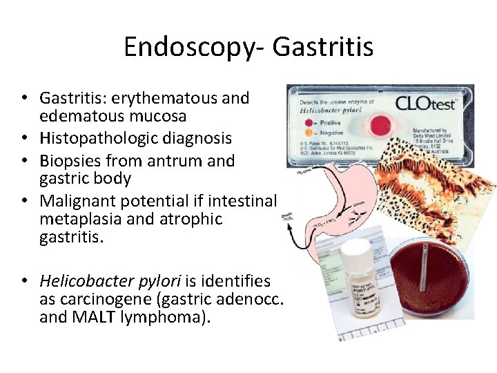
Endoscopy- Gastritis • Gastritis: erythematous and edematous mucosa • Histopathologic diagnosis • Biopsies from antrum and gastric body • Malignant potential if intestinal metaplasia and atrophic gastritis. • Helicobacter pylori is identifies as carcinogene (gastric adenocc. and MALT lymphoma).
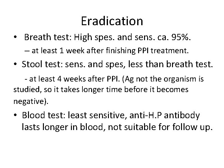
Eradication • Breath test: High spes. and sens. ca. 95%. – at least 1 week after finishing PPI treatment. • Stool test: sens. and spes, less than breath test. - at least 4 weeks after PPI. (Ag not the organism is studied, so it takes longer time before it becomes negative). • Blood test: least sensitive, anti-H. P antibody lasts longer in blood, not suitable for follow up.
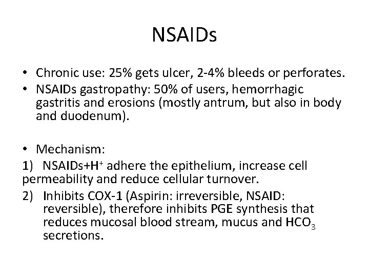
NSAIDs • Chronic use: 25% gets ulcer, 2 -4% bleeds or perforates. • NSAIDs gastropathy: 50% of users, hemorrhagic gastritis and erosions (mostly antrum, but also in body and duodenum). • Mechanism: 1) NSAIDs+H+ adhere the epithelium, increase cell permeability and reduce cellular turnover. 2) Inhibits COX-1 (Aspirin: irreversible, NSAID: reversible), therefore inhibits PGE synthesis that reduces mucosal blood stream, mucus and HCO 3 secretions.
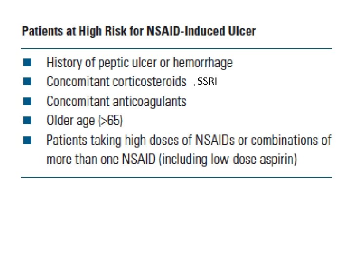
, SSRI
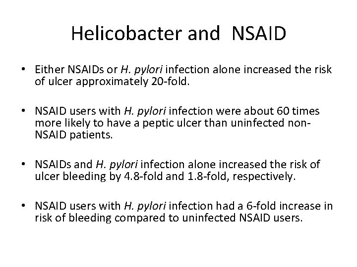
Helicobacter and NSAID • Either NSAIDs or H. pylori infection alone increased the risk of ulcer approximately 20 -fold. • NSAID users with H. pylori infection were about 60 times more likely to have a peptic ulcer than uninfected non. NSAID patients. • NSAIDs and H. pylori infection alone increased the risk of ulcer bleeding by 4. 8 -fold and 1. 8 -fold, respectively. • NSAID users with H. pylori infection had a 6 -fold increase in risk of bleeding compared to uninfected NSAID users.
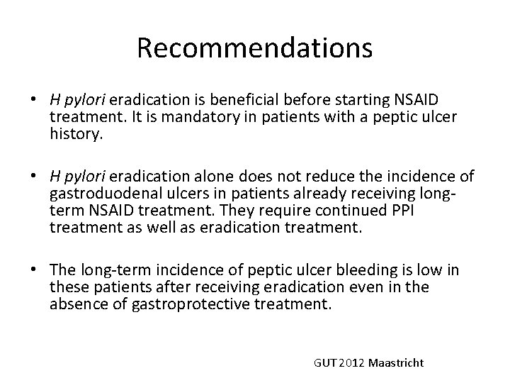
Recommendations • H pylori eradication is beneficial before starting NSAID treatment. It is mandatory in patients with a peptic ulcer history. • H pylori eradication alone does not reduce the incidence of gastroduodenal ulcers in patients already receiving longterm NSAID treatment. They require continued PPI treatment as well as eradication treatment. • The long-term incidence of peptic ulcer bleeding is low in these patients after receiving eradication even in the absence of gastroprotective treatment. GUT 2012 Maastricht
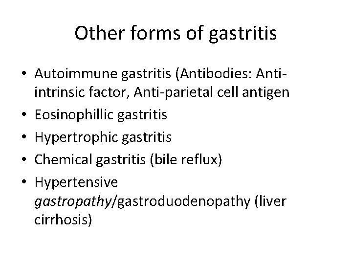
Other forms of gastritis • Autoimmune gastritis (Antibodies: Antiintrinsic factor, Anti-parietal cell antigen • Eosinophillic gastritis • Hypertrophic gastritis • Chemical gastritis (bile reflux) • Hypertensive gastropathy/gastroduodenopathy (liver cirrhosis)
Complications • • Bleeding, Perforation, Penetration, Obstruction.
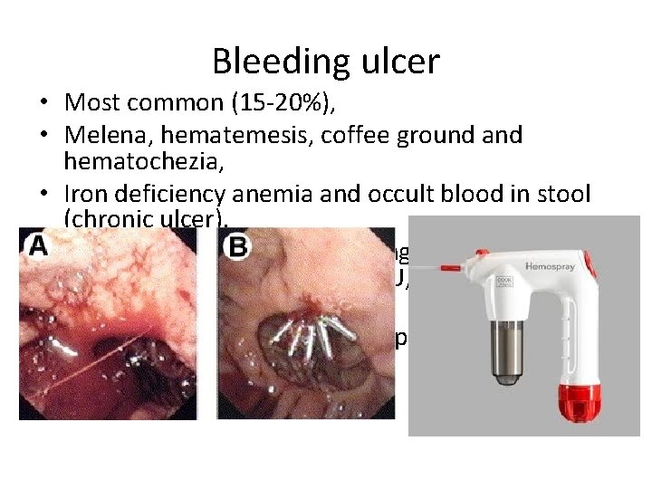
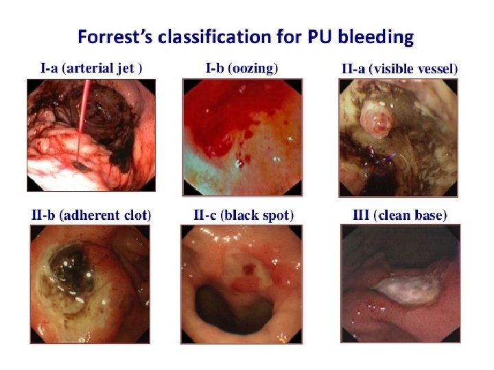
Bleeding ulcer • Most common (15 -20%), • Melena, hematemesis, coffee ground and hematochezia, • Iron deficiency anemia and occult blood in stool (chronic ulcer). • Rebleeding risk: active bleeding at gastoscopy, size >1 -2 cm, posterior wall DU, location at minor curvature. • PPI inf. , Adrenalin inj, hemoclips, gold probe, hemospray
Perforation Acute severe pain Circulatory shock Peritonitis P. Exam. : tenderness, muscular defense, no liver dullness, bowel sounds are first hyperactive then disappear upon clinical worsening. • Plain abd. X-ray: free air under diaphragm • Gastroscopy is contraindicated >> Gastrosurgery. • •
Gastric outlet obstruction • Obstruction due to ulcer at pylorus or bulbus duodeni. • N/V (30 -60 min after meals), • Early satiety and bloating, • Weight loss. • Gastroscopy and CT abdomen. • N-G tube if gastric retention. • PPI tx. (ulcer heals and edema disappears)
Penetration • Til pancreas, liver, biliary ducts, colon. • Treatment: NG catheter, PPI.
Gastroscopy • Stabilize the patient (iv. fluids, P-RBC) • NG catheter? • Give Erythromycin? to increase gastric emptying (if needed) • Biopsy from GU edges, outlooks dysplasia (D. U has less risk for malignity) • Control gastroscopy after 4 -8 weeks.
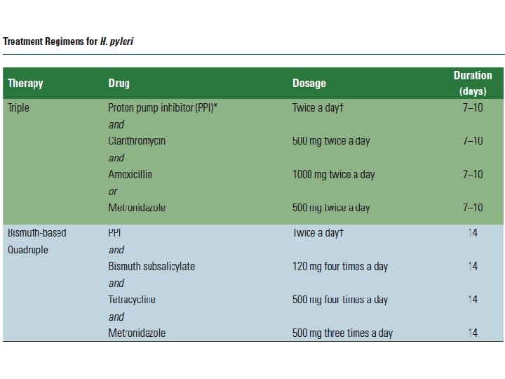
Treatments • D/C NSAIDs. H. pylori status? • PPI tablets (esomeprazole, rabeprazole, pantoprazole. . . etc), - inf. 72 hrs if bleeding ulcer - Superior to sucralfate, H 2 blokkere og PGE (misoprostol prophylaxis with voltaren). - Cardiac patients. : Aspirin/Plavix continue within 1 week (risk for new MI).
Thanks for your attention