
Radiologic Dx of GI Tract Dong Ho LEE












- Slides: 48
Radiologic Dx of GI Tract Dong Ho LEE, M. D. Department of Diagnostic Radiology Kyung Hee University Medical Center
• • • Simple abdomen Barium study US CT MRI
Simple abdomen view SB obstruction Pneumoperitoneum
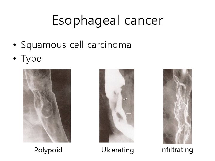
Esophageal cancer • Squamous cell carcinoma • Type Polypoid Ulcerating Infiltrating
Esophageal cancer • Staging CT EUS
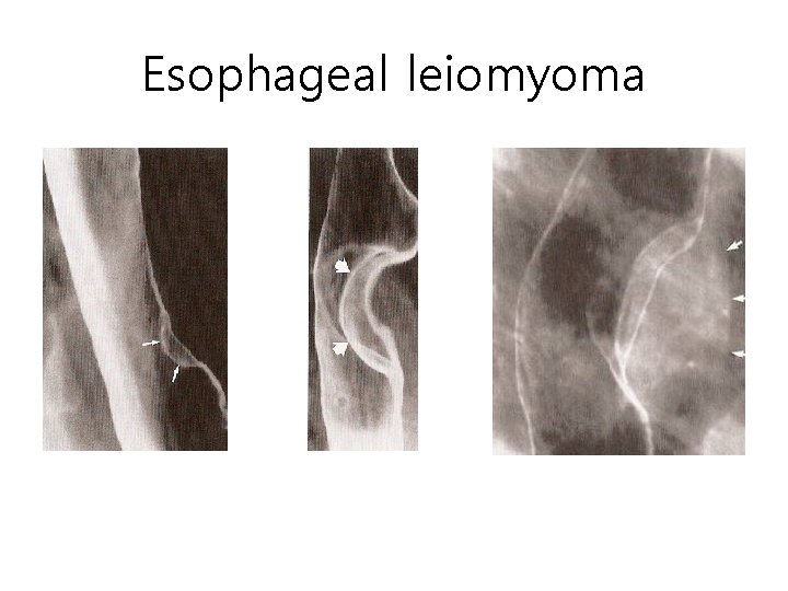
Esophageal leiomyoma • Most common benign tumor • Location – Distal third – Middle third – Proximal third 60% 30% 10% • Sharply outlined, discrete intraluminal mass • Right or obtuse angle with esophageal wall
Esophageal leiomyoma
Reflux esophagitis • Decreased lower esophageal sphincter pressure • Gastroesophageal reflux • Radiologic findings – Abnormal motility – Mucosal nodularity – Ulceration – Thickened fold Ulcer Fold thickening
Barrett esophagus • Long-standing GE reflux and reflux esophagitis • Progressive columnar metaplasia of distal esophagus • Radiologic findings – Midesophageal stricture – High ulcer – Sliding hiatal hernia – GE reflux
Barrett esophagus Midesophageal stricture High ulcer
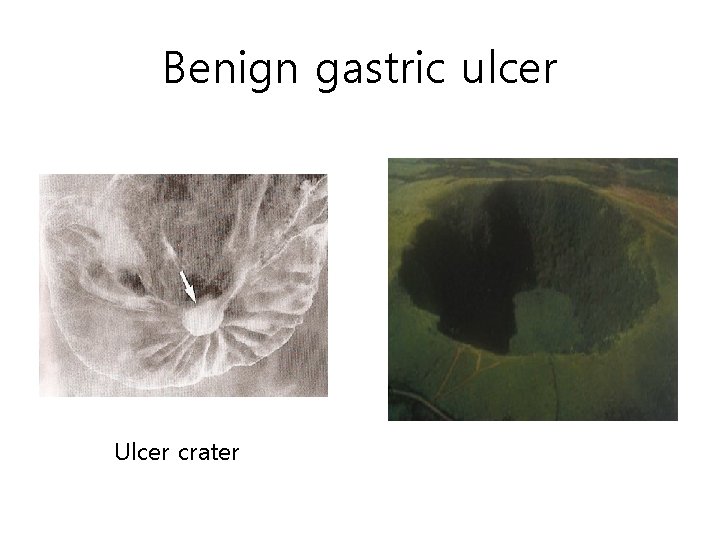
Benign gastric ulcer Ulcer crater
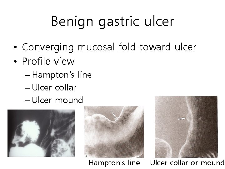
Benign gastric ulcer • Converging mucosal fold toward ulcer • Profile view – Hampton’s line – Ulcer collar – Ulcer mound Hampton’s line Ulcer collar or mound
EGC
EGC IIa EGC IIc
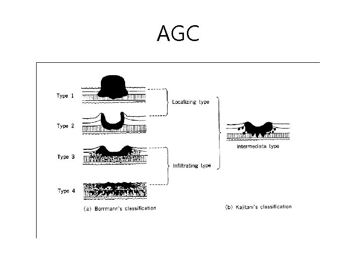
AGC
AGC B 1 B 2
AGC B 3 B 4
CT of AGC
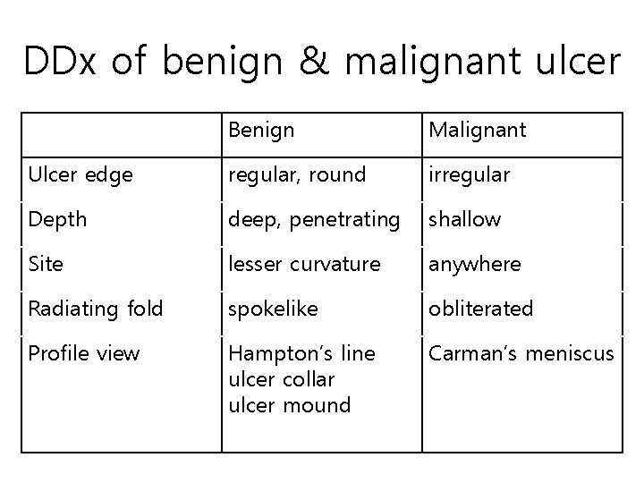
DDx of benign & malignant ulcer Benign Malignant Ulcer edge regular, round irregular Depth deep, penetrating shallow Site lesser curvature anywhere Radiating fold spokelike obliterated Profile view Hampton’s line ulcer collar ulcer mound Carman’s meniscus
Gastric submucosal tumor • Benign GIST • Malignant GIST • Lymphoma
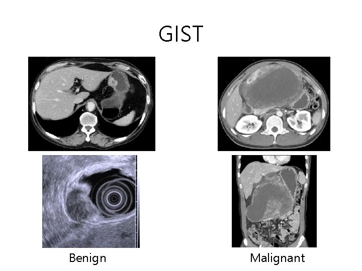
GIST Benign Malignant
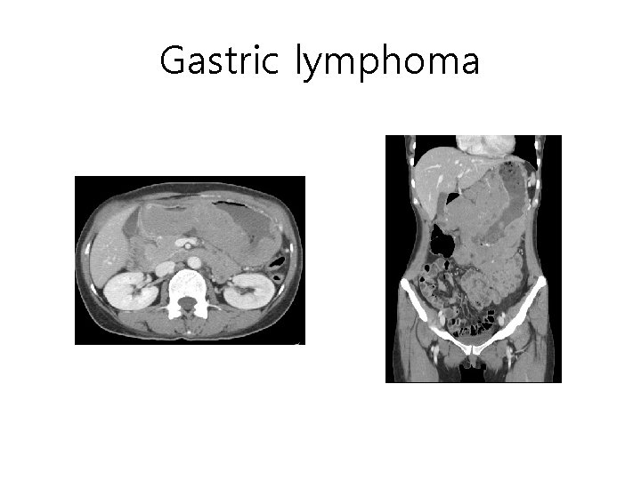
Gastric lymphoma
Duodenal ulcer • Benign • Posterior wall of duodenal bulb
Intestinal tuberculosis • Inflammatory disease of terminal ileum and colon by Mycobacterium tuberculosis • Radiologic findings – Fold thickening – Transverse ulcer – Inflammatory spasm – Accelerated transit time – Cecal retraction – Shortening of A-colon – Stricture
Intestinal tuberculosis Cecal retraction Transverse ulcer A-colon shortening Symmetric involvement
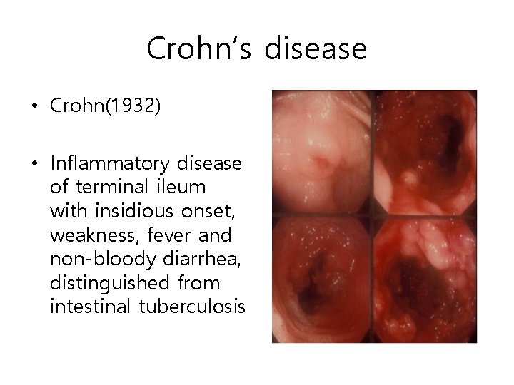
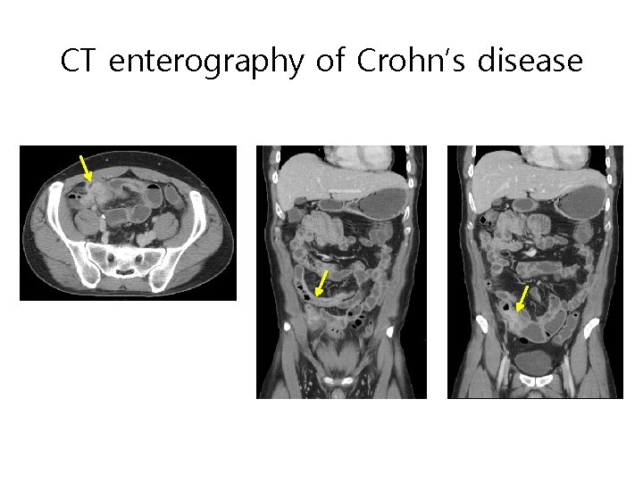
Crohn’s disease • Crohn(1932) • Inflammatory disease of terminal ileum with insidious onset, weakness, fever and non-bloody diarrhea, distinguished from intestinal tuberculosis
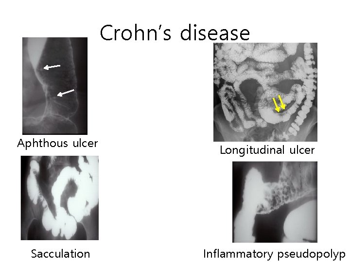
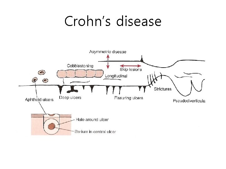
Crohn’s disease Aphthous ulcer Sacculation Longitudinal ulcer Inflammatory pseudopolyp
Crohn’s disease Deep ulcer Fissure Cobblestone appearance
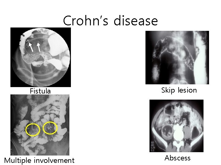
Crohn’s disease Fistula Skip lesion Multiple involvement Abscess
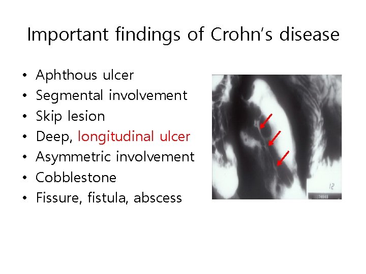
Important findings of Crohn’s disease • • Aphthous ulcer Segmental involvement Skip lesion Deep, longitudinal ulcer Asymmetric involvement Cobblestone Fissure, fistula, abscess
CT enterography of Crohn’s disease
Crohn’s disease
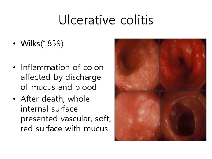
Ulcerative colitis • Wilks(1859) • Inflammation of colon affected by discharge of mucus and blood • After death, whole internal surface presented vascular, soft, red surface with mucus
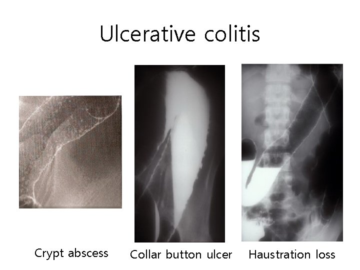
Ulcerative colitis • Radiologic findings – Mucosal granule – Crypt abscess – Collar button ulcer – Loss of haustration – Inflammatory pseudopolyp – Continuous involvement
Ulcerative colitis Crypt abscess Collar button ulcer Haustration loss
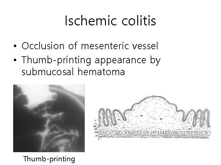
Ischemic colitis • Occlusion of mesenteric vessel • Thumb-printing appearance by submucosal hematoma Thumb-printing
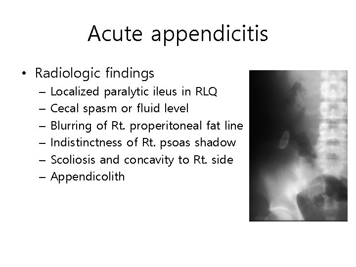
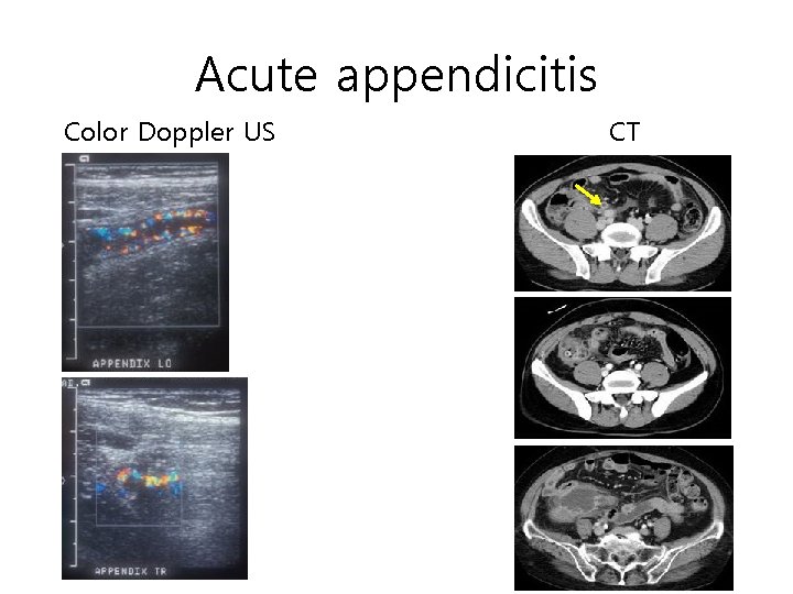
Acute appendicitis • Radiologic findings – – – Localized paralytic ileus in RLQ Cecal spasm or fluid level Blurring of Rt. properitoneal fat line Indistinctness of Rt. psoas shadow Scoliosis and concavity to Rt. side Appendicolith
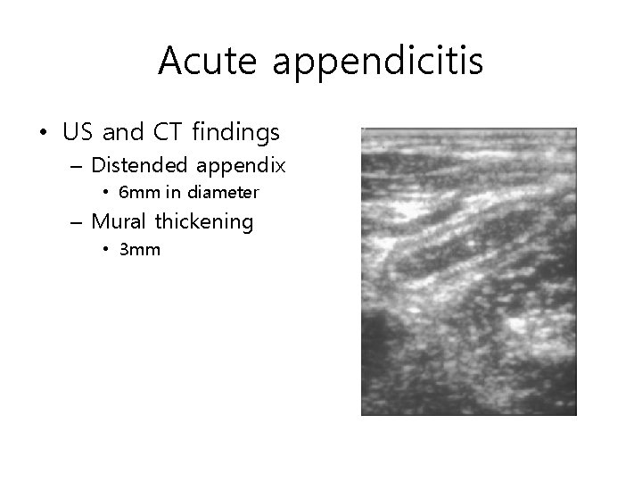
Acute appendicitis • US and CT findings – Distended appendix • 6 mm in diameter – Mural thickening • 3 mm
Acute appendicitis Color Doppler US CT
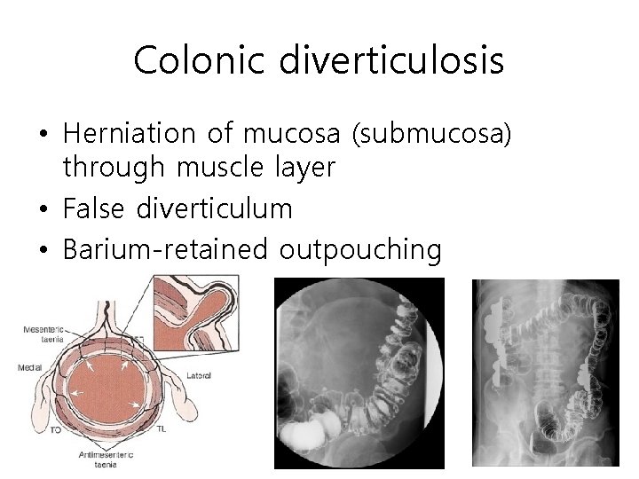
Colonic diverticulosis • Herniation of mucosa (submucosa) through muscle layer • False diverticulum • Barium-retained outpouching
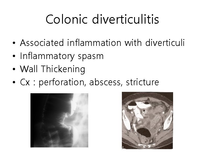
Colonic diverticulitis • • Associated inflammation with diverticuli Inflammatory spasm Wall Thickening Cx : perforation, abscess, stricture
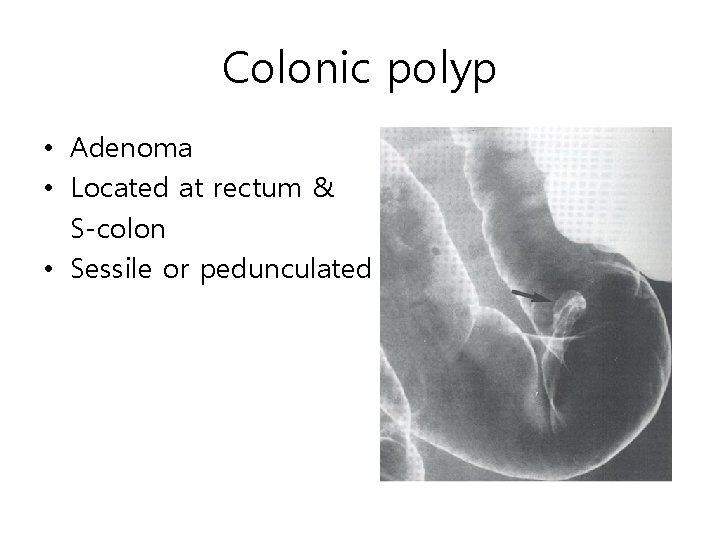
Colonic polyp • Adenoma • Located at rectum & S-colon • Sessile or pedunculated
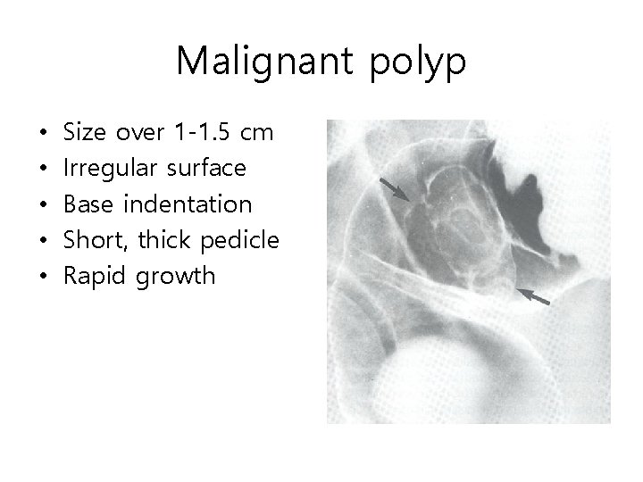
Malignant polyp • • • Size over 1 -1. 5 cm Irregular surface Base indentation Short, thick pedicle Rapid growth
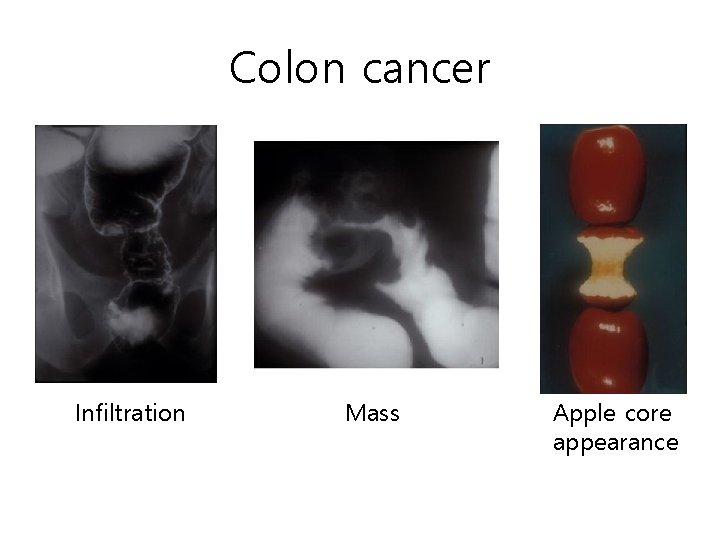
Colon cancer Infiltration Mass Apple core appearance
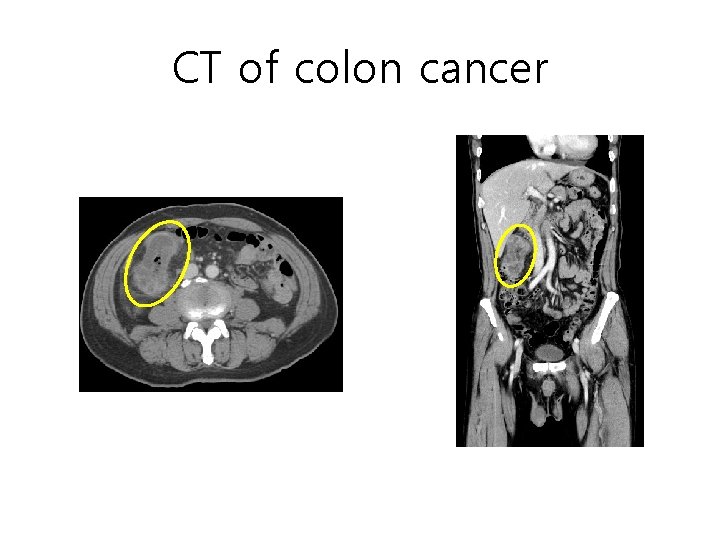
CT of colon cancer
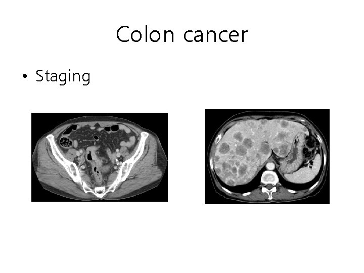
Colon cancer • Staging
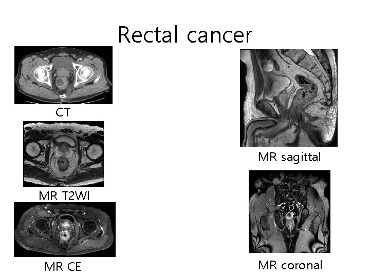
Rectal cancer CT MR sagittal MR T 2 WI MR CE MR coronal
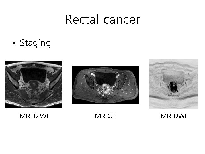
Rectal cancer • Staging MR T 2 WI MR CE MR DWI