Peer Edit Day Peer Edit Guidelines Everyone needs


 �Digests and absorbs food �Coiled, hollow, muscular tube with")


: folds in stomach to")
: ring of muscle where esophagus and stomach")


 Deglutition Mastication Peristalsis Churning Defecation Segmentation Mechanical Process Mastication")

- Slides: 44
Peer Edit Day!
Peer Edit Guidelines �Everyone needs a pink peer edit/self-check sheet PSA �Focus only on the peer edit sheet today! This isn’t about bashing your partner’s paper. �Swap papers and your peer edit sheet with a partner compliments and positive you Give trust tothem edit your paper thoroughly. feedback, too! Duh! �Utilize the highlighters and pens provided, mark all over that paper! Constructive criticism is key, make telling someone Give notes, write helpful information, corrections, etc. their paper is awful is not helpful. Be kind, be � Follow the peer edit sheet, mark each level appropriate for be each paper that requirement provide professional, aware you areand editing a notes/suggestions paper worth 100 points of this person’s grade.
Self-Check and Final Reminders �The self-check should be filled out on Friday when you submit your final paper! Check all levels to ensure you have your paper completed fully, sign it! This signature assures me your paper is an original work of beautiful writing done by you and only you and that there is absolutely positively no plagiarism/cheating that got you to this final draft �Your paper is due on Friday by the time I leave this school… I highly recommend you submit it during your allotted class time. Hard-copy form, no electronic submissions � Save an electronic copy to your One. Drive for your portfolio submission Order: Final draft, peer edit sheet, rough draft The paper on the very top of what you submit is what I am grading.
Digestive Articles Group Work Article 1: Intestinal Snake Robot Article 2: Discovery of the Mesentery as an Organ Article 3: Paneth Cell (Rutgers University) Article 4: Backtrack on the Mesentery �Every person will need a “Read, Pair, Share” Article Summary sheet. �Each person will need one article of their choice, there are 4 articles total… just make sure your group covers all four. �If you have a group of 4+ people, double up or split the longer article. �If you have less than 4 in your group, make sure you find someone from another group to help you summarize the article you’re missing, or double up readings!
The Digestive System ADVANCED HUMAN ANATOMY
Introduction General function: physical /chemical breakdown of food so it can be absorbed into the bloodstream and used by the cells/ tissues and eliminate non-digestible substances produced during metabolism
Digestive Processes �Ingestion: process of taking food into the digestive tract �Propulsion: process of moving food through the alimentary canal (swallowing, peristalsis) �Mechanical digestion: physical preparation of food for chemical digestion; mastication, mixing of food with saliva by tongue, churning and mixing of food in stomach, segmentation in intestine �Chemical digestion: catabolic process in which large food molecules are broken down into smaller molecules by enzymatic hydrolysis �Absorption: transport of digested end products from the GI tract into the capillaries and lymph vessels �Defecation: elimination of indigestible materials and waste from the body
Alimentary Canal �Gastrointestinal Tract (GI) �Digests and absorbs food �Coiled, hollow, muscular tube with 2 openings �Approximately 30 feet long �Includes: mouth, pharynx, esophagus, stomach, small intestine, large intestine
Four Layers of the Alimentary Canal �Mucosa layer: Formed by epithelium and some connective tissue, comes into direct contact with food passing through the canal; secretes mucus that protects and lubricates the lining �Submucosa layer: Composed of loose connective tissue, blood vessels, and many nerve endings; blood vessels carry away the nutrients that are absorbed, and the nerve endings stimulate the muscle fibers so that the food is continually moving by peristalsis �Muscular layer: Consists of a circular and longitudinal band of visceral muscles; is the thickest of the four layers. Main function: peristalsis �Serous layer: Continuous with the mesentery, the connective tissues that attach to the posterior body wall and hold the digestive organs in their proper position
Peristalsis • Wavelike ripple of smooth muscle • Ring of contraction occurs where GI wall is stretched, pushing bolus forward • Stimulated by CCK – secreted by endocrine cells in presence of chyme Segmentation • Mixing movement • Forward and backward movement that mechanically breaks down food and mixes it with digestive juices
Oral Cavity �Mouth: Receives and tastes food Physical breakdown of food Partial digestion by saliva Lubrication of food Deglutition: the act of swallowing �Hard palate: bony structure, roof of mouth, separates mouth from nasal cavity
Pharynx/Esophagus Passageways approximately 10 inches long; food only here 4 -8 seconds; no chemical changes take place �Pharynx: throat, tube that carries food and air �Epiglottis: flap that covers the trachea when food or water is swallowed �Esophagus: muscular tube dorsal to the trachea; carries food to the stomach by peristalsis
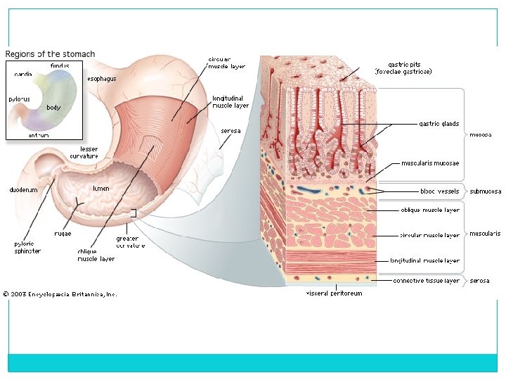
Stomach �Food stays 3 -4 hours for physical and chemical breakdown �Stomach is located in the Left Upper Quadrant (LUQ) of abdomen � 4 layers (much like AC) Mucosa Submucosa Muscular (3 layers of muscle) � Oblique, Serous circular, longitudinal
� 3 parts: Fundus, body, antrum � Rugae (gastric folds): folds in stomach to increase surface area � Stomach acid: hydrochloric acid, potassium chloride, and sodium chloride Key role in digestion and denaturing of enzymes Gastric acid is produced by cells in the lining of the stomach Other cells in the stomach produce bicarbonate to buffer the fluid, ensuring that it does not become too acidic. � These cells also produce mucus which forms a viscous physical barrier to prevent gastric acid from damaging the stomach � Gastric enzymes: pepsin (protein), rennin, lipase (fats), HCl (kills bacteria, helps in absorption of Fe, activates pepsin) � Chyme: gastric juices plus digested food Stomach (ctd. )
Stomach Sphincters �Sphincters: prevent reflux Cardiac (gastroesophageal): ring of muscle where esophagus and stomach join; keeps stomach contents from moving up into the esophagus Pyloric: ring like muscle between the stomach and small intestine; keeps food in stomach 3 -4 hours so that digestion can occur
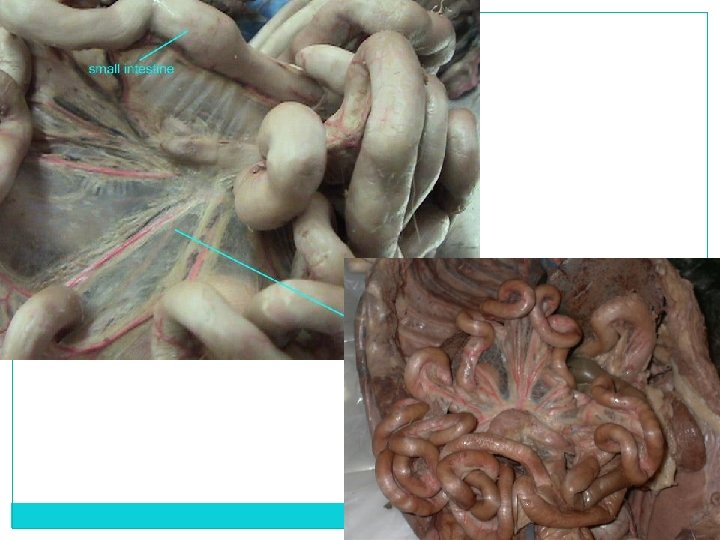
Small Intestine �Approximately 21 feet long � 1 inch in diameter � 80% of absorption occurs here � 3 sections Duodenum, Jejunum, Ileum �Enzymes stimulate intestinal secretions �Hormones inhibit intestinal secretions �Peyer’s patches: lymph nodes in intestine to aid in defense/protection
Duodenum � 1 st 10 -12 inches �Ducts from pancreas and gallbladder (sphincter of Oddi) enter here �Receives chyme Pancreatic juices: amylopsin (sugars), trypsin (proteins), lipase (fats) Gallbladder: bile (emulsifies and breaks down fats) Intestinal juices: maltase/sucrase/lactase (breakdown sugars)
�Jejunum Middle portion; 8 feet partially responsible for absorbing nutrients into the bloodstream. It is lined with finger-like projections that are called villi, that move nutrients, vitamins and minerals from the intestine to the bloodstream �Ileum Final 12 feet Connects to large intestine at cecum Process of digestion completed Absorbs bile acids, which are returned to the liver to be made into more bile, then stored in the gallbladder for future use in the duodenum. Absorbs vitamin B 12 , which the body uses to make nerve cells and red blood cells. Jejunum and Ileum
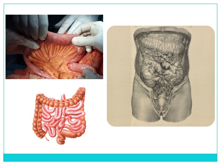
Protective Linings �Peritoneum: silk-like membrane that lines your inner abdominal wall and covers the organs within your abdomen, decreases friction of organs Inflammation from bacterial or fungal infection: peritonitis �Mesentery: greater and lesser omentum forms protective covering that insulates organs and holds them in place
The Digestive System ADVANCED HUMAN ANATOMY – DAY 2… SORT OF
Large Intestine � Ileocecal valve: circular, sphincter muscle; prevents food from returning to ileum (small intestine) � Cecum: pouch at first portion of the large intestines; lower end is the appendix � Colon: absorbs water, remaining nutrients, electrolytes; storage of indigestible materials until elimination; transport waste out of the body Ascending (right side) Transverse Descending (left side) Sigmoid Rectum (final 6 -8 inches): storage of feces
Anal Canal �Anal canal: anus; outlet of rectum Fecal matter: non-digestible waste products and bacteria Defecation: emptying of fecal matter from rectum � 2 Sphincters Internal anal sphincter: involuntary, smooth muscle External anal sphincter: voluntary, skeletal muscle
Accessory Organs Liver � Largest gland/solid organ of body; � Located in the RUQ under diaphragm; 3 pounds � Secretes 1 liter of bile /24 hours � Biliary tree: composed of hepatic duct, cystic duct, and common bile duct Functions � Manufactures blood proteins (i. e. antibodies), blood clotting factors � Stores iron, copper, vitamin A/D/B 12, glycogen � Produces bile for fat digestion � Detoxifies blood
Gallbladder � Pear-shaped muscular sac under the liver � Stores bile (approx. 500 -600 ml. stored) � Fatty foods enter duodenum stimulates CCK hormone contracts GB to release bile emulsifies fats and stimulates peristalsis of small intestine muscular tissue � Bile pigments: bilirubin (formed from breakdown of RBCs) � Bile helps soften stools, thus helping to speed up the movement of feces in the colon � Bile helps destroy bacteria and other microbes that can be present in foods � Obstruction of gallbladder icterus and jaundice � Cholelithiasis: gallstones = cholesterol and bile salts Caused mainly by dietary choices Cholecystectomy: removal of GB
HOLD UP �Let’s talk about CCK �Cholecystokinin is secreted by cells of the upper small intestine. Its secretion is stimulated by the introduction of hydrochloric acid, amino acids, or fatty acids into the stomach or duodenum. �Just mentioned Cholecystokinin stimulates the gallbladder to contract and release stored bile into the intestine. � It also stimulates the secretion of pancreatic juice and may induce satiety. Meal-induced secretion of cholecystokinin activates the satiety centre of the hypothalamus in the brain so that the person feels full and stops eating. Because cholecystokinin inhibits emptying of the stomach, the sensation of satiety may be the result of distension of the stomach.
Pancreas �Behind the stomach; head attached to duodenum, tail reaching to spleen �Exocrine functions: acini cells secrete digestive juices and bicarbonate ions (help adjust p. H) �Endocrine functions: carbohydrate metabolism Insulin and Glucagon: secreted from the islets of Langerhans for carbohydrate metabolism �Diabetes: decreased secretion of insulin or insufficient insulin, therefore glucose is increased in blood �Pancreatitis: inflammation caused by overproduction of pancreatic juices
Appendix �Sits at the junction of the small intestine and large intestine. About 4 inches long. Sits in the lower right abdomen. �Function not fully known; but thought to provide safe place for good bacteria Reboots digestive system after diarrheal illnesses �Appendicitis inflammation of appendix not common in elderly Appendectomy – surgical removal of appendix
Oral Cavity Accessory Organs Salivary Glands �Saliva: lubricates food for swallowing; body produces 1500 ml in 24 hours Lubricates mouth during speech and chewing Moistens food for swallowing Contains enzyme (salivary amylase) which begins chemical breakdown of complex carbohydrates and starches into sugars Teeth � 20 primary form 6 months to 2 years � 32 permanent: incisors, canines, premolars/molars, cuspids/bicuspids Mastication: chewing Bolus: mass of chewed food
Rest of the Year… � Monday 4/24: Urinary System � Wednesday 4/26: Labeling, Vocabulary � Friday 4/28: Urinary System � Tuesday 5/2: Kidney Dissection � Thursday 5/4: Reproductive System (B 3 Juniors, ODW) � MWF 5/8 – 5/12: Cat Dissection Grades posted seniors on Thursday 18 th @ 8: 00 MONDAY May 15 th A-day TUESDAY May 16 th B-day WEDNESDAY May 17 th A-day THURSDAY May 18 th B-day FRIDAY May 19 th A-day 8: 25 – 9: 10 A 1 class B 1 class A 2 class B 2 class A 1 - As normal No time change 9: 15 – 11: 15 A 2 FINAL B 2 FINAL A 1 FINAL B 1 FINAL A 2 - As normal No time change 11: 20 – 1: 10 A 3 class B 3 class A 4 class B 4 class 1: 15 – 3: 15 A 4 FINAL B 4 FINAL A 3 FINAL B 3 FINAL A 3 - As normal No time change A 4 - As normal No time change
The Digestive System ADVANCED HUMAN ANATOMY – DAY 3
Rumble in the Tummy? � Stomach growling is the result of moving food through the stomach and into the small intestine. � Pockets of air and gas also get squeezed into chyme and create the noises we hear. � Stomach growling can happen at any time -- not just when you're hungry -- but if there's food in your stomach or small intestine, the growling becomes quieter. Hungry? Grab a Snickers. � About two hours after your stomach empties itself, it begins to produce hormones that stimulate local nerves to send a message to the brain to prep for more food. � The brain replies by signaling for the digestive muscles to restart the process of peristalsis. � Two results occur: First, the contractions sweep up any remaining food that was missed the first time around. Second, the vibrations of an empty stomach make you hungry. Muscle contractions will come and go about every hour, generally lasting 10 to 20 minutes, until you eat again.
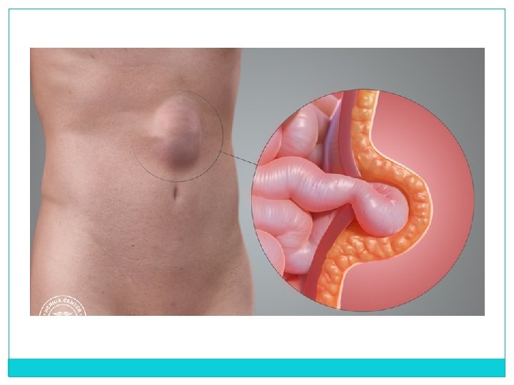
Heartburn, GERD, Acid Reflux, Oh my! �Heartburn is sometimes used interchangeably with acid reflux �Mainly caused by acid splashing up and out of the stomach. Food is a key trigger to heartburn! Relaxation of esophageal sphincter or over production of stomach acid �GERD is usually caused by changes in the barrier between the stomach and the esophagus Abnormal relaxation of the lower esophageal sphincter Impaired expulsion of gastric reflux from the esophagus Hiatal Hernia – stomach pushes through diaphragm
Crohn’s Disease �Crohn’s disease belongs to a group of conditions known as Inflammatory Bowel Diseases (IBD). �Crohn’s disease is a chronic inflammatory condition of the gastrointestinal tract. Crohn’s most commonly affects the ileum and the beginning of the colon �Symptoms: persistent diarrhea, rectal bleeding, abdominal cramps and pain, sensation of incomplete evacuation, constipation (can lead to bowel obstruction) �Treatments: immunosuppressants, steroids, corrective surgery, diet
Stomach Ulcers, Esophageal Varices �Ulcers: occur when the thick layer of mucus that protects your stomach from digestive juices is reduced enables digestive acids to eat away at the lining tissues of the stomach �Esophageal varices: dilated sub- mucosal veins in the lower third of the esophagus Diagnosed with EGD, medicines to help with bleeding, banded to prevent further bleeds
Why’s my stomach going crazy? �Diarrheal illnesses and quick evacuation of the digestive system can be caused by what you eat High fat/oily foods can upset the stomach, along with lactose
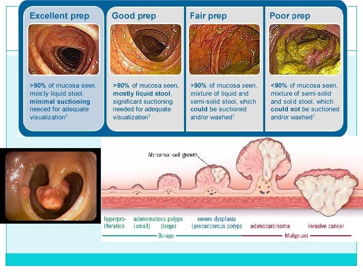
Colonoscopy �Test that allows your doctor to look at the inner lining of your large intestine (rectum and colon) �Uses a thin, flexible tube called a colonoscope to look at the colon �Helps find ulcers, colon polyps, tumors, and areas of inflammation or bleeding
Colectomy �Colectomy is a surgical procedure to remove all or part of your colon. Total colectomy involves removing the entire colon. Partial colectomy involves removing part of the colon and may also be called subtotal colectomy. Hemicolectomy involves removing the right or left portion of the colon. Proctocolectomy involves removing both the colon and rectum.
Organ Mouth (teeth and tongue) Deglutition Mastication Peristalsis Churning Defecation Segmentation Mechanical Process Mastication Deglutition Pharynx Esophagus Deglutition Peristalsis Stomach Churning Peristalsis Segmentation Peristalsis Small Intestine Ascending and Transverse Colon Descending Colon Rectum Segmentation Peristalsis Defecation
Article Questions �Answer the following questions on the back of the article! 1. 2. 3. 4. 5. What is the role of the Paneth cell in your digestive system? What types of things can occur when the Paneth cells activity is weak or non-existant? What does IBD do to a person’s digestive system? Explain Nan Gao’s role in the research of this particular type of cell. What type of benefits could come of Gao and Yap’s research in this particular field?