Pathophysiology L 11 Female Genital System Prof Fakhir


- Slides: 35

Pathophysiology L 11 Female Genital System Prof. Fakhir Al-Ani fakeralani 2000@yahoo. com

Include ü Vulva ü Vagina ü Cervix ü Body of Uterus ü Fallopian Tubes ü Ovaries

Vulva Diseases of vulva: ü Vulvitis (Common but not serious). ü Painful bartholin cysts (obstruction of glands). ü Imperforate hymen. ü Non-Neoplastic epithelial disorders. ü Carcinomas (uncommon but life threatening).

Vulvitis 1. Most important forms is related to STDs: - ü HSV 1 or 2 (Herpes genitalis): Vesicular eruption. ü Gonococcal suppurative infection ü Syphilis: Primary chancre at site of inoculation. ü Candidal vulvitis.

Alergic Vulvitis 2. Contact dermatitis: Most common causes of vulvar pruritus. It is a reactive inflammation to an exogenous stimulus (Irritant or an allergen). - Contact irritant dermatitis: Erythema & crusting papules & plaques. May be a reaction to urine, sops, detergents, antiseptics, or alcohol. - Contact allergic dermatitis: Same gross appearance. Result from allergy to perfumes, additives in creams, lotions, soaps, chemical treatments on clothing.

Painful bartholin cysts Obstruction of glands: Pain when walking, sitting, or having sex. - It is rare for a Bartholin’s abscess to get better on its own. - Usually, the abscess needs to be drained through surgery.

Imperforate hymen It is a congenital disorder. The hymen without an opening. Primary amenorrhea

Non-Neoplasic epithelial disorders The epithelium of vulvar mucosa may undergo: 1. Atrophic thinning = Lichen sclerosus 2. Hyperplastic thickening = Lichen simplex chronicus Both: Exist in different areas in the same person. Appear as Leukoplakia (Depigmented white lesions).

Non-Neoplastic epithelial disorders Lichen Sclerosus: - Atrophic epithelium (with dermal fibrosis). - Pathogenesis is uncertain (may be autoimmune) - Carries risk of developing squamous cell carcinoma Lichen Simplex Chronicus: - Hyperplastic thickening, hyperkeratosis. - Pathogenesis chronic inflammatory reaction. - Not predisposing to cancer, but suspiciously present at margins of established cancer of the vulva.

Tumors Condylomas: - Two distinctive forms: - Condylomata lata: (not commonly seen today). moist, minimally elevated lesions. Occur in secondary syphilis - Condylomata accuminata: (more common) Papillary & distinctly elevated. Occur anywhere on the anogenital surface. Not pre-cancerous. Human papillomavirus (HPV) Carcinoma of the vuvla Rare; 3% of all genital tract cancers in women.

Vaginal diseases - It is usually secondary to infection or cancer arising in cervix, vulva, bladder, or rectum. - It is seldom the site of primary disease The secondary lesion due to: - Vaginitis. - Primary tumors. The primary disorders are: - Few congenital anomalies: Total absence of vagina Separate or double vagina

Vaginitis - Common, Often Transient & not serious. - Infection: Candida Albicans, Trichomonas, Gonococcus - Nonspecific atrophic vaginitis: - Postmenopausal women Atrophy & thinning of squamous vaginal mucosa. Candidal vaginitis 5% of normal women Asymptomatic Curdy white discharge Flashy meat bad smell Microscopic Exam. T. vaginalis 10% of normal Asymptomatic watery green Putrid odor often Microscopic Gonococcus Sexual transmited dis. Often are asymptomatic Mucopurulent discharge Pelvic inflammation Microscopic

Cervicitis - Extremely common - Associated with purulent vaginal discharge. The inflammation: 1. Infectious. 2. Noninfectious cervicitis. Microorganisms: 1. Aerobic. 2. Anaerobic. T. vaginlais (40%), Candida, Neisseria gonorrheae, herpes simplex genitalis, Streptococci, staphylococci, enterococci, E. coli, Chlamydia trachomatis, , HPV. Many of them are STDs.

Tumors of the cervix - Major causes of cancer-related deaths in women. - Pap smear reduces the incidence of cervical cancer. But The incidence of cervical intraepithelial neoplasia has increased > 50, 000 cases annually Because: - Precursor of cervical intraepithelial neoplasia is not diagnosed by Pap smear.

Invasive carcinoma of the cervix 75% = Sqamous cell carcinoma 20% =Adenocarcinoma & adenosquamous carcinoma. 5% = Small cell neuro-ednocrine carcinoma. - Adenocarcinoma has been increasing in recent years because glandular lesions are not detected well by Pap smear. - The only reliable way to monitor the course of the disease is with careful follow-up and repeat biopsies.

Body of Uterus - Common. - Chronic & recurrent & sometimes disastrous. The more frequent & significant are: ü Endometritis ü Adenomyosis ü Endometriosis ü Dysfunctional uterine bleeding & endometrial hyperplasia ü Tumors

Endometritis - Associated with: - pelvic inflammatory disease. Foreign bodies or retained tissue Miscarriage or delivery. They act as a nidus for infection: - removal= resolution Endometritis: - Acute or - chronic Depending on the predominant WBCs - Neutrophilic = Acute - Lymphoplasmacytic = Chronic endometritis Acute endometritis is frequently due to N. gonorrhoeae or C. trachomonus.

Endometritis S&S: - Fever. - Abdominal pain. - Menstrual abnormalities. - Infertility & ectopic pregnancy due to damage to the fallopian tubes. Granulomatous endometritis: Seen in immunocompromised individuals in U. S, and in other countries where T. B. is endemic.

Adenomyosis Ø Growth of basal layer of the endometrium down into the myometrium. - Endometrial stroma, glands are found in uterine wall. - So uterine wall becomes thickened & enlarged uterus Adenomyosis do not undergo cyclical bleeding: Because they are derived from stratum basalis of the endometrium, . But may produce menorrhagia, dysmenorrhea, & pelvic pain before the onset of menstruation.

Endometriosis Patho. Ø Characterized by: Endometrial glands & stroma in a located outside the endomyometrium. In pelvis cause mass filled with blood. Most accepted theory of endometriosis is: Regurgitation theory: - Menstrual backflow through the fallopian tubes with subsequent implantation. - Because menstural endometrium is viable & survives even when injected into the anterior abdominal wall.

Endometriosis Clinical Manifestations: Depend on the distribution of the lesions. - Sterility due to extensive scaring of the oviducts & ovaries produces. - Discomfort in the lower abdominal quadrants. - Pain on defecation reflects rectal wall involvement. - Dyspareunia (painful intercourse). - Dysuria reflect involvement of the bladder serosa. - Almost in all cases, there is severe dysmenorrhea & pelvic pain as a result of intra-pelvic bleeding and periuterine adhesions.

Dysfunctional uterine bleeding Ø Abnormal bleeding in the absence of a well-defined organic lesion in the uterus. Various causes segregated into 4 groups depending on the age of women: 1. Failure of ovulation: Estrogen relative to progesterone. 2. Inadequate luteal phase: Prgesterone due to failure of corpus maturation. 3. Contraceptive-induced bleeding: 4. Endomyometrial disorders: After menopause

Endometrial hyperplasia Estrogen relative to progestin hyperplasia, Classification: - Simple hyperplasia. - Complex hyperplasia. - Atypical hyperplasia. Endometrial hyperplasia is pre-neoplastic. The risk dependent on: - The severity hyperplesia. - Failure of ovulation. - Prolonged administration of estrogen. - Polycystic ovaries - Obesity.

Tumors Endometrial polyps: - Composed of endometrium resembling the basalis, frequently with small muscular arteries. - More often they have cystic dilated glands, glands but some have normal endometrial architecture. - They may occur at any age, age but more commonly at time of menopause. Clinical significance: - Production of abnormal uterine bleeding. - Risk of giving rise to a cancer (rare).

Tumors Leiomyoma: - The most common benign tumor in females. - Found in 30% to 50% of women during reproductive life. - In Blacks > than in Whites. - They are firm. - Estrogens & oral contraceptives stimulate their growth; conversely, so shrink postmenopausally. - They may be asymptomatic, discovered on routine pelvic examination. - Frequently manifested as menorrrhagia with or without metrorrhagia.

Tumors Liomyosarcomas: - Almost always solitary tumors. - They are frequently soft, hemorrhagic & necrotic. - Diagnostic features include tumor necrosis & mitotic activity. - They present a wide range of differentiation - Recurrence after removal is common with these cancers. - Many metastasize, typically to the lungs. Yielding a 5 years survival rate of about 40%.

Tumors § Endometrial carcinoma: - Most frequent cancer. - Appears mostly between the ages of 55 & 65 years. There are 2 clinical stetting's: - In peri-menopausal women with estrogen excess. - In older women with endometrial atrophy. Risk factors: - Obesity - Diabetes. - Hypertension. - Infertility All are associated with increased estrogen stimulation.

Fallopian tubes Salpingitis: Most common disease of the fallopian tubes. Always a component of pelvic inflammatory disease. It is almost always microbial in origin. Non-gonococcal infections are more invasive: - Salpingitis risk of tubal ectopic pregnancy. All forms of salpingitis may produce: - Fever. - Lower abdominal. - Pelvic pain. - Pelvic masses. - They may result in tubo-ovarian abscess.

Fallopian tubes Primary adenocarcinomas: may be of: - Papillary serous - Endometrioid type.

Ovaries Follicle and luteal cysts Polycystic ovaries Tumors of the ovary Surface epithelial-stromal tumors - Serous tumors - Mucinous tumors - Endometrioid tumors - Brenner tumor Other tumors - Teratomas *Benign (mature) cystic teratomas *Immature malignant teratomas

Ovaries Follicle & luteal cysts: Ø Originate from un-ruptured graafian follicles. Ø Or from immediately sealed ruptured follicles. - Produce pelvic pain. - Variable size, they may become palpable masses when they achieve diameters of 4 -5 cm - Sometimes these cysts rupture, producing intraperitoneal bleeding & acute abdomin. - When small they are lined by granulosa lining cells or luteal cells, but as the fluid accumulates, Pr. may cause atrophy of these cells.

Ovaries Polycystic ovaries: Excessive high concentration of androgens, LH, and low concentration of FSH. Causing: - Multiple cystic follicles in the ovaries. - Oligomenorrhea, hirsutism, infertility. - Sometimes obesity appears in girls after menarche - Ovaries usually increase in size (twice normal size).

Tumors of the ovary Ovarian cancer: - Common: (5 th most common cancer in US women). (5 th leading cause of cancer death in women). - 90 % of ovarian cancers are originated from the surface epithelial.

Tumors of the ovary Risk factors: - Nulliparity. - Family history. - Prolonged use of oral contraceptives reduce the risk. - Mutations in: BRCA 1 & BRCA 2 genes. P 53 in 50% of all ovarian cancers.

Teratomas • • Neoplasms of germ-cell origin. Constitute 15 -20% of ovarian tumors. They arise in the first two decades of life. More than 90% of these germ-cell are benign mature cystic teratomas. • The immature malignant variant is rare.
 Female sex anatomy diagram
 L
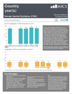
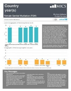
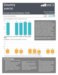
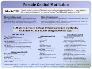
L Female genital mutilation
 Angle of anteflexion
 Female genital
 Function of fallopian tube
 Figure of reproductive system
 Chlamydia curable
 Male genital variation
 Genital hijyen
 La etapa falica
 Monogenia
 Genital hijyen
 Genital hijyen nedir
 Historia natural de herpes genital
 Hsv-1 genital recurrence rate
 Etapas de la familia segun duvall
 What class do sharks belong to
 Espace svt ac rennes
 Appareil génital souris femelle
 Etapa incorporativa
 Etapa locomotora genital
 Clitoris structure
 Taenia solium class
 External genitalia of female
 Function of male
 Parenchimul testicular
 Sigmund freud theory of psychosexual development
 Anal fixation
 Google imagens
 Tubuli rekti
 Prolapsplastik
 L'appareil génital féminin en coupe frontale
 Male genital
 Salphagitis
 Enterotest

