OCULAR ISCHEMIC SYNDROME Kiana Kamrava MD Vitreoretinal Fellowship

 is a rare, but visionthreatening, condition associated with severe")


 60% • Prolonged")


- Slides: 26
OCULAR ISCHEMIC SYNDROME Kiana Kamrava MD. Vitreoretinal Fellowship
Introduction Ocular ischemic syndrome (OIS) is a rare, but visionthreatening, condition associated with severe carotid artery occlusive disease leading to ocular hypoperfusion. Atherosclerosis is the main cause of OIS. Other causes include dissecting aneurysm of the carotid artery, giant cell arteritis, fibrovascular dysplasia, Takayasu arteritis, aortic arch syndrome, Behçet’s disease, trauma or inflammation causing stenosis of the carotid arteries and complications after intravitreal anti-VEGF injections and after radiotherapy for nasopharyngeal carcinoma and vasospasm
The patient with OIS is often elderly, but can range in age from 50 to 80. The incidence of OIS in men is twice that in women. Generally, these patients will have co-existing systemic morbidities, such as diabetes, hypertension, peripheral vascular disease or cardiovascular disease
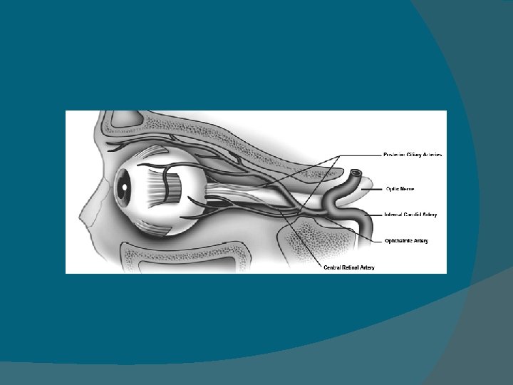
OIS develops especially in patients with poor collateral circulation between the ICA and external carotid artery systems or between the two ICAs. Patients with a healthy collateral circulation may not develop OIS even with total occlusion of the ICA, whereas in those with poor collaterals an ICA stenosis <50% may be sufficient to develop OIS.
Patients who develop OIS show decreased blood flow in the retrobulbar vessels and reversal of blood flow in the ophthalmic artery. The OA may behave as a steal artery shunting blood flow away.
Ophthalmic signs Decrease in visual acuity in OIS may be severe, with acute or subacute presentation (90%). Ischemic pain (50%) begins gradually over hours to days and is described as a dull, constant ache in the affected eye, over the orbit, upper face, and temple, and may worsen when the patient is upright. Lying down relieves or lessens pain. Approximately 80% of cases are unilateral.
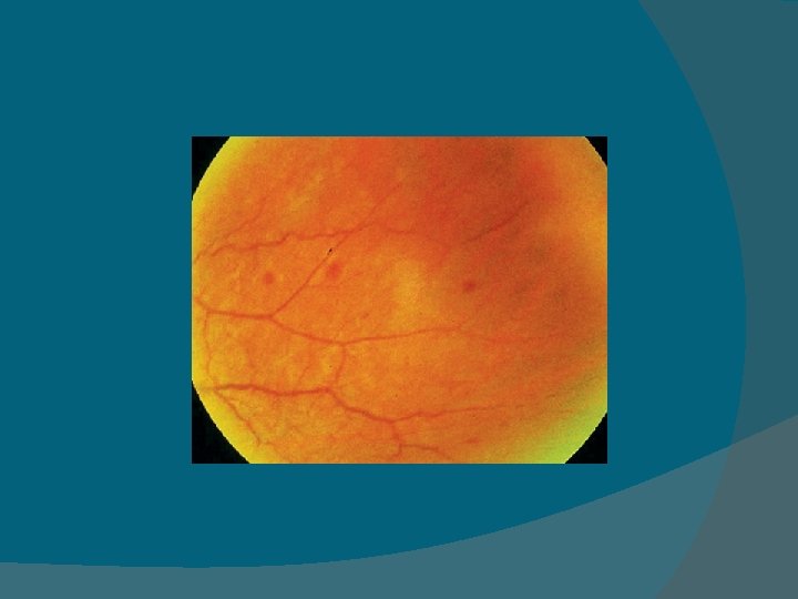
Some other important signs of OIS include red eye; unusual or asymmetric cataracts; Anterior segment ischemic signs include a low-grade uveitis or a sluggish, minimally reactive pupil and ocular hypotony. Dilated but not tortuous retinal veins and mid-peripheral dot and blot hemorrhages or neovascularization either on the retina or on the disc in some cases. The eye pressure may become high due to associated neovascular glaucoma. An ischemic optic neuropathy may eventually occur
In some cases, the recovery of vision following exposure to bright lights is slow due to hypoxia interfering with the regeneration of visual pigment. Conditions that either increase retinal metabolic demands or decrease perfusion pressure can precipitate transient visual loss. This has been reported following exposure to bright light, postural change, or after eating a meal.
Differential diagnosis Diabetic retinopathy and CRVO are the two most likely conditions to be confused with OIS. The presence of optociliary shunt vessels, presence of disc edema, no report of decreased vision after bright light exposure and absence of orbital and periorbital pain help differentiate CRVO from ocular ischemic syndrome. The differential diagnosis of OIS should also include the hyperviscosity syndromes. A basic workup should therefore include a complete blood cell count with differential, serum protein electrophoresis, and immunoelectrophoresis.
Fluorescein angiography • Delayed choroidal filling time (most specific angiographic sign) 60% • Prolonged arteriovenous (AV) transit time (most sensitive angiographic sign) 95% • Retinal vascular staining in 85% • Macular edema (non cystic 15%) • Retinal capillary non-perfusion
Indocyanine green angiography The arm-to-choroid circulation time , and the intra-choroidal circulation time are both prolonged in OIS.
In CRAO, where there is ischemia of the inner retina, the amplitude of the b-wave is decreased. In contrast, in eyes with OIS where both the retinal and the choroidal circulation are compromised, there is ischemia of the inner and outer retina that results in decreased amplitude of both a and b waves. Reduction in the amplitude of the oscillatory potential of the b-wave has been demonstrated in eyes with carotid artery stenosis even if the fluorescein angiography is normal.
Visual-evoked potentials Photostress induces transient VEP changes consisting of an increase in response latency and a decrease in amplitude. The time it takes the VEP to recover to the baseline status ranges in normal subjects between 68 and 78 seconds. This recovery time after photostress is prolonged in patients with severe carotid artery stenosis and improves following endarterectomy surgery.
Carotid Duplex Ultrasound Duplex carotid ultrasonography is the most commonly used noninvasive test and combines B-mode ultrasound and Doppler ultrasound, providing both anatomical imaging of the vessel and flow velocity information. Compared to conventional intra-arterial digital substraction angiography (DSA) for detection of high-grade symptomatic carotid artery stenosis, duplex ultrasound has a sensitivity of 89% and a specificity of 84%. For detecting occlusion, duplex ultrasound has a sensitivity of 96% and a specificity of 100%.
MRA & CTA For the diagnosis of 70– 99% carotid stenosis, MRA had a pooled sensitivity of 95% and a pooled specificity of 90%. For detection of complete occlusions, MRA yielded a sensitivity of 98% and a specificity of 100%
Limitations of MRA include claustrophobia, pacemakers and metallic stents or implantable defibrillators, and obesity. Disadvantages of CTA are the necessity of administrating a nephrotoxic iodinated contrast agent and ionizing radiation and/or artifacts related to heavily or circumferentially calcified arterial walls. The combined use of MRA, CTA, and doppler ultrasound improves diagnostic accuracy for high-grade symptomatic carotid stenosis and minimizes the need for invasive carotid arteriography.
Conventional intra-arterial digital substraction angiography has been considered as the gold standard for imaging the cerebrovascular system Nevertheless, it is not ideal for screening and follow-up both because of the risks of disabling cerebral infarction, systemic complications and high cost. Duplex ultrasound should be chosen as the first-line investigation of patients suspected of having carotid stenosis. If surgically significant stenosis is identified or in equivocal cases, further imaging with either MRA or CTA should be performed. Only if results are contradictory or inconclusive, should DSA be performed.
Management The management of OIS involves a multidisciplinary approach. The aim is threefold, firstly to treat the ocular complications and prevent further damage, secondly to investigate and treat the associated vascular risk factors, and thirdly to perform vascular surgery whenever indicated
The ocular treatment is directed toward control of anterior segment inflammation, retinal ischemia, increased IOP and neovascular glaucoma the underlying carotid occlusive disease would need to be treated, either through anti-coagulants or anti-platelet therapy, possibly carotid endarterectomy surgery. carotid endarterectomy should be considered in cases of 60% or greater carotid obstruction However, approximately 30% of the patients will improve, and a third will stay the same. A third will worsen.
Carotid Artery Endarterectomy Carotid artery surgery therefore can reduce ocular ischemia and improve hypotensive retinopathy as well as reduce the risk of stroke. However it is important to note that the presence of iris neovascularization implies a greater degree of ocular ischemia and damage, making reversal of ischemia and visual recovery unlikely and limiting any beneficial effect of CEA on visual acuity.
Carotid Artery Stenting CAS has been used for patients who are considered to be at highrisk for complications after CEA including those with anatomic conditions rendering surgery technically difficult, such as previous neck irradiation or radical neck surgery, recurrent stenosis after CEA, tracheostomy, and carotid stenosis above the C 2 vetebral body. Medical conditions that increase the risk of surgery, such as unstable angina, recent myocardial infarction, multivessel coronary disease, congestive heart failure, are also indications for CAS.
Extracranial–Intracranial Arterial Bypass Surgery EC-IC bypass surgery involves the surgical anastomosis of the superficial temporal artery with a branch of the middle cerebral artery. It is indicated when there is complete occlusion of the ICA or the CCA or when ICA stenosis is inaccessible (at or above the C 2 vertebral body) to CEA
Prognosis The overall mortality rate for patients with OIS is 40% at 5 years with the leading cause of death being cardiovascular disease, usually myocardial infarction, (67%) followed by cerebral infarction. (19%)
Thanks for your attention