Vitreoretinal Evidence in Practice Group Department of Ophthalmology

- Slides: 24
Vitreo-retinal Evidence in Practice Group, Department of Ophthalmology, University Hospital. When the evidence base is low, is the clinical librarian compromised? Making the role work for the patient. Jacqueline Verschuere Clinical Librarian Conference, York, June 2007
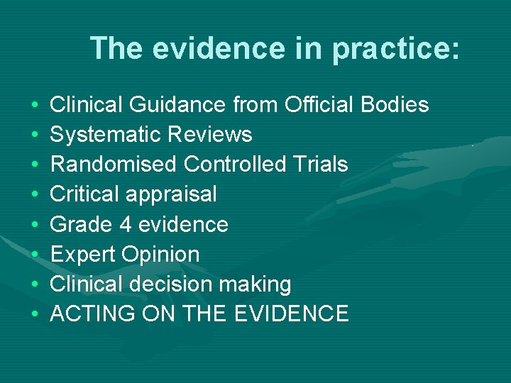
The evidence in practice: • • Clinical Guidance from Official Bodies Systematic Reviews Randomised Controlled Trials Critical appraisal Grade 4 evidence Expert Opinion Clinical decision making ACTING ON THE EVIDENCE
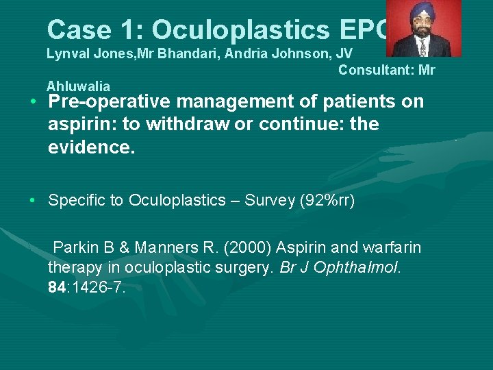
Case 1: Oculoplastics EPG Lynval Jones, Mr Bhandari, Andria Johnson, JV Consultant: Mr Ahluwalia • Pre-operative management of patients on aspirin: to withdraw or continue: the evidence. • Specific to Oculoplastics – Survey (92%rr) Parkin B & Manners R. (2000) Aspirin and warfarin therapy in oculoplastic surgery. Br J Ophthalmol. 84: 1426 -7.
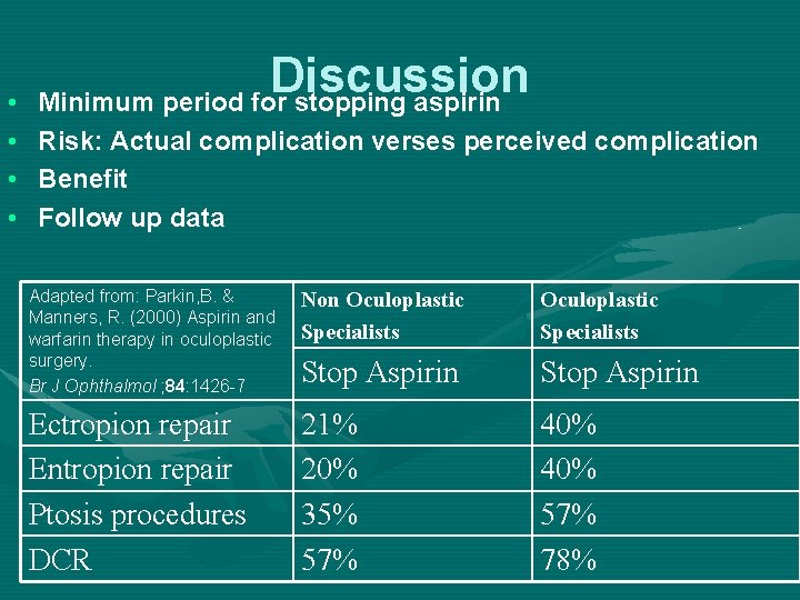
• • Discussion Minimum period for stopping aspirin Risk: Actual complication verses perceived complication Benefit Follow up data Adapted from: Parkin, B. & Manners, R. (2000) Aspirin and warfarin therapy in oculoplastic surgery. Br J Ophthalmol ; 84: 1426 -7 Non Oculoplastic Specialists Stop Aspirin Ectropion repair Entropion repair Ptosis procedures DCR 21% 20% 35% 57% 40% 57% 78%
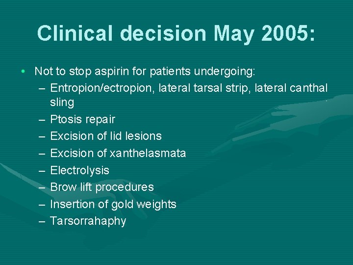
Clinical decision May 2005: • Not to stop aspirin for patients undergoing: – Entropion/ectropion, lateral tarsal strip, lateral canthal sling – Ptosis repair – Excision of lid lesions – Excision of xanthelasmata – Electrolysis – Brow lift procedures – Insertion of gold weights – Tarsorrahaphy
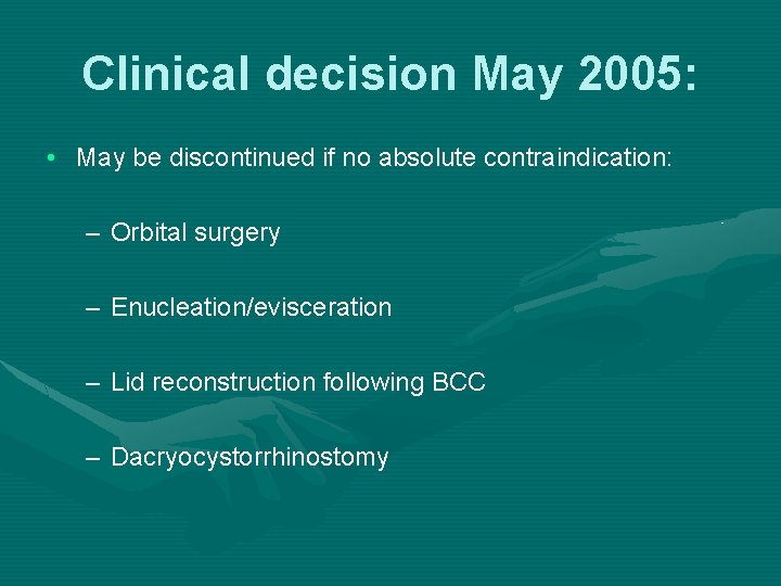
Clinical decision May 2005: • May be discontinued if no absolute contraindication: – Orbital surgery – Enucleation/evisceration – Lid reconstruction following BCC – Dacryocystorrhinostomy
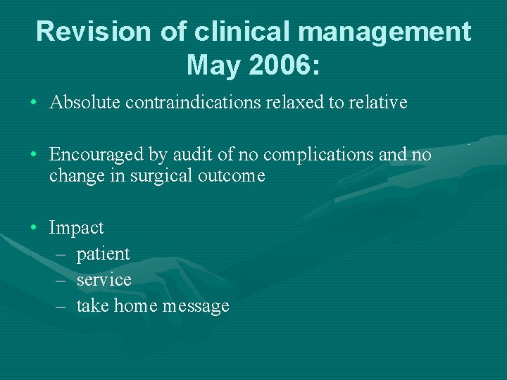
Revision of clinical management May 2006: • Absolute contraindications relaxed to relative • Encouraged by audit of no complications and no change in surgical outcome • Impact – patient – service – take home message
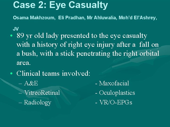
Case 2: Eye Casualty Osama Makhzoum, Eli Pradhan, Mr Ahluwalia, Moh’d El’Ashrey, JV • 89 yr old lady presented to the eye casualty with a history of right eye injury after a fall on a bush, with a stick penetrating the right orbital area. • Clinical teams involved: – A&E – Vitreo. Retinal – Radiology - Maxofacial - Oculoplastics - VR/O-EPGs
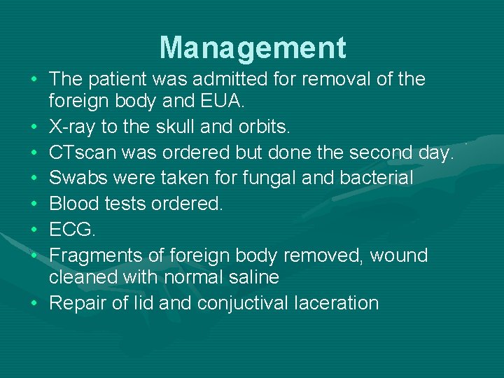
Management • The patient was admitted for removal of the foreign body and EUA. • X-ray to the skull and orbits. • CTscan was ordered but done the second day. • Swabs were taken for fungal and bacterial • Blood tests ordered. • ECG. • Fragments of foreign body removed, wound cleaned with normal saline • Repair of lid and conjuctival laceration
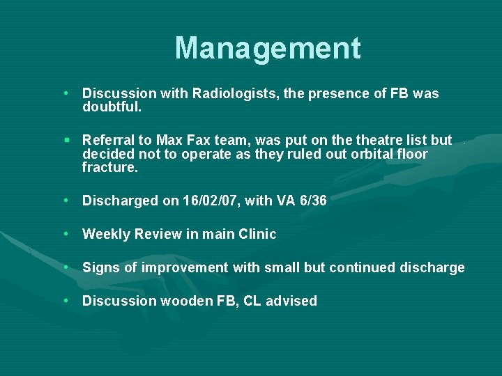
Management • Discussion with Radiologists, the presence of FB was doubtful. § Referral to Max Fax team, was put on theatre list but decided not to operate as they ruled out orbital floor fracture. • Discharged on 16/02/07, with VA 6/36 • Weekly Review in main Clinic • Signs of improvement with small but continued discharge • Discussion wooden FB, CL advised
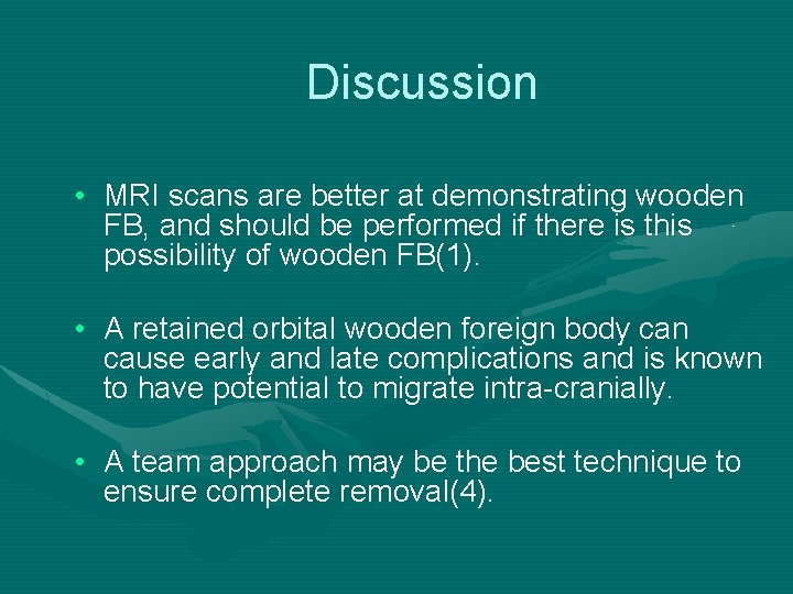
Discussion • MRI scans are better at demonstrating wooden FB, and should be performed if there is this possibility of wooden FB(1). • A retained orbital wooden foreign body can cause early and late complications and is known to have potential to migrate intra-cranially. • A team approach may be the best technique to ensure complete removal(4).
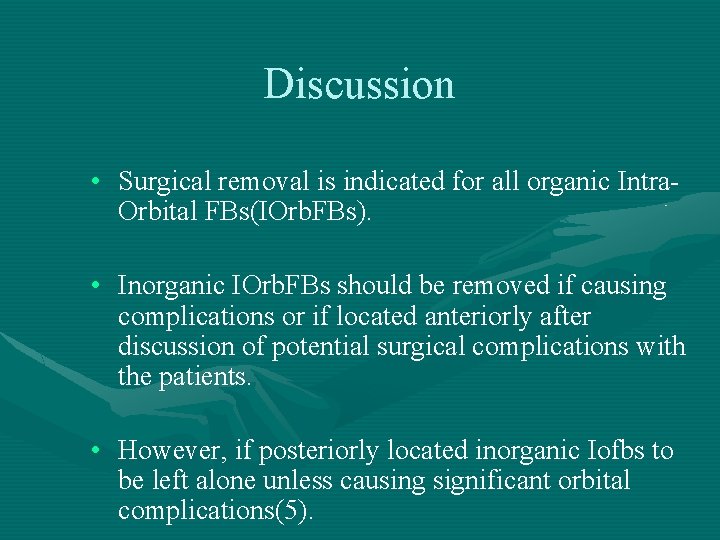
Discussion • Surgical removal is indicated for all organic Intra. Orbital FBs(IOrb. FBs). • Inorganic IOrb. FBs should be removed if causing complications or if located anteriorly after discussion of potential surgical complications with the patients. • However, if posteriorly located inorganic Iofbs to be left alone unless causing significant orbital complications(5).
Discussion • It depends on • degree of wood hydration • location of the wood • the extent of the collateral inflammation(1, 2). • It can misinterpreted as air (2, 3).
Further Management • Debate about presence of foreign body as the size of lesion in MRI is seen less than in CT • Clinically there is improvement following initial removal and systemic antibiotic therapy, with the decrease in proptosis and regaining of EOMs which was completely lost at the time of admission. • Finally decided to re-explore
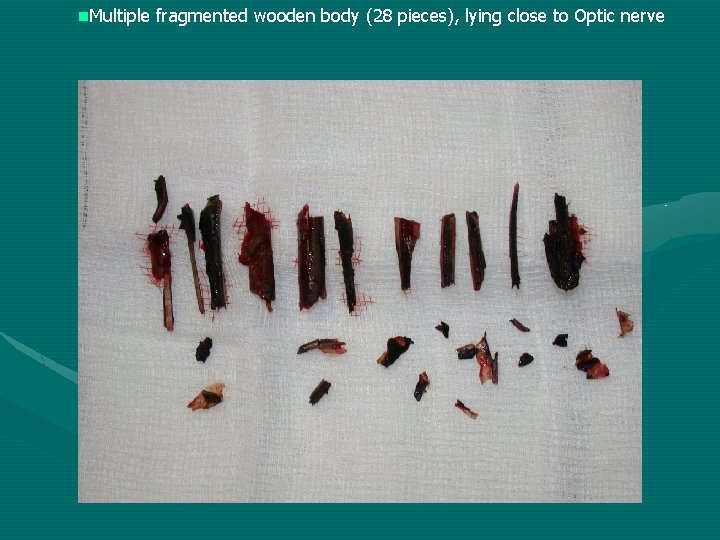
n. Multiple fragmented wooden body (28 pieces), lying close to Optic nerve
Take home message • Intra-orbital wooden foreign bodies can be difficult to detect • Radiographic studies and CT may fail to identify their presence.
References: • • • 1. Green B. E. Kraft. SP, Carter KD et al. Intraorbital wood : Detection by Magnetic resonance imaging. Ophthalmology 1990; 97608 -611. 2. Roberts CF, Leehey PJ 3 rd. Intraorbital wooden foreign body mimicking air at CT. Radiology 1992; 185: 507 -508. 3. Adesanya-O-O, Dawkins-Denise-M. Intraorbital wooden foreign body ( IOFB) mimicking air at CT. Emergency Radiology; 31 January 2007 (epub: 31 1 2007), ISSN: 1070 -3004. 4. Liu-Don, Al-Shail-Essam. Retained orbital wooden foreign body: surgical technique and rationale. Ophthalmology; Feb 2002, Vol. 109, no. 2, p 393 -9, ISSN: 0161 -6420. 5. Fulcher et al. Clinical Features and Management of Intraorbital Foreign Bodies. Ophthalmology; Vol 109, Nov 3, March 2002.
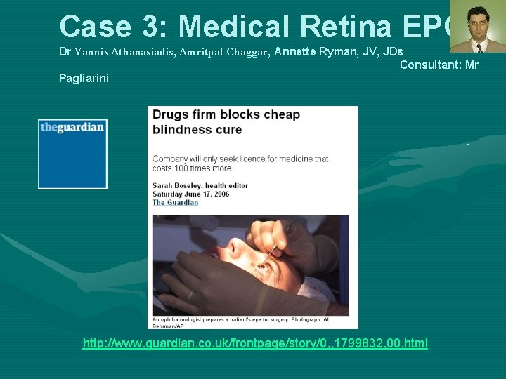
Case 3: Medical Retina EPG Dr Yannis Athanasiadis, Amritpal Chaggar, Annette Ryman, JV, JDs Consultant: Mr Pagliarini http: //www. guardian. co. uk/frontpage/story/0, , 1799832, 00. html
Is intravitreal Avastin a safe and effective treatment for patients with wet age related macular degeneration? • Avastin is unlicensed for use in AMD • 4 published case series and 12 unpublished conference case series. • Use of this evidence for the benefit of patients – Overall change in practice – Individual patient care
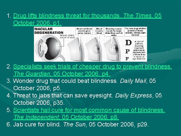
1. Drug lifts blindness threat for thousands. The Times, 05 October 2006, p 1. 2. Specialists seek trials of cheaper drug to prevent blindness. The Guardian, 05 October 2006, p 4. 3. Wonder drug that could beat blindness. Daily Mail, 05 October 2006, p 5. 4. Threat to jabs that can save eyesight. Daily Express, 05 October 2006, p 35. 5. Scientists hail cure for most common cause of blindness. The Independent, 05 October 2006, p 8. 6. Jab cure for blind. The Sun, 05 October 2006, p 29.
Is Ranibizumab (Lucentis®) for the treatment of wet age-related macular degeneration? • Lucentis is licensed in Europe Jan 2007 – awaiting NICE appraisal • 3 published RCTs, 23 located ongoing studies • Use of this evidence for the benefit of patients – Overall change in practice – Individual patient care
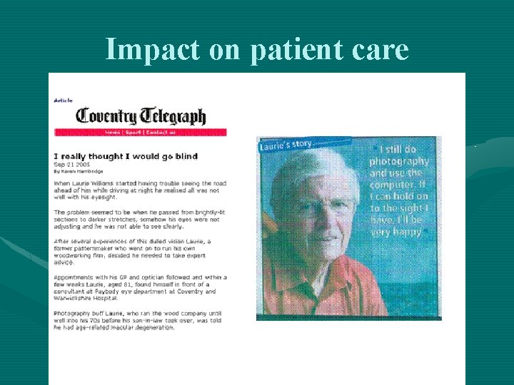
Impact on patient care
TAKE HOME MESSAGE: • Never assume low base evidence equates to ‘not enough evidence’ to either change routine practice or impact on individual patient management. • The work of the CL can be a pivotal point in the delivery of quality patient care
Contact Details • Jacqueline Verschuere – jacqueline. verschuere@uhcw. nhs. uk – 02476 96 8838