NEUROMUSCULAR JUNCTION DISORDERS Sindhu Ramchandren MD MS Assistant


 The Acetylcholine receptor (ACh. R) is a sodium channel")


+ normal DLco = Extrinsic")

 Synaptic vesicles 5, 000 -10, 000 molecules of Ach (quantum) Each")


 ○ Starts working in 30 -60 minutes")


 Botulism Amyotrophic Lateral Sclerosis (ALS) Dermatomyositis Multiple Sclerosis Sarcoidosis")

 Presynaptic disorder of neuromuscular transmission >92% with antibodies against P/Q-type")


- Slides: 38
NEUROMUSCULAR JUNCTION DISORDERS Sindhu Ramchandren, MD, MS Assistant Professor of Neurology Associate Program Director of Clinical Neurophysiology (EMG track) sramchan@med. wayne. edu
Lecture Objectives Clinical features of NMJ disorder NMJ Anatomy- normal and abnormal MG Pathophysiology Autoimmune dysfunction Epidemiology Clinical Presentation Work-up Electrodiagnostic studies Treatment Differential Diagnosis LEMS
Clinical Features of NMJ disorder Involve cranial, limb girdle, proximal muscles +/- respiratory muscles Normal sensation If presynaptic: +ve autonomic signs and post-exercise increase in strength If post-synaptic: fatiguability
NMJ Anatomy Neuromuscular Junction (NMJ) The Acetylcholine receptor (ACh. R) is a sodium channel that opens when bound by ACh ○ There is a partial depolarization of the postsynaptic membrane and this causes an excitatory postsynaptic potential (EPSP) ○ If enough sodium channels open and a threshold potential is reached, a muscle action potential is generated in the postsynaptic membrane
Myasthenia Gravis Pathophysiology In MG, antibodies are directed toward the acetylcholine receptor at the neuromuscular junction of skeletal muscles Results in: Decreased number of nicotinic acetylcholine receptors at the motor end-plate Reduced postsynaptic membrane folds Widened synaptic cleft
Autoimmune Dysfunction in MG Evidence of B-cell mediated disease: ○ Anti-ACh. R antibody is found in 80 -90% of patients with MG ○ Proven with passive transfer experiments Evidence of dysfunction of T- cell mediated immunity: ○ Thymic hyperplasia and thymomas
Epidemiology Frequency Annual incidence in US- 2/1, 000 Worldwide prevalence 1/10, 000 Mortality/morbidity Recent decrease in mortality rate due to advances in treatment ○ 3 -4% (as high as 30 -40%) Risk factors ○ Age > 40 ○ Short history of disease ○ Thymoma Sex F-M (6: 4) Mean age of onset (M-42, F-28) Incidence peaks: M- 6 -7 th decade F- 3 rd decade
Clinical Presentation Fluctuating weakness increased by exertion Weakness increases during the day and improves with rest 40% have ONLY ocular symptoms Only 16% of those with ocular symptoms at onset remain exclusively ocular at the end of 2 years Extraocular muscle weakness Ptosis is present initially in 50% of patients and during the course of disease in 90% of patients The disease remains ocular in 16% of patients Head extension and flexion weakness Respiratory fatigue (counting) Sensory examination and DTR’s are normal
Clinical presentation Facial muscle weakness is almost always present, usually symmetric Ocular muscle weakness Asymmetric ○ Usually affects more than one extraocular muscle and is not limited to muscles innervated by one cranial nerve ○ Weakness of lateral and medial recti may produce a pseudointranuclear ophthalmoplegia Limited adduction of one eye with nystagmus of the abducting eye on attempted lateral gaze Ptosis caused by eyelid weakness Diplopia is very common
Clinical presentation Bulbar muscle weakness Palatal muscles ○ “Nasal voice”, nasal regurgitation ○ Chewing may become difficult ○ Severe jaw weakness may cause jaw to hang open ○ Swallowing may be difficult and aspiration may occur with fluids—coughing and choking while drinking Neck muscles ○ Neck flexors affected more than extensors
Clinical presentation Limb muscle weakness Upper limbs more common than lower limbs Upper Extremities Lower Extremities Deltoids Wrist extensors Finger extensors Triceps > Biceps Hip flexors (most common) Quadriceps Hamstrings Foot dorsiflexors Plantar flexors
Clinical presentation-ER CN: pseudo-INO, ptosis, facial weakness Respiratory muscle weakness Weakness of the intercostal muscles and the diaphragm may result in CO 2 retention due to hypoventilation ○ Bedside counts in 1 breath-rough estimate of VC (~100 cc per count) Weakness of pharyngeal muscles may collapse the upper airway ○ Monitor negative inspiratory force(< -25 cm H 20), vital capacity (< 10 -15 m. L/kg) ○ Do NOT rely on pulse oximetry Arterial O 2 may be normal while CO 2 is retained
Signs of impending respiratory failure Tachypnea, sinus tachycardia Staccato speech Inability to count to 20 Profound weakness of neck flexion Use of accessory muscles (visible, palpable) • Orthopnea • Paradoxical breathing pattern • Signs of bulbar dysfunction (nasal voice, accumulation of saliva, weak cough) • • • – think about aspiration!
Work-up Bedside: Ice pack, tensilon test Edrophonium is a short acting Acetylcholine Esterase Inhibitor that improves muscle weakness ONLY worthwhile with ptosis, pseudo-INO) Lab studies Anti-acetylcholine receptor antibody ○ 85% in generalized myasthenia ○ 50% of patients with pure ocular myasthenia Anti-muscle-specific tyrosine kinase (MUSK) antibody ○ Present in one-half to two-thirds of patients with “seronegative” generalized myasthenia ○ Unknown (but lower) frequency in pure ocular myasthenia
Work-up Co-existing autoimmune diseases Hyperthyroidism ○ Occurs in 10 -15% MG patients Exophthalmos and tachycardia point to hyperthyroidism Weakness may not improve with treatment of MG alone in patients with co-existing hyperthyroidism Rheumatoid arthritis Scleroderma Lupus
Work-up PFT’s: Restrictive pattern (normal to high FEV 1/FVC ratio)+ normal DLco = Extrinsic restrictive disease (seen with scoliosis or myasthenia gravis) Imaging studies High resolution Chest CT scan (with and without contrast) is mandatory to identify thymoma MRI of the brain and orbits may help to rule out other causes of cranial nerve deficits but should not be used routinely
Electrodiagnostic studies Repetitive nerve stimulation ○ Several factors can affect RNS results Lower temperature increases the amplitude of the compound muscle action potential Many patients report clinically significant improvement in cold temperatures ACh. E inhibitors prior to testing may mask the abnormalities and should be avoided for at least 1 day prior to testing Single fiber electromyography (SFEMG) ○ SFEMG is more sensitive than RNS in MG
Neuromuscular Junction (NMJ) Synaptic vesicles 5, 000 -10, 000 molecules of Ach (quantum) Each vesicle released causes a 1 m. V change in the post-synaptic membrane. This occurs spontaneously and is the MEP (motor end plate potential) m=pn m= the number of vesicles released after a nerve action potential reaches the NMJ p= the probability of release ○ Proportional to the [Ca 2+], typically 20% n= the number of vesicles available
Vesicle stores Primary store- immediately available 1000 vesicles Secondary store- mobilization, replenishes primary store after 1 -2 seconds- 10, 000 vesicles Tertiary store- reserve, found in the axon and cell body- 100, 000 vesicles
Safety Factor The difference between the EPP and the threshold required for initiating a muscle fiber action potential. Influenced by vesicle release, Ach. R conduction properties/density, postsynaptic folds that focus end plate current on voltage gated sodium channels
Role of calcium Pre-synaptic depolarization leads to calcium influx that leads to vesicular docking and Ach release Calcium diffuses slowly out of the presynaptic terminal at a rate of 100 -200 ms. RNS < 5 Hz -calcium’s role in vesicular release is not enhanced RNS > 10 Hz increased calcium greatly increases the probability that a vesicle will be released
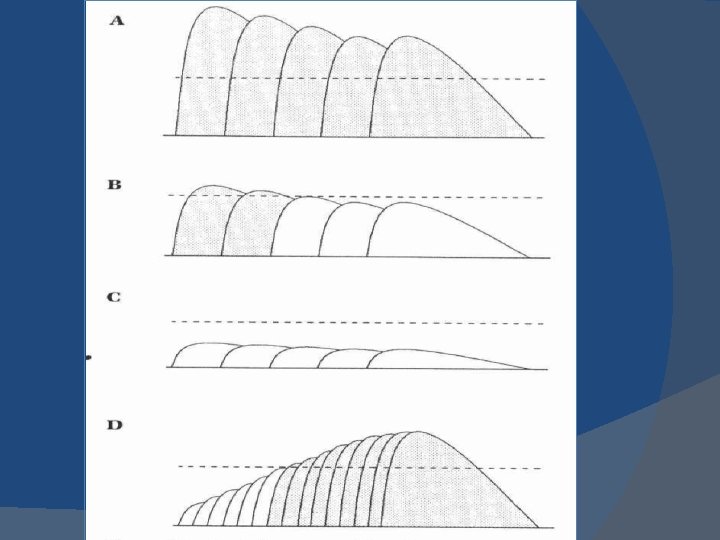
Normal RNS In normals, with slow RNS, the EPP falls as vesicles are slowly depleted, but because of a normal safety factor, threshold is achieved and a muscle fiber action potential is generated With maximal exercise (similar to Fast RNS), the depletion of vesicles is counterbalanced by an accumulation of calcium, and there is an increase in the EPP
Post-Synaptic Disorders The baseline EPP and safety factor are reduced, however threshold is still reached and the CMAP is normal Slow RNS- The EPP falls further, some become subthreshold, muscle fiber action potentials fail and the CMAP drops (10%) The greatest drop is seen between stimulus 1 & 4. By stimulus 5 the CMAP may increase with mobilization of the 2 nd store There may be a repair of the decrement after 10 seconds of exercise- “Post Exercise Facilitation” Without a decrement, slow RNS should be repeated every minute after one minute of exercise- “Post Exercise Exhaustion”
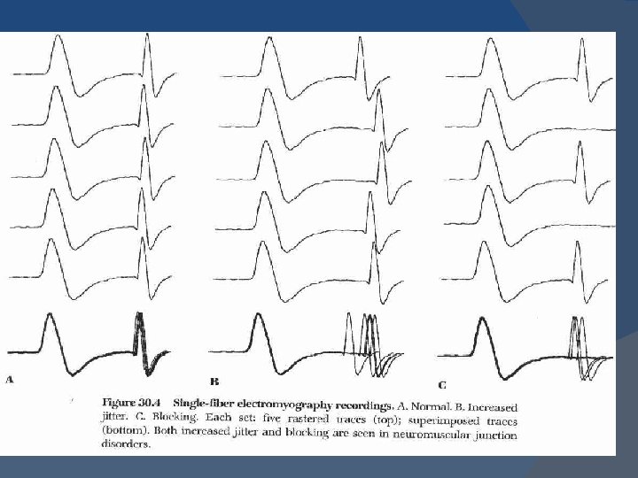
Single-fiber electromyography Jitter- variability of the time interval between two muscle fiber action potentials innervated by the same motor unit; calculated by MCD (mean consecutive difference of 20 pairs, each recorded 100 consecutive times) Blocking- the failure of transmission leading to an absent muscle fiber action potential in a pair; correlates with weakness Generalized MG ○ Abnormal Extensor Digiti Communis found in 87% ○ Second abnormal muscle increases sensitivity to 99% Ocular MG ○ Frontalis muscle is abnormal in almost 100%
Jitter and block
Approach to treating MG Spontaneous remissions are rare Most remissions with treatment occur within the first three years Treat any exacerbating factors Infections, medication effect, endocrine disease Thymectomy- thymoma Standard RX: Acetylcholinesterase inhibitors Immunosuppressants Prednisone Azathioprine; Cyclosporine Plasma exchange Intravenous immunoglobulin Monitor: clinical improvement (respiratory, strength, ptosis), rather than fatigue/ ACh. Rantibody levels
Treatment- Remove exacerbating Drugs
Treatment ACh. E inhibitor Pyridostigmine bromide (Mestinon) ○ Starts working in 30 -60 minutes and lasts 3 -6 hours; individualize dose ○ Average dose: 60 -90 mg TID-QID, plus 180 Timespan QHS ○ May cause cholinergic crisis, which can exacerbate pulmonary symptoms during acute crisis
Immunosuppressants Prednisone ○ Significant improvement is often seen after a decreased antibody titer which is usually 1 -4 months ○ No single dose regimen is accepted Outpatient regimen: start at 10 -15 mg QD; increase by 5 mg every 3 days, to a maximum of 50 mg QD Inpatient- can start slow at 20 and increase by 5 mg q 3 d to max of 50; personally, if patient already intubated, will start higher. ○ Transient initial severe exacerbation, usually after 1 to 3 weeks (2% to 5%) Steroid Sparing agents: ○ Azathioprine- Maintenance dose 2 -3 mg/kg; average dose: 140 and 210 mg qd; can take between 6 -24 months for onset of action ○ Cyclosporine- more toxic; onset between 1 -3 months
Acute therapies Plasma exchange: ○ Originally based on positive outcome in Qureshi study (Neurology, 1999) ○ However, AAN’s 2011 position stated “. . insufficient evidence to support or refute the efficacy of plasmapheresis in the treatment of myasthenic crisis. . due to lack of RCTs” ○ Result: Insurance denial, however still most likely used treatment in ICU settings IVIG ○ Total of 2 grams/kg, divided over 5 days (0. 4 grams/kg/day)
Differentials Lambert-Eaton Myasthenic Syndrome (LEMS) Botulism Amyotrophic Lateral Sclerosis (ALS) Dermatomyositis Multiple Sclerosis Sarcoidosis Thyroid disease Basilar Artery Thrombosis Brainstem gliomas Cavernous sinus syndromes Oculopharyngeal muscular dystrophy Brainstem syndromes
Pre-Synaptic Disorders The baseline EPP is reduced and some are sub- threshold The baseline CMAP is reduced Slow RNS- the EPP falls further, fewer muscle fibers generate action potentials and the CMAP is reduced more Fast RNS- pre-synaptic calcium increases, more Ach is released, larger EPPs generate muscle fiber action potentials and the CMAP is increased LEMS- Increment often > 100200% Botulism- smaller increment 30100%
Lambert-Eaton Myasthenic Syndrome (LEMS) Presynaptic disorder of neuromuscular transmission >92% with antibodies against P/Q-type voltagegated calcium channels (presynaptic) Impaired influx of calcium into nerve terminal with reduced neuromuscular junction transmission Facilitation with sustained contraction >100% CMAP increase with repetitive stimulation
LEMS Clinical Features Mimics MG, often affects older males Proximal weakness (initially LE, then UE), areflexia or hyporeflexia; Respiratory and craniobulbar involvement rare Autonomic dysfunction prominent dry mouth, dry eyes, impotence, orthostatic hypotension, hyperhidrosis Associated with underlying carcinoma; precedes tumor diagnosis by several years: 45% to 60%: SCLC; also renal cell, breast & lymphoma
LEMS- Treatment Unlike most paraneoplastic syndromes LEMS usually responds to: Plasmapheresis/ intravenous immunoglobin Corticosteroids 3, 4 Diaminopyridine: 20 mg tid (orphan drug; can obtain as compassionate use from Jacobus Pharmaceutical Company, NJ; Phone: 609 -9217447; Fax: 609 -799 -1176) Pyridostigmine Immunosuppressants Long-term treatment often needed