Management of Neck Lumps David Howe Consultant ENT


- Slides: 36
Management of Neck Lumps David Howe Consultant ENT Surgeon Heart of England Foundation Trust Spire Parkway
What to cover? • Common neck lumps • What the likely pathology is • Initial investigations and management • Red Flags
How to approach assessment • History - consider age of patient • Duration of mass, has it changed? • Pain ? referred • Associated symptoms eg swallowing/voice issues • Risk factors eg smoking, immunosuppression, previous RT
Location of key structures • Thyroid • Major salivary glands • Lymphatic groups • Thyroglossal duct cysts • Branchial cysts • Dental sepsis • Direct spread of tumours
Mandatory Anatomy slide!!
Salivary lesions • Glands in order of frequency • Parotid • Submandular then Sublingual • Minor salivary • Rates of malignancy affected by gland type • Parotid lesions approximately 80% benign • Submandibular gland others 50% risk of malignancy
• Parotid • Commonly pleomorphic adenoma circa 60% • Has small risk of malignant potential • Hence patients offered surgery (also to confirm pathology) • Warthins tumours - entirely benign, common in smokers and often bilateral, tail of parotid. • Malignancy, primary or met (often from skin e. g. scc) • Prognosis and treatment dependant on grade of tumour
Submandibular masses • Most common site for a calculus • Often show up on uss but sometimes requires ct • Treatment options - conservative, endoscopic, open • Tumours, benign generally a PA, remember 50% rate of malignancy. • Assess with USS and probably FNA • Surgery generally primary treatment.
Minor salivary lesions • Generally noticed on per oral inspection - often by the patient. • I would suggest refer in as they need biopsy (60 -80% malignant) • Floor of mouth swelling around submandibular duct could be a calculus or a sublingual tumour or ranula.
Dental Sepsis • Increasingly common!! Dearth of NHS dentistry • Patients presenting with deep space neck infections and swellings under mandible • Particularly consider this in submental swellings and those with discharge • Look at teeth, consider OPG and dental review • Often need tooth removing to resolve problems
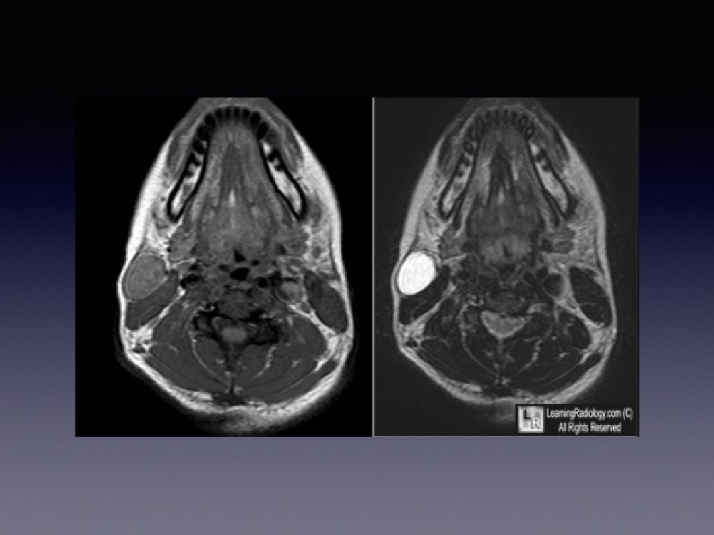
Branchial cyst • Common presentation of sudden swelling in level 2/3 of neck • Occurs in late teens and early 20’s • Can occur later in life but in age over 40 consider possible sinister causes. • Recommend referral and removal if indicated or wanted.
• Younger age group, benign lesion ? cystic degeneration of lymph node. • Over 40’s likely to be benign, but possibility of cystic metastasis from a SCC • Therefore more extensive workup in higher risk group and often pet scan undertaken • Objective to identify potential primary lesion and avoid surgery to neck unless therapeutic. • Definitely consider 2 WW referral for these, particularly in over 40’s • Most turn out to be benign, very rarely a cystic metastasis from a thyroid cancer
Lymph nodes • Can be difficult sometimes to prove there is nothing of concern • Worries about over/under investigating • Possible risk of not identifying a low grade/quiescent lymphoma • Limitations of both imaging/biopsy/clinical assessment • Risks of lymph node biopsy not inconsiderable
• Consider • Age • Duration and associated symptoms • Risk factors • Size definitely matters!!!! • Location of nodes is important, particularly supraclavicuar and posterior triangle • Careful assessment of throat and ask about pain/swallowing issues etc as patient may not volunteer them
• Investigations • Could simply refer • If low risk/clinical concern • Baseline bloods/USS • How long to allow to improve? ? • Patient anxiety
• USS of lymph nodes • Operator dependant!! • Description sometimes difficult to lead management. • But - a reactive node should retain a fatty hilum, be ovoid in shape and have a normal vascular pattern despite being enlarged • Morphologically abnormal nodes need further investigation, sometimes reactive nodes can look like this and subsequently improve.
• Role of referral • Allows nasoendoscopy and comprehensive assessment of throat • Consideration of fna • May require open lymph node biopsy • Limitations of fna in neck nodes, may give an indication of high grade lymphoma but not diagnostic and either a core biopsy or open biopsy needed. • Can infer if likely to be granulomatous e. g. TB • Node maybe related to either a systemic metastasis or unknown primary lesion
• Unknown primary • Carcinoma in lymph node with no primary evident • May be head and neck primary levels 1 -4 • Distant primary more likely in supraclavicular node - lung/bowel/renal etc • Role of cross sectional imaging and PET scan • Head and neck primary often an occult tonsillar tumour or tongue base primary
Thyroid Nodules are very common circa 45% of female population age over 50 have thyroid nodules Malignancy relatively rare. 2010 Annual incidence in the UK 5. 1 per 100, 000 women cf 2. 3 in 1971 -95 (Cancer Registry) 900 new cases and 250 deaths recorded in England Wales per year
Most common endocrine malignancy Only 1% of all malignancies Increasing incidence of PTC - much of this incidental micro papillary carcinoma (<1 cm) or lower stage cancers Overall mortality from thyroid cancer unchanged for many years
In Primary Care - What do USS reports mean USS report should hopefully recommend referral to a specialist if there are concerning thyroid lesions. Potential difficulty if report has no explicit U classification and simply descriptive text regarding echogenicity of nodules - what does that mean? ? ? Strong family history/previous radiation exposure High TSH - association with increased risk of cancer Suggest - U 2 nodules and euthyroid probably don't require investigation -subjective U 3 upwards need referral as per 2 WW Compressive symptoms/goitre - consideration for decompressing surgery
Does size matter? ? Historically as the size of the nodule increased in size the reliability of biopsies and scans reduced. Probably minimal increased risk of cancer just uncertainty This still applies
Imaging is key to investigation of nodules
New Guidelines Aim of guidelines - to reduce unnecessary investigation cost - lots of nodules, most benign Big cost implication for healthcare system Thyroid Nodules are now classified based on USS Appearance Low risk (benign lesions) - U 1/U 2 considered benign No further investigation needed Higher risk U 3/U 4/U 5 progressive increasing risk of cancer Require a biopsy and possibly surgery Introduced in 2014 New system - entirely dependant on radiology opinion. Subjective interpretation of USS findings - need to consider thyroid specialist radiologist Reclassification of USS appearance frequently at mdt - important as this can determine if
Indications for Surgery Concern regarding malignant nature of nodule Compressive consequences of a large goitre - normal tracheal compression Thyrotoxicosis Cosmetic - not generally recommended although often requested!!!
Case Study 40 year lady Neck lump incidental No family history What next?
History Often of minimal influence in thyroid management Strong FHx/radiation exposure Voice change very rare even with malignancy Haemoptysis -very rare Compressive symptoms? Symptoms of thyroid dysfunction
Investigations Bloods TFTs - if toxic consider endocrine referral - maybe a toxic nodule (low chance of malignancy) Thyroid antibodies - helpful for interpretation of uss and biopsy findings (pseudo tumour)
Ultrasound USS finding 3 cm hypo echoic, well defined nodule, some intralesional vascular flow (U 3). Rest of thyroid normal What next?
Management - will require biopsy - therefore refer USS FNA scored Thy 1 -5 -needs MDT review This lesion thy 3 f (unable to refine between benign and malignant lesions) Therefore offer diagnostic lobectomy Risk of malignancy approximately 25% in this situation
Thyroglossal duct cyst • Commonest in childhood and young adults • Can be complicated by infection • Malignancy very rare but reported • Investigation of choice - use • Check for normal thyroid tissue • Surgery not essential
Acute swellings • Lymph nodes - probably inflammatory, can become necrotic and form an abcess particularly in infants ? I&D • Thyroid cysts - spontaneous bleed and swell ? coughing fit • Deep neck infection, potential life threatening!!! From quinsy, fb or dental source typically • Often need ITU or tracheostomy and agressive Abx therapy
Summary • Position of neck lump informs likely pathology • Risk factors key • Consider red flags - pain, voice change, swallowing, B symptoms • Early referral suggested if concern (2 WW) • USS often helpful first line investigation