Head and neck The head neck The pharyngeal


 gives rise to styloid process, lesser horn,")


- Slides: 15
Head and neck •
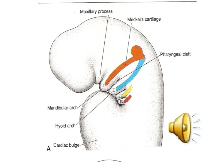
• The head & neck: • The pharyngeal or branchial arches appear in the 4 th-5 th weeks. • The mesenchyme of the pharyngeal arches is derived from neural crest cells. • The pharyngeal arches consist of bars of mesenchymal tissues, separated by pharyngeal pouches and pharyngeal clefts. • Each arch has its own artery, cranial nerve, muscles, and cartilage or skeletal elements.
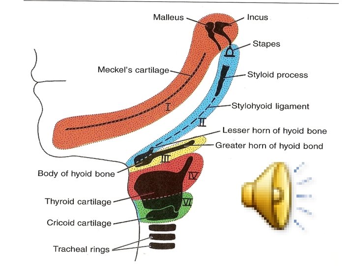
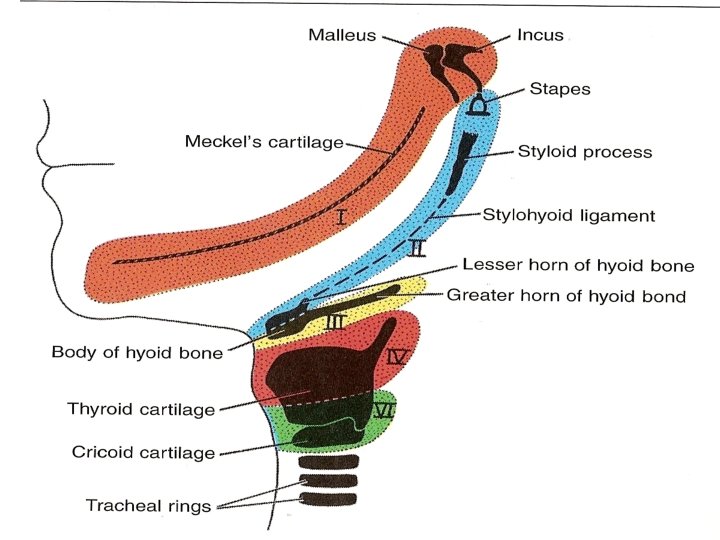
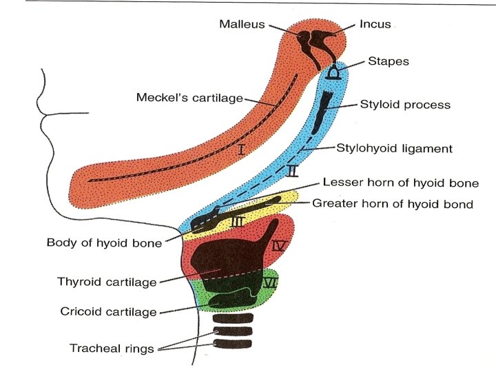
• First arch gives rise to zygomatic bone, part of temporal bone, incus and malleus, • muscles of mastication, tensor tympani and tensor palate muscles. • The bone of the mandible develops by membranous ossification of the mesenchyme covering the Meckel's cartilage.
• 2 nd arch (hyoid arch) gives rise to styloid process, lesser horn, upper part of body of hyoid bone • muscles of facial expression.
• 3 th arch gives rise to: • Cartilage: that produces lower part and greater horn of hyoid bone, • muscles supplied by 9 th cranial nerve ( stylopharyngeus m. ).
• The 4 th & 6 th arches: • Cartilages; that produce thyroid, cricoid, arytenoid, corniculate and cuneiform cartilages of larynx. • Muscles from 4 th arch that are supplied by superior laryngeal N. (cricothyryoid, levator palate, and constrictors of pharynx).
• muscels of 6 th arch are supplied by recurrent laryngeal branch of vagus including intrinsic muscles of larynx.
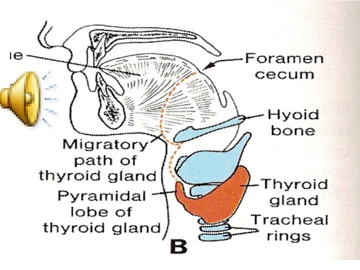
• The thyroid gland: • It develops from an epithelial proliferation between tuberculum impar & copula at position of foramen cecum at apex of sulcus terminalis. This thyroid tissue descends anterior to pharynx. During this descend, thyroid remains connected to tongue by a canal called thyroglossal duct that disappears later on. Thyroid function begins in the 3 rd month.
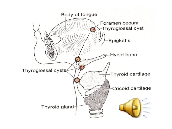
• Clinical notes: • Remnant of thyroglossal duct forms the thyroglossal cyst that may lie in any point along descending pathway of thyroid. This cyst may be opened to the out side by thyroglossal fistula.