Male genital system 1 Testes 2 epididymis 3


- Slides: 22


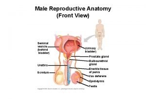
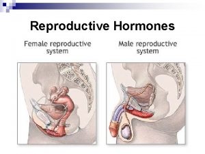
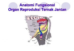
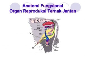
Male genital system 1 - Testes 2 - epididymis 3 - vas deferens 4 - spermatic cord 5 - seminal vesicle 6 - ejaculatory duct 7 - prostate gland 8 - penis

Scrotum. ** Layers of the scrotum, from outside inwards, 1 - Skin. 2 - Dartos muscle, involuntary muscle to corrugate the scrotum (genital branch of genitofemoral N) (No superficial fatty layer). 3 - Membranous layer of superficial fascia. 4 - External spermatic fascia. 5 - Cremasteric muscle and fascia. 6 - Internal spermatic. ** Contents of the scrotum: 1 - Testis. 2 - Spermatic cord and its contents. 3 - Tunica vaginalis (peritoneal covering the testis) parietal & visceral layers. (Ext. Abd. Ob. ) (Int. Abd. Ob. ) (Fascia transversalis. )

Cremasteric muscle ** N. B; It presents only in the male. ** Origin, internal abdominal oblique. ** Insertion; It is in the form of loops around the testis and inserted into the pubic tubercle. ** Nerve supply; genital branch of the genitofemoral nerve. ** Action; elevates the testis towards the superficial inguinal ring. - It is concerned with temperature regulation. ** Cremasteric reflex; • Scratching of the upper part of the medial side of the thigh leads to upward retraction of the testis. It is much more active in children.

Upper pole Vasa Efferentia Lower pole

-Testes - It is the male primary sex organ. - Functions: a- Formation of the sperms. b- Secretion of testosterone hormone. * Site; in the scrotum ** Shape, it is oval in shape. ** Size, It is 1. 5 inches long, 1 inch broad and 1/2 inch thick. - External features, a- 2 poles (upper and lower) - Upper related to the head of epidydimes. - Lower related to the tail of epidydimes. - The Left testis is Lower than the right. b- 2 borders (anterior and posterior), c- 2 surfaces (lateral and medial). ** Structure; - The testis has a thick capsule called the Tunica albuginea. - It is divided into lobules by septa. Each lobule contains 1 -3 semeniferous tubules.

Testis • Arterial supply: Testicular artery from the abdominal aorta. • Venous drainage pamipiniform plexus of vein that form testicular vein: • ** Termination of the testicular vein: a. Right vein ends in the inferior vena cava. b. Left vein ends in the left renal vein. • Why the varicocele is common in the left side 1. Left testicular vein is Longer than right 2. It opens in the left renal vein by right angle. 3. Compression by a full sigmoid colon in chronic constipation. 4. Vascular spasm by adrenaline coming from left suprarenal gland.

** How to identify that a given testis are right or left 1 - Epididymis is posterior to the testis. 2 - Vas deferens arises from the lower end or tail of epididymis. 3 - Sinus of epididymis (recess of tunica vaginalis) is directed laterally. ** Applied anatomy, - undescended testes leading to arrest of the spermatogenesis. - In bilateral cases occurs permanent sterility. - If neglected, the testis transforms into malignant.

Spermatic cord ** Coverings of the spermatic cord from outside inwards, I- External spermatic fascia. 2 - Cremasteric muscle and fascia. 3 - Internal spermatic fascia.

** Contents of the spermatic cord; 1. Vas deferens. 2. Artery of the vas deferens. 3. Testicular artery from the abdominal aorta. 4. Pampiniform plexus of veins that form testicular veins. 5. Sympathetic and parasympathetic nerve fibers. 6. Lymph vessels. 7. Cremasteric artery. 8. Genital branch of the genitofemoral nerve. 9. Remnant of process vaginalis.

Prostate Pelvic fascia

** measurements - 2 cm A-P diameter. - 3 cm Vertical diameter: - 4 cm Transverse diameter. ** Relations 1 - Base: directed upwards and related to the neck of the urinary bladder. - It is pierced by the urethra. 2 - Apex: directed inferiorly and rests on the pelvic fascia. 3 - Anterior surface: - lies 2 cm behind the lower part of the symphysis pubis separated from it by fat. - The urethra exits from this surface nearer to the apex. 4 - Posterior surface: - This surface is related to the rectum. - The two ejaculatory ducts penetrate its upper part. 5 - Inferolateral surfaces: related to the anterior part of the levator ani.

Lobes of the prostate - The urethra and 2 ejaculatory ducts traverse the prostate dividing it into; 1 - Median lobe: between prostatic urethra and two ejaculatory ducts. - This lobe projects into the interior of the bladder forming the uvula. 2 - Right and left lateral lobes; on each side of the urethra. - They are directly continuous with each other behind the urethra. - They are connected together in front of the urethra by a narrow band called Isthmus (devoid of glandular substance). Prostate Urethra Sagittal S. Right lobe Left lobe Urethra Coronal S.

** Arterial supply: 1) inferior vesical. 2) Middle rectal. 3) Internal pudendal. ** Venous drainage: The veins form a prostatic venous plexus. ** Lymphatic drainage: into 1) internal iliac. 2) Sacral lymph nodes. ** Nerve supply: from the pelvic plexus. • Senile enlargement affects the median lobe and causes retention of urine. • Cancer prostate commonly spreads to the vertebrae because The prostatic venous plexus is connected to the internal vertebral venous plexuses by valveless veins.

- It is a thick cord-like tube, about 45 cm long. - It carries and stores the sperms. - It begins from the tail of the epididymis - runs in the inguinal canal through the spermatic cord, - curves around the inferior epigastric artery. - Then, it descends on the lateral wall of the pelvis crossing the following structures; 1 - External iliac vessels. 2 Superior vesical (obliterated umbilical) artery. 3 - Obturator nerve. 4 - Obturator vessels. Vas (ductus) deferens

• Vas (ductus)Deferens - Then, It curves medially crossing above the ureter then behind the base of the urinary bladder. ** End by forming the ampulla which joins the seminal vesicle to form the ejaculatory duct. • Seminal Vesicles - It is a sacculated and coiled pouch Behind the base of the urinary bladder, ** Function; they are glands which contract during ejaculation. - Their secretions constitute the greater amount of the seminal fluid. • Ejaculatory Ducts - It is a very narrow duct, 2 cm long which immediately passes through the base of the prostate gland. - It opens into the seminal colliculus of the prostatic urethra.

Prostatic urethra

With Deep artery of the penis Urethra

Transverse section in the penis - Tunica albuginea (5) is the fibrous envelope of the corpora cavernosa. It is directly involved in maintaining an erection - Buck's fascia (4) constricting the erection veins of the penis, preventing blood from leaving and thus sustaining the erect state. The erection veins include the deep dorsal vein, two cavernosal veins, and four para-arterial veins.

** Structures of the penis - The penis is formed of three elongated bodies a- 2 corpora cavernosa, each one is continuous posteriorly with the crus of the root of the penis. - The deep artery of the penis runs in the center of the corpus cavernosum. b- One corpus spongiosum, is continuous posteriorly with the bulb of the root of the penis. - It is traversed by the penile urethra. The penis

Penis - This is the male copulatory organ. It is formed of two main parts. 1 - Root of the penis consists of bulb and 2 crura. 2 - Body of the penis this is the free pendulous part. - The end of the body is enlarged called the glans penis. - The tip of the glans carries the external urethral orifice. - The constriction between the body and the glans called neck of the penis (corona). - Prepuce (Foreskin), skin folds cover the glans penis that removed during circumcision - The superficial fascia of the penis is devoid of fat. • Erection of the penis - Erection of the penis (or clitoris) is a purely vascular mechanism. Parasympathetic stimulation leading to rapid inflow of blood and filling of the erectile tissue of the corpora cavernosa and thus pressure over the draining veins, leading to erection of the penis (or clitoris). - Ejaculation is sympathetic stimulation.

Th ank Qu you est ion s I/Azzam - 2004
 Frimbae
 Male genital variation
 Reproductive hygiene
 Functions of cowpers gland
 Function of prostate gland
 I ii iii
 How cats mate
 Facies urethralis penis
 Umbilical arteries
 The epididymis is a long coiled tube that sits atop the
 Bulbourethralis mirigy
 Plica genitalis
 Womens anatomy diagram
 Psicometria significado
 Frog testis labeled
 Testes neuropsicopedagógicos
 Orchidodesis adalah
 Absennya satu atau kedua buah pelir adalah
 Testes regressivos
 Fertilization and implantation
 Male reproductive system
 Testes post hoc
 Testes


