Lymphoreticular system Assistant Professor Dr Maysem Mouayad Alwash
- Slides: 72
Lymphoreticular system Assistant Professor : Dr. Maysem Mouayad Alwash
Lymphocytes are the immunologically competent cells that assist the phagocytes in defence of the body against infection and other foreign invasion.
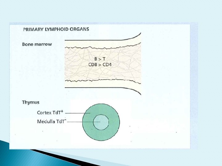
-The primary lymphoid organs: include the bone marrow and thymus , where T & B lymphocytes develop in the postnatal life. - The secondary lymphoid organs in which specific immune responses are generated are the lymph nodes, spleen and lymphoid tissues of the alimentary and respiratory tracts.
Mucosa-associated lymphoid tissue (MALT) includes : (i)discrete lymphoid structures such as the appendix, Peyer’s patches in the submucosa of the intestine and the tonsils and adenoids (collectively referred to as Waldeyer’s ring) in the pharynx. (ii) lymphocytes in the submucosa of various organs.
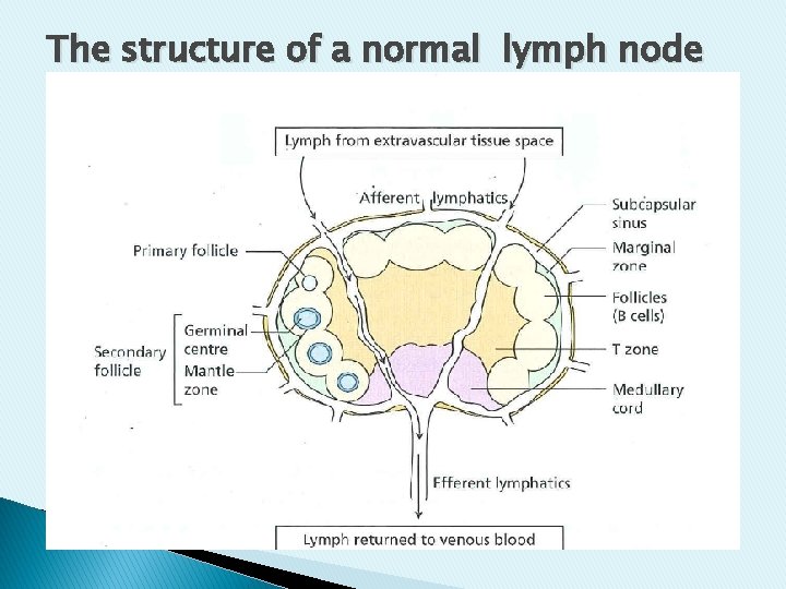
The structure of a normal lymph node
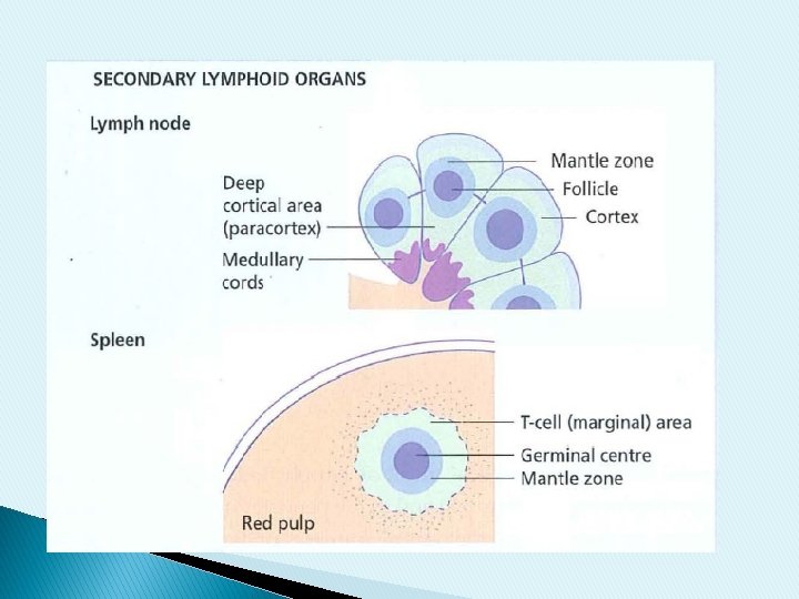
Lymph nodes are divided into : 1. Cortex : composed of B lymphocytes and follicular dendritic cells. -The primary follicle -The secondary follicle comprising a germinal centre surrounded by a mantle zone of small B lymphocytes. Outside the mantle zone some lymph node germinal centres have a marginal zone, also composed of B lymphocytes.
2. paracortex : T cells occupy the paracortex. 3. medulla. In the centre of the lymph node , it composed of medullary cords and sinuses. The medullary cords are occupied by B and T lymphocytes, plasma cells and macrophages.
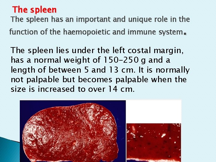
The spleen has an important and unique role in the . function of the haemopoietic and immune system The spleen lies under the left costal margin, has a normal weight of 150 -250 g and a length of between 5 and 13 cm. It is normally not palpable but becomes palpable when the size is increased to over 14 cm.
Extramedullary haemopoiesis The spleen is an important site of haemopoiesis in utero and retains the ability to reactivate this process after birth. This can occur : in severe anaemia, such as chronic haemolysis , megaloblastic anaemia and thalassaemia major, or myelofibrosis or malignant disorders in the bone marrow.
Causes of splenomegaly -Leukaemia -lymphomas -myeloproliferative disorders -haemolytic anaemias -glandular fever -portal hypertension -malaria -Leishmaniasis -schistosomiasis.
Causes of massive splenomegally: 1. Chronic myeloid leukaemia 2. lymphoma � 3 -Primary myelofibrosis � 4 -Thalassaemia � 5 - major or intermedia leishmaniasis and schistosomiasis � 6 - Gaucher’s disease �
Hypersplenism: Hypersplenism is a clinical syndrome that can be seen in any form of splenomegaly (it does not imply a specific causal mechanism).
-Enlargement of the spleen; -Reduction of at least one cell line in the blood in the presence of normal bone marrow ftmction; -Evidence of increased release of premature cells, such as reticulocytes or immature platelets, from the bone marrow into the blood.
Normally, only approximately 5% (3070 m. L) of the total red cell mass is present in the spleen and up to half of the total marginating neutrophil pool and 30% of the platelet mass may be located there.
As the spleen enlarges, the proportion of haemopoietic cells within the organ increases such that up to 40% of the red cell mass, and 90% of platelets , may be pooled in an enlarged spleen.
Hyposplenism Patients with functional hyposplenism have impaired immunity to blood borne bacterial and protozoal infections, and persistent thrombocytosis. Hyposplenism (excluding that induced by medical or surgical intervention) occurs in a wide range of conditions:
-sickle cell disease -gluten - induced enteropathy (coeliac syndrome). -dermatitis herpetiformis. -Crohn’s disease and ulcerative colitis -essential thrombocythaemia -Congenital absence -Elderly
Reactive Lymphadenitis Any immune response against foreign antigens is often associated with lymph node enlargement (lymphadenopathy). The infections that cause lymphadenitis are numerous and varied and may be acute or chronic. In most instances, the histologic appearance of the nodes is entirely nonspecific.
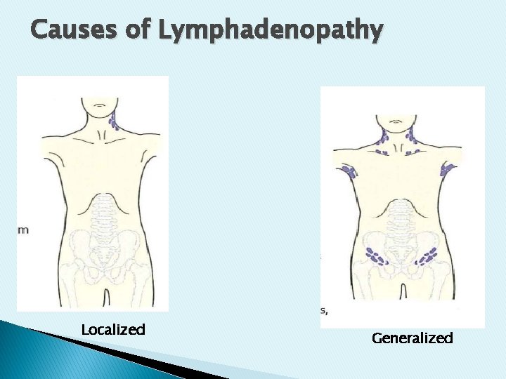
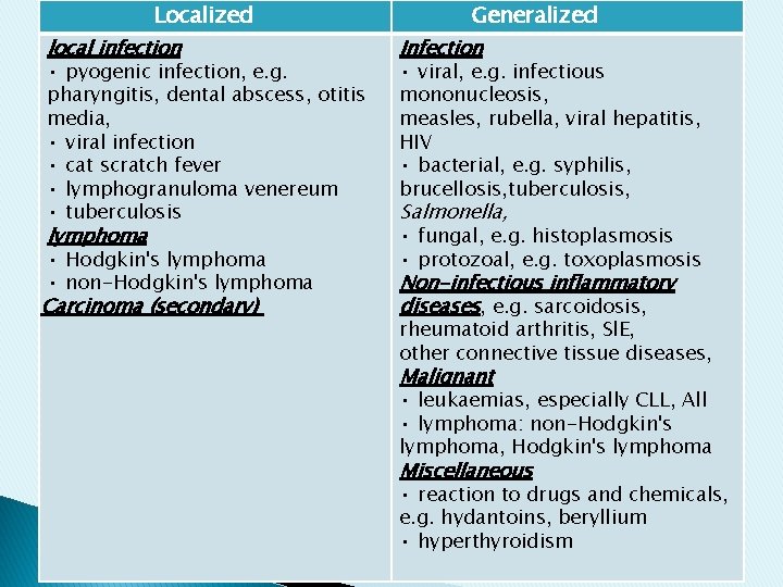
Causes of Lymphadenopathy Localized Generalized
Localized local infection • pyogenic infection, e. g. pharyngitis, dental abscess, otitis media, • viral infection • cat scratch fever • lymphogranuloma venereum • tuberculosis lymphoma • Hodgkin's lymphoma • non-Hodgkin's lymphoma Carcinoma (secondary) Generalized Infection • viral, e. g. infectious mononucleosis, measles, rubella, viral hepatitis, HIV • bacterial, e. g. syphilis, brucellosis, tuberculosis, Salmonella, • fungal, e. g. histoplasmosis • protozoal, e. g. toxoplasmosis Non-infectious inflammatory diseases, e. g. sarcoidosis, rheumatoid arthritis, Sl. E, other connective tissue diseases, Malignant • leukaemias, especially CLL, All • lymphoma: non-Hodgkin's lymphoma, Hodgkin's lymphoma Miscellaneous • reaction to drugs and chemicals, e. g. hydantoins, beryllium • hyperthyroidism
■Acute Nonspecific Lymphadenitis : This form of lymphadenitis may be confined to a local group of nodes draining a focal infection, or be generalized in systemic bacterial or viral infections. Morphology: Macroscopically, inflamed nodes are swollen, gray-red, and engorged. Microscopically , there are large germinal centers containing numerous mitotic figures. -When the cause is a pyogenic organism, a neutrophilic infiltrate is seen. . -With severe infections, there is necrosis and abscess formation.
The overlying skin is frequently red, and sometimes penetration of the skin can produce draining sinuses. -With control of the infection, the lymph nodes can revert to their normal appearance or, if damaged by the immune response, undergo scarring.
■Chronic Nonspecific Lymphadenitis : This condition can assume one of three patterns, depending on the causative agent: follicular hyperplasia, paracortical hyperplasia, or sinus histiocytosis.
●Morphology: -Follicular Hyperplasia There is follicular (or germinal center) reaction. Causes : rheumatoid arthritis, toxoplasmosis, and the early stages of HIV infection.
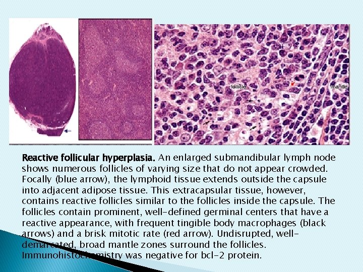
Reactive follicular hyperplasia. An enlarged submandibular lymph node shows numerous follicles of varying size that do not appear crowded. Focally (blue arrow), the lymphoid tissue extends outside the capsule into adjacent adipose tissue. This extracapsular tissue, however, contains reactive follicles similar to the follicles inside the capsule. The follicles contain prominent, well-defined germinal centers that have a reactive appearance, with frequent tingible body macrophages (black arrows) and a brisk mitotic rate (red arrow). Undisrupted, welldemarcated, broad mantle zones surround the follicles. Immunohistochemistry was negative for bcl-2 protein.
- Paracortical Hyperplasia. This pattern is characterized by reactive changes within the T-cell regions of the lymph node. It is encountered in viral infections (such as EBV), following certain vaccinations (e. g. , smallpox), and in immune reactions induced by certain drugs (especially phenytoin ).
-Sinus Histiocytosis. This reactive pattern is characterized by distention and prominence of the lymphatic sinusoids, owing to a marked hypertrophy of lining endothelial cells and an infiltrate of macrophages (histiocytes). Sinus histiocytosis is often encountered in lymph nodes draining cancers ( may represent an immune response to the tumor or its products).
Cat scratch disease �: -A self-limited lymphadenitis caused by the bacterium Bartonella henselae. -It is primarily a disease of childhood; 90% of the patients are younger than 18 years of age. -It presents as regional lymphadenopathy, most frequently in the axilla and neck. The nodal enlargement appears approximately 2 weeks after a feline scratch or, uncommonly, after a splinter or thorn injury.
A raised, inflammatory nodule or vesicle, is sometimes visible at the site of skin injury In most patients the lymph node enlargement regresses over the next 2 to 4 months. Rarely, patients develop encephalitis, osteomyelitis, or thrombocytopenia Morphology Sarcoid-like granulomas
Glandular fever is a general term for a disease characterized by : fever , sore throat , lymphadenopathy and atypical lymphocytes in the blood.
It may be caused by primary infection with Epstein-Barr virus (EBV), cytomegalovirus , human immunodeficiency virus (HIV) or toxoplasma. EBV infection, otherwise known as infectious mononucleosis, is the most common cause.
Infectious mononucleosis This is caused by primary infection with EBV. In most cases infection is subclinical. The diagnosis depends on the Following findings: In increasing order of specificity: (1) lymphocytosis with the characteristic atypical lymphocytes in the peripheral blood. (2) a positive heterophil reaction (monospot test). (3) a rising titer of antibodies specific for EBV antigens
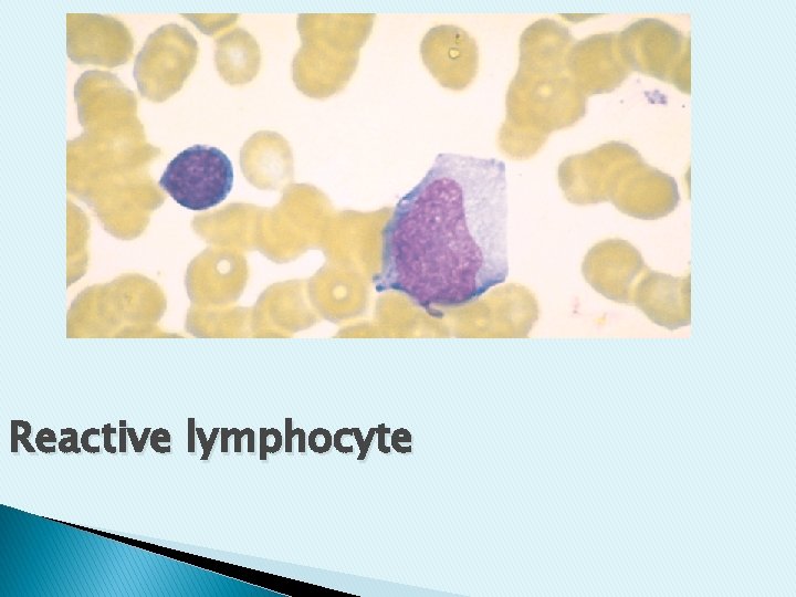
Reactive lymphocyte
Lymphomas: Lymphomas are a group of diseases caused by malignant lymphocytes(T, B, or NK) that accumulate in lymph nodes and cause the characteristic clinical features of lymphadenopathy - Although having different characteristics from their normal counterparts, the neoplastic cells of many lymphomas have the features of lymphoid cells at a particular stage of differentiation.
Lymphomas arise as a result of a series of mutations in a single lymphoid cell. The major subdivision of lymphomas is into Hodgkin's lymphoma and non-Hodgkin's lymphoma and this is based on the histological presenceof Reed-Sternberg (RS) cells in Hodgkin's lymphoma
Hodgkin's Lymphoma: Hodgkin lymphoma include distinctive group of neoplasms that arise almost invariably in a single lymph node or chain of lymph nodes and spread characteristically in a stepwise fashion to the anatomically contiguous nodes.
Morphology ■ It is characterized morphologically by the presence of distinctive neoplastic giant cells called Reed-Sternberg (RS) cells, which are mixed with reactive, nonmalignant inflammatory cells. . ■Molecular studies have shown that it is a tumor of B-cell origin
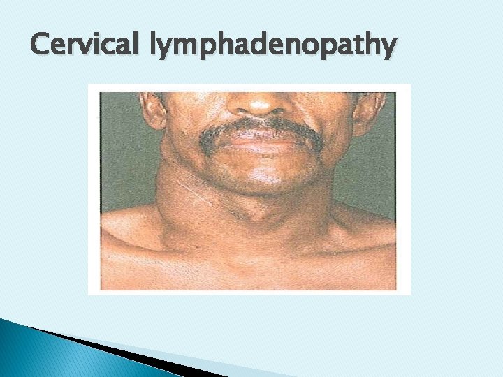
■The disease can present at any age but is rare in children and has a peak incidence in young adults. There is an almost 2 : 1 male predominance . ■The usual clinical presentation is with painless asymmetrical lymphadenopathy –most commonly in the neck. Typically the disease is localized initially to a single peripheral lymph node region and its subsequent progression is by contiguity within the lymphatic system
Cervical lymphadenopathy
Constitutional symptoms of fever , weight loss & sweating are prominent in patients with widespread disease. Alcohol-induced pain in the areas where disease is present occurs in some patients. ■Blood tests may show anaemia, neutrophilia , eosinophilia & raised erythrocyte sedimentation rate (ESR) or lactate dehydrogenase(LDH). �
Diagnosis and histological classification: -■The diagnosis is made by histological examination of an excised lymph node. -The histologic diagnosis of Hodgkin lymphoma rests on the definitive identification of Reed-Sterberg cells or their variants in the appropriate background of reactive cells.
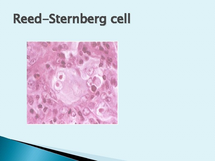
Morphology: The Reed-Sternberg (RS) cell is a large cell with an enlarged multilobated nucleus, prominent nucleoli, and abundant, usually slightly eosinophilic, cytoplasm. Particularly characteristic are cells with two mirror-image nuclei or nuclear lobes, each containing a large (inclusion-like) acidophilic nucleolus surrounded by a distinctive clear zone; together they give an owl-eye appearance. The. nuclear membrane is distinct.
Such "classic" RS cells are common in the mixed-cellularity subtype, uncommon in the nodular sclerosis subtype, and rare in the lymphocyte-predominance subtype
Reed-Sternberg cell
Classification : Five subtypes of Hodgkin lymphoma are recognized: (1) nodular sclerosis. (2) mixed cellularity. (3) lymphocyte predominance. (4) lymphocyte rich. (5) lymphocyte depletion. Uncommon
♦WHO classifi cation of Hodgkin lymphoma. -Nodular lymphocyte - predominant Hodgkin lymphoma -Classical Hodgkin lymphoma Nodular sclerosis classical Hodgkin lymphoma Mixed- cellularity classical Hodgkin lymphoma Lymphocyte- rich classical Hodgkin lymphoma Lymphocyte- depleted classical Hodgkin lymphoma.
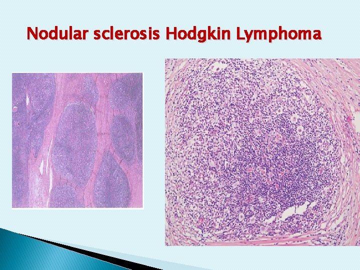
Nodular Sclerosis Hodgkin Lymphoma : - The most common form. - It is equally frequent in men and women. - Most of the patients are adolescents or young adults. - The overall prognosis is excellent.
Morphology: A variant of the RS cell, the lacunar cell. This cell is large and has a single multilobate nucleus with multiple small nucleoli and an abundant, palestaining cytoplasm. -In formalin-fixed tissue, the cytoplasm often retracts, giving rise to the appearance of cells lying in empty spaces, or lacunae. There are varying proportions of lymphocytes, eosinophils, histiocytes. - Classic RS cells are infrequent. -There are collagen bands that divide the lymphoid tissue into circumscribed nodules.
Nodular sclerosis Hodgkin Lymphoma
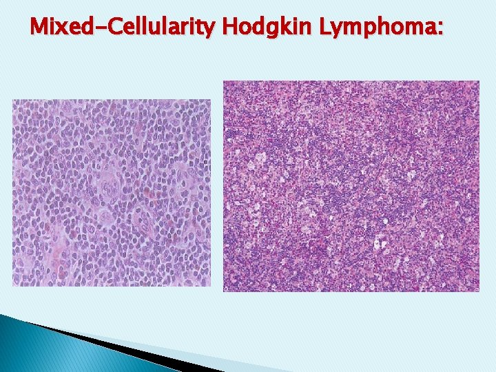
Mixed-Cellularity Hodgkin Lymphoma: - Patients older than the age of 50 year. -Male predominance. - Classic RS cells are plentiful within cellular infiltrate of small lymphocytes, eosinophils, plasma cells, and benign histiocytes. - More patients with mixed cellularity have disseminated disease and systemic manifestations. �
Mixed-Cellularity Hodgkin Lymphoma:
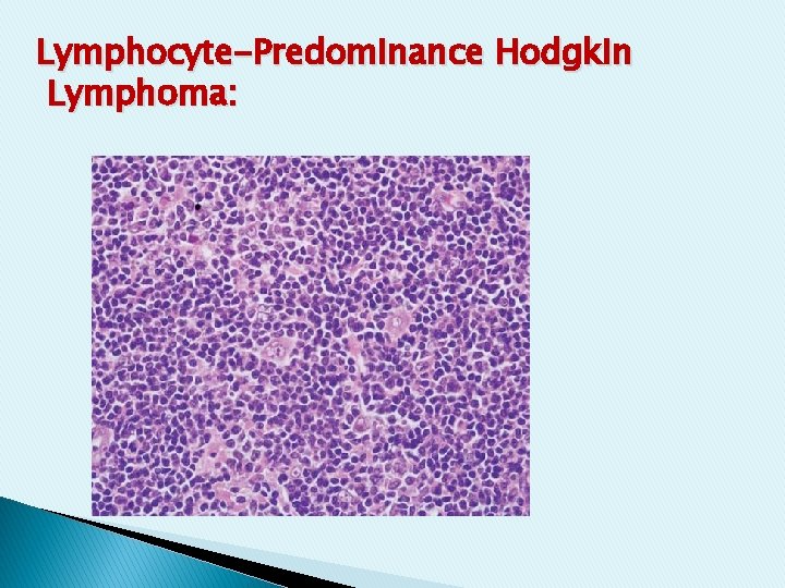
Lymphocyte-Predominance Hodgkin Lymphoma: - This subgroup, comprising about 5% of Hodgkin lymphoma. -It is characterized by a large number of small resting lymphocytes admixed with a variable number of benign histiocytes. -Eosinophils, neutrophils, and plasma cells, are scanty or absent, and classic RS cells are extremely difficult to find. - Lymphohistiocytic (L&H) variant RS cells that have a delicate multilobed, puffy nucleus that has been likened in appearance to popcorn ("popcorn cell").
Lymphocyte-Predominance Hodgkin Lymphoma:
■ In all forms, involvement of the spleen, liver, bone marrow, and other organs may appear in the course of the disease. �
Non-Hodgkin's lymphomas These are a large group of clonal lymphoid tumours, about 85% of B cell origin and 15% of T or NK(natural killer) cell origin. They are characterized by an irregular pattern of spread and a significant proportion of patients develop extranodal disease. -
For many years clinicians have subdivided lymphomas into low and high-grade disease. -In general terms, the low grade disorders are relatively indolent, respond well to chemotherapy but are very difficult to cure whereas high-grade lymphomas are aggressive and need urgent treatment but are often curable.
Clinical features of non-Hodgkin's lymphomas: 1. Superficial lymphadenopathy: asymmetric painless enlargement of lymph nodes in one or more peripheral lymph node regions. 2. Constitutional symptoms Fever, night sweats and weight loss occur less frequently than in Hodgkin's disease. 3. Oropharyngeal involvement.
4. Features due to Anaemia, neutropenia or thrombocytopenia. 5. organs involvement , the liver and spleen are often enlarged. The gastrointestinal tract is the most commonly involved extranodal site after the bone marrow. Also Skin, brain, testis or thyroid can be involved.
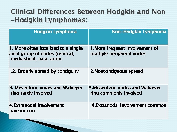
Clinical Differences Between Hodgkin and Non -Hodgkin Lymphomas: Hodgkin Lymphoma 1. More often localized to a single axial group of nodes (cervical, mediastinal, para-aortic. 2. Orderly spread by contiguity 3. Mesenteric nodes and Waldeyer ring rarely involved 4. Extranodal involvement uncommon Non-Hodgkin Lymphoma 1. More frequent involvement of multiple peripheral nodes 2. Noncontiguous spread 3. Mesenteric nodes and Waldeyer ring commonly involved 4. Extranodal involvement common
Investigations --Histology. . Lymph node biopsy is the definitive investigation. . . Immunophenotypic and genetic analysis. . . A fine needle aspiration may be performed to exclude another cause of lymphadenopathy (e. g. tuberculosis, carcinoma) but is not useful in establishing a diagnosis of lymphoma.
Laboratory investigations: 1. In advanced disease with marrow involvement , there may be anaemia, neutropenia or thrombocytopenia. 2. Lymphoma cells may be found in the peripheral blood in some patients. 3. Trephine biopsy of marrow to see if there is bone marrow involvement. 4. increase LDH and uric acid. 5. Immunoglobulin electrophoresis may reveal a paraprotein. �
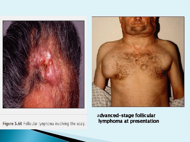
■ Specific subtypes of non-Hodgkin's lymphoma: ● Low-grade non-Hodgkin's lymphoma: 1♦ Follicular lymphoma: 40% of adult lymphomas. It is often characterized by a benign course for many years. It is associated with the t(14, 18) translocation. 2♦ Mantle cell lymphoma: it is associated with increased expression of the cyclin D 1. It is of aggressive behavior.
advanced-stage follicular lymphoma at presentation
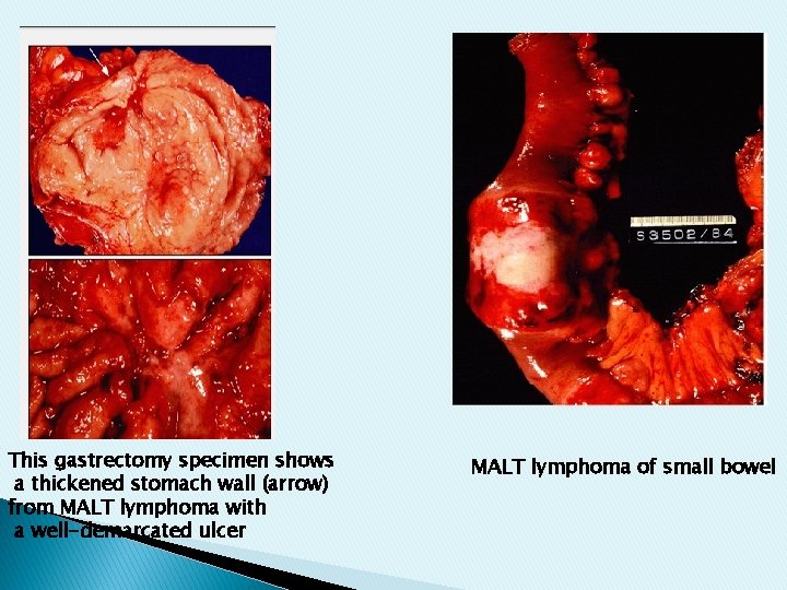
3♦Marginal zone lymphomas: are typically extranodal. Mucosa associated lymphoid tissue (MALT) lymphomas come into this category at sites such as the stomach or thyroid. Gastric MALT lymphoma is the most common form and is preceded by Helicobncter (H. ) pylori infection. 4♦Lymphocytic lymphomas: Lymphocytic lymphomas are closely related to CLL
This gastrectomy specimen shows a thickened stomach wall (arrow) from MALT lymphoma with a well-demarcated ulcer MALT lymphoma of small bowel
●High Grade Lymphoma: 1♦Diffuse Large B-Cell Lymphoma : It is the most common type of NHL lymphoma and has aggressive clinical behavior. 2♦Burkitt's lymphoma: is one of the most highly proliferative subtypes of any tumours. It is associated with t (8, 14).
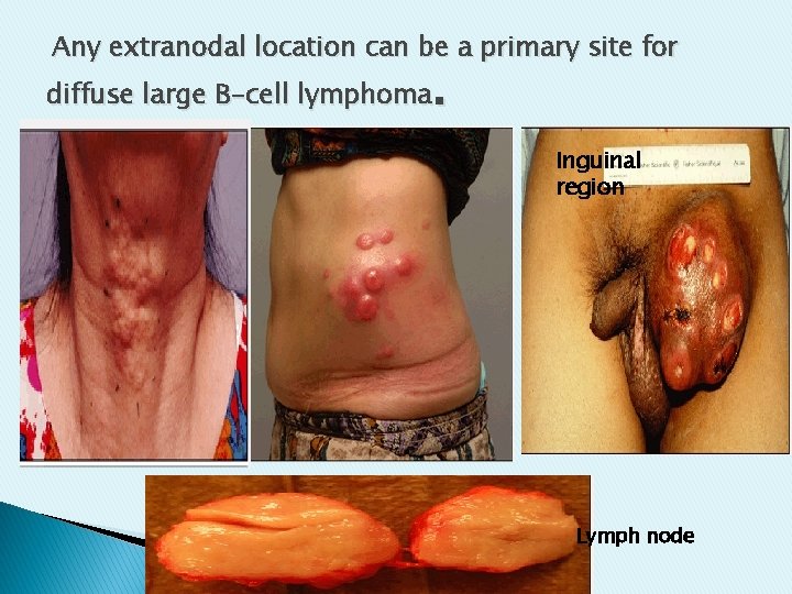
Any extranodal location can be a primary site for . diffuse large B-cell lymphoma Inguinal region Lymph node
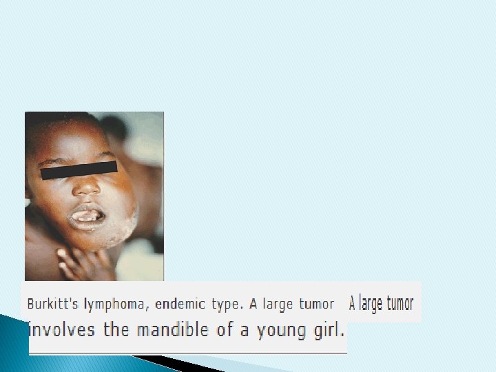
Endemic (African) Burkitt's lymphoma is seen in areas with chronic malaria exposure and is associated with Epstein-Barr virus (EBV) infection. Sporadic cases may occur elsewhere in the world. ◙T-cell lymphoma are less common and � include: mycosis fungoides, peripheral T-cell lymphoma and anaplastic large cell lymphoma �
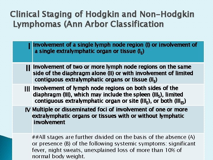
Clinical Staging of Hodgkin and Non-Hodgkin Lymphomas (Ann Arbor Classification I II Involvement of a single lymph node region (I) or involvement of a single extralymphatic organ or tissue (IE) Involvement of two or more lymph node regions on the same side of the diaphragm alone (II) or with involvement of limited contiguous extralymphatic organs or tissue (IIE) Involvement of lymph node regions on both sides of the diaphragm (III), which may include the spleen (IIIS), limited contiguous extralymphatic organ or site (IIIE), or both (IIIES) IV Multiple or disseminated foci of involvement of one or more extralymphatic organs or tissues with or without lymphatic involvement ##All stages are further divided on the basis of the absence (A) or presence (B) of the following systemic symptoms: significant fever, night sweats, unexplained loss of more than 10% of normal body weight.