Linteressamento del sistema nervoso centrale nelle malattie reumatiche


 ≥ 2 clinicamente attivo ORGANI COINVOLTI MANIFESTAZIONI SNC")
 0 - 42 , dove 0 = assenza di")


 MRI The recommended MRI protocol (brain and spinal cord)")


 DISEASE RELATED RISK FACTORS Who")


 Prevalence Per 100, 000 population UK : Scotland / Yorkshire")


,")
 criteria for Neuro Behçet Disease (NBD) diagnosis (2014) DEFINITE NBD")


 Comuni (10 - 40%) Rari (< 5%)")


 Afasia/disartria")


- Slides: 63
L’interessamento del sistema nervoso centrale nelle malattie reumatiche sistemiche Corso opzionale AA 2017 -2018 Marcello Govoni gvl@unife. it Rheumatology Unit Department of Medical Sciences ITALY
Why rheumatologist should be aware of CNS involvement ? The nervous system is a major target organ in most rheumatological diseases
Rheumatic diseases with potential CNS involvement Connective tissue diseases § § § § Antiphospholipid syndrome Systemic lupus erythematosus Sjogren’s syndrome Mixed connective tissue disease Scleroderma Dermatomyositis/polymyositis Rheumatoid arthritis Infectious diseases § Lyme disease § Whipple’s disease Systemic vasculitis § § Takayasu’s arteritis Giant cell arteritis Wegener’s granulomatosis Polyarteritis Nodosa Miscellanea § Behçet’s disease § Sarcoidosis PACNSV : Primary Central Nervous System Vasculitis
Don’t forget drug induced neuro-psychiatric syndrome/symtoms Antimalarials Psychosis Corticosteroids Psychosis, euphoria, depression Cyclosporin/Tacrolimus Leukoencephalopathy, Headache, insomnia, paraesthesia, confusion, depression, dizziness, convulsions, incoordination, psychosis, Cytokine inhibitors Demyelinating disorders, seizures, drowsiness, dizziness, paraesthesia, PML Leflunomide Headache, dizziness, paraesthesia Methotrexate Dizziness, drowsiness, malaise, headache, mood changes Mycophenolate Insomnia, headache, tremor NSAIDs Headache, dizziness, depression, aseptic meningitis Sulfasalazine Ataxia, aseptic meningitis, vertigo, insomnia, depression, hallucinations
2 scenarios in every day clinical practice • New neuropsychiatric sign or symptoms during the course of an already diagnosed rheumatic disease – Proceed with attribution : related or unrelated • Neurolpsychiatric signs or symptoms in the absence of known rheumatological diseases – Start work-up for DD – Don’t forget rheumatic diseases ! (*) (in about 11 % - Olsen ML et al. Am J Med 1991) (*) NP symptoms may represent the heralding manifestation of a rheumatic disease.
Main pathogenetic pathways for CNS damage § § § § Ischemia, thrombosis Different clinical pictures Microangiopathy/vasculopathy & pathogenetic pathways may coexist Vessel inflammation (vasculitis) Brain tissue or meningeal inflammation Neurotoxicity Autoantibodies BBB dysfunction or damage Cytokines Conditions which can open or damage BBB : § Acute stress § Infections § Disease activity § Drugs …
L’interessamento del sistema nervoso centrale nel LES
Neuropsychiatric lupus § CNS involvement affects patient’s quality of life and is associated with poor prognosis Hanly JG et al. J Rheumatol 2009 NPSLE § NP involvement represents one of the main causes of morbidity and mortality in SLE Predictors of mortality in SLE (meta-regression) Neuropsychiatric damage Renal damage Mak A, et al. Semin Arthritis Rheum. 2012
The burden of NPSLE Range of reported prevalence : 14 - 95 % Sem Arthritis Rheum 2011 According to 10 high-quality prospective studies, which included 2, 049 SLE patients The overall prevalence of NP syndromes in SLE patients was estimated to be 56. 3 % 93. 1 % CNS vs 6. 9 % PNS i. e. one out of two !
A milestone in the NPSLE research www. rheumatology. org/at/1999/aprilappendix. html 10
Focal Major CNS Diffuse Minor PNS CNS Aseptic meningitis CVD Demyelinating sd. Headache Movement disorder Myelopathy Seizure disorders Acute confusional state Anxiety disorder Cognitive dysfunction Mood disorder Psychosis PNS Guillain Barré syndrome Autonomic neuropathy Mononeuropathy Myasthenia gravis Cranial neuropathy Plexopathy Polyneuropathy (no ENG) According to ACR According to EULAR According to 1999 Ainiala et al. 2001 Rec. 2010 case definition 11
NP syndromes as disease classification criteria New SLICC criteria 2012 ARA 1982 & ACR 1997 criteria § Psychosis § Seizures § § Myelopathy Seizure disorders Acute confusional state Psychosis § Mononeuropathy § Cranial neuropathy § Polyneuropathy
SLEDAI-2 K (da 0 a 105) ≥ 2 clinicamente attivo ORGANI COINVOLTI MANIFESTAZIONI SNC (LES correlato) Fenomeni epilettici Psicosi Demenza Disturbi visivi Neuropatia nervi cronici Cefalea lupica Vasculite cerebrale 8 8 8 8 VASCOLARE RENE MUSCOLO-SCHELETRICO SIEROSE ALTERAZIONI IMMUNOLOGICHE CUTE SINTOMI GENERALI Vasculiti Alterazione sedimento (cilindri) Ematuria (GR > 5 per campo) Leucocituria (GB > 5 per campo) Proteinuria (0. 5 g/24 ore) Artrite (> 2 articolazioni) Miosite Pleurite Pericardite Riduzione C 3 o C 4 Anticorpi anti-DNA Rash recenti Alopecia Ulcere mucose Febbre 8 4 4 4 2 2 2 2 1 SCORE
SLICC - Disease Index (SDI) 0 - 42 , dove 0 = assenza di danno ORGANI COINVOLTI SNC OCCHIO RENE POLMONE MANIFESTAZIONI (parte 1) Disturbi cognitivi Psicosi Epilessia (che richieda terapia per almeno 6 mesi) Accidenti vascolari cerebrali (score 2 se > 1) Neuropatia cranica o periferica (esclusa neuropatia ottica) Mielite trasversa Cataratta Alterazioni retiniche o atrofia ottica Filtrato glomerulare renale > 50 % Proteinuria > 3. 5 g/24 ore IR terminale Ipertensione polmonare Fibrosi polmonare Shrinking lung Fibrosi pleurica Infarto polmonare o resezione per patologia non neoplastica CUORE SIST. VASCOLARE Angina o by-pass coronarico IMA (score 2 > 1) Cardiomiopatia Pericardite per 6 mesi o pericardiotomia Claudicatio per 6 mesi Perdita minima di tessuto SCORE 1 1 (2) 1 1 1 3 1 1 1 (2) 1 1 1
FREQUENCY Frequency of neuropsychiatric syndromes as reported in cohort studies using the ACR nomenclature and case definitions for NPSLE SPECIFICITY
One of the major challenges in diagnosing NP involvement, is that in NPSLE nothing is specific ! • • • No specific clinical pictures No specific patterns on neuroimaging No specific lab or serologic biomarkers No specific changes in CSF High prevalence of some NP pictures in GP 16
ACR 1999 Definition of NPSLE “Neuropsychiatric lupus erythematosus includes the neurologic syndromes of the central, peripheral and autonomic nervous system and the psychiatric syndromes observed in patients with SLE in which other causes have been excluded” In clinical practice, NPSLE is a diagnosis «per exclusionem» ATTRIBUTION of NP events in SLE patients, remains one of the most challenging issue LUPUS-RELATED MANIFESTATIONS Lupus HEADACHE Lupus DEPRESSION Lupus STROKE Lupus SEIZURE ? ? … and so on
How to approach attribution in NPSLE ? The Italian Study Group for NP-Lupus (Research project : on behalf of Italian Society of Reumatology) Items TIME onset of NP event Tipology of NP event Result Score Items Before 0 After 2 CONFOUNDING FACTORS Concomitant Major 3 Minor 0 Normalized score from 0 to 10 3 FAVOURING FACTORS Result YES > 1 YES at least 1 None or NA YES at least 1 YES > 1 Score 0 1 2 A score ≥ 7 is highly suggestive for NPSLE 18
WHAT ABOUT NEUROIMAGING ? Morphology Functional MRI SPECT, PET, f. MRI, PWI Anatomy Macro-architecture Metabolism Perfusion Quantitative MRS, MTI, DWI Micro-architecture Metabolism No single imaging technique covers all brain NPSLE pathology 19
Magnetic Resonance Imaging (EULAR 2010) MRI The recommended MRI protocol (brain and spinal cord) includes : § conventional MRI sequences (T 1/T 2, FLAIR) § Diffusion weighted imaging (DWI) § gadolinium-enhanced T 1 sequences (T 1 -gd) In FOCAL presentation c. MRI provides useful information In DIFFUSE presentation c. MRI can be frequently negative 20
The pattern of MRI abnormalities in NP-SLE Cortical atrophy Brain infarcts 8
The most frequent Small punctate hyperintense T 2 -weighted focal lesions in subcortical and periventricular WM microinfarcts, demyelination, gliosis 9
axial-FLAIR scan highlights multiple punctate WMHIs in semioval centres; B: multiple confluent lesions in periventricular WM
Small punctate hyperintense T 2 -weighted focal lesions in subcortical and periventricular WM Clinical correlations Common in chronic NP-SLE (25 -60 %) correlate with : - clinical severity - past history of CNS involvement - cognitive dysfunction - APLA They are NOT SPECIFIC Can be seen in SLE without NP symptoms, in patients without SLE Increase with aging, heart valvular disease & hypertension De Leeuw et al, Brain 2002 De Leeuw et al, Stroke 2004 Roldan et al, Am J Cardiol 2005 Roldan et al, Cardiology 2007 Sibbitt et al, Arthritis Rheum 1999 Sanna et al, J Rheumatol 2003 Sibbitt et al, J Rheumatol 2003 Ainiala et al, Scand J Rheumatol 2005 Ovbiagele & Saver, Cerebrovasc Dis 2006
T 2 -scan shows 2 hyperintense focuses of myelitis (at C 6 -T 1 and D 7 -D 8 levels; arrows) in a 45 year old SLE female, with associated cord swelling; B: after administration of IV gadolinium, an inhomogeneous pattern of contrast enhancement is remarked; C: after 3 months of immunosuppressive therapy, a reduction in cord swelling, a complete regression of D 7 -D 8 lesion and a subtotal regression of C 6 -T 1 lesion were demonstrated.
T 2 -scan highlights a bilateral and symmetrical mild hyperintensity in cerebellar hemispheres; B: low hyperintensity on DWI; C: increase in ADC; D: complete reversibility of lesions on axial T 2 -scan after 2 months; E: normalization of ADC.
EULAR Recommendations for the Management of NPSLE (ARD 2010) DISEASE RELATED RISK FACTORS Who is at risk for (primary) NPSLE? SLE disease activity SLE damage Previous or concurrent NPSLE a. PL abs (Evidence: 2/B) Nat Rev Rheumatol. 2010; 6: 358 - 27
326 patients with NP involvement 469 NP events 28
Ischemic-thrombotic-vascular phenotype § § § § Usually focal presentation Cerebrovascular disease Seizures Frequent MRI abnormalities Movement disorder Usually acute in onset Myelopathy High prevalence of a. PL antibodies Cranial neuropathy Brain vasculopathy (or vasculitis) PNS involvement Usually anatomical-structural damage No clear relationships with underlying SLE disease activity Refractory to therapy Main clinical phenotypes Inflammatory-neurotoxic phenotype § § § § Usually diffuse presentation Frequent normal MRI Usually insidious, develop slowly over time anti-NR 2, anti-P-Rib antibodies & cytokines Usually no structural pathology is detectable Associated mostly with cognitive dysfunctions SLE high disease activity may be relevant Usually reversible with therapy Aseptic meningitis Demyelinating syndrome Headache Acute confusional state Anxiety disorder Cognitive dysfunction Mood disorder Psychosis
If you want to know more …
L’interessamento del sistema nervoso centrale nel m. di Behçet
Ocular Oral aphtae Genital ulcers
Epidemiology • BD is rare among children No case among 450, 000 in one survey in Turkey Ozen S, et al. J Rheumatol 1998; 25: 2445 • More common in men in the Mediterranean populations • The sex ratio is reversed in Asian populations • Familial disease is uncommon in Caucasians • Positive family history in 12% of non-caucasoid patients
World Prevalence Country (reference) Prevalence Per 100, 000 population UK : Scotland / Yorkshire 0. 27 / 0. 64 USA : Olmsted County, MN 0. 33 Sweden 1. 18 Portugal 1. 53 Germany 2. 26 Italy : RE / PZ 3. 8 / 15. 9 Spain 7. 5 Japan 13. 5 Iran 16. 7 Saudi Arabia 20. 0 Turkey North-Eastern Ankara region Istanbul 370 110 420
The geografical distribution of BD is distinctive: 45º N it is most prevalent along the Silk Route Kazakhstan Italy Bulgaria Uzbekistan Portugal Azerbaijan Spain Greece Turkey Turkmenistan Tunisia Iran Afghanistan Syri Morocco Israel Jordan a Pakistan Egypt Algeria Libya Saudi Arabia Mongolia China Korea Japan 30º N
Distribution of HLA-B 51 in the world
Although BD is a well-defined disease with well-established criteria for its diagnosis (1, 2), the same is not true for NBD Criteri ISG 1 Criteri ICBD 2 International Study Group for Behcet’s Disease (1990) Lancet International Team for the Revision of the International Criteria for Behcet’s Disease (ITRICBD) (2014) JEADV. Manifestazioni Aftosi orale ricorrente (major or minor : almeno 3 episodi in 12 mesi) Punti Oculari 2 Aftosi orale 2 Ulcere genitali 2 Cutanee 1 Lesioni cutanee Neurogiche 1 (EN, pseudofollicolite, noduli acneiformi) Vascolari 1 Patergy test + Patergia 1 + 2 delle seguenti Ulcere genitali ricorrenti Manifestazioni oculari (UA, UP, vitreite, vasculite retinica) Per la diagnosi: ≥ 4 punti Sens Spec Accur 82. 4 96. 0 86. 7 94. 8 90. 5 93. 9
International Consensus Recommendation (ICR) criteria for Neuro Behçet Disease (NBD) diagnosis (2014) DEFINITE NBD (all of the following) 1. BD satisfying International Study Group (ISG) criteria 2. Neurological syndrome (with objective neurological signs) recognised to be caused by BD (attribution) and supported by Neuroimaging and/or CSF 3. No better explanation for the neurological findings PROBABLE NBD (1 of the following) 1. Neurological syndrome as in definite NBD, with systemic BD features but not satisfying the ISG criteria 2. A non-characteristic neurological syndrome occurring in the context of ISG criteria-supported BD in the absence of a better explanation for the neurological findings
Epidemiologia del coinvolgimento neurologico nel m. di Behçet Frequenza: 5. 3 - 38 % dei pazienti 4. 8 - 5. 3 % studi clinici Akman-Demir G et al. Brain 1999 Siva A et al. J Neurol 2001 20 % studi autoptici Lakhanpal S et al. Hum Pathol 1985 Sex ratio : M/F=3: 1 Esordio: in genere 4 - 6 aa dopo l’esordio della malattia; nel 10% circa prima o all’esordio della malattia Età media all’esordio: terza decade 31. 5 8. 9 (Akman-Demir G et al. Brain 1999) 32. 0 8. 7 (Siva A et al. J Neurol 2001) 30. 2 8. 5 (Koçer n et al. AJNR 1999)
CNS involvement : 5 % Clin Experiment Rheu 2009
Classificazione Neuro-Behcet Subtypes which differ by clinical, neuro-radiological, pathological, and prognostic characteristics Sistema Nervoso Centrale Parenchimale Non-Parenchimale (Extra-assiale) LARGE VESSEL (Intra-assiale) SMALL VESSEL • • • Troncoencefalico Midollo spinale Cerebrale Asintomatico Neurite ottica ~ 80 % • Trombosi venosa cerebrale ~ 20 • Aneurismi intracranici/dissecazione • Aneurismi extracranici/dissecazione Sistema Nervoso Periferico • Neuropatie periferiche e mononeurite multipla • Miopatie e miositi Altre sindromi • Tumor like neuro-BD • Sintomi psichiatrici % uncommon Akmar-Demir et al. Brain 1999 Siva A. et al. J Neurol 2001 Siva A Saip S, J Neurol 2009 Kalra S et al. J Neurol 2014
Clinical phenotypes Parenchymal NBD § usually presents with : ü a sub-acute onset of brainstem syndrome (with/without other features) ü cerebral hemispheric or spinal cord syndrome (multifocal) § features : pyramidal weakness, behavioural changes, headaches, ophthalmoplegia, optic neuropathy and sphincter changes. § usually follows a relapsing-remitting pattern or a primary/secondary progressive course Non-parenchymal NBD (vasculo-NBD) § commonly presents with headache and visual features secondary to intracranial hypertension, usually due to cerebral venous thrombosis. § It can present as an acute stroke related to arterial thrombosis, dissection, or aneurysm (uncommon), acute meningeal syndrome § can be monophasic, but recurrences may occur A mixed parenchymal and non-parenchymal disease presentation can occur
Principali segni neurologici Molto frequenti (> 50%) Comuni (10 - 40%) Rari (< 5%) § Segni piramidali § Emiparesi § Disturbi comportamento § S. troncoencefaliche § S. piramido-cerebellari § Disturbi sensitivi § Paraparesi § Febbre § Disturbi movimento § Meningismo § Ipersonnia/iperfagia § Crisi comiziali § Disturbi psichiatrici § S. cerebellari pure § Ipo-anacusia § Neuropatia ottica § Emianopsia § Afasia § Disturbi sfinterici/impotenza § Cefalea Akmar-Demir G et al. Brain 1999
Pricipali sintomi neurologici all’esordio CEFALEA 61. 6% S. motori 53. 7% S. cerebellari 29. 9% S. tronco-encefaliche 29. 3% Disartria 22. 6% S. sensitivi 11% Alterazioni coscienza 7. 3% S. cognitivi 2. 4% altri 9. 4% Esordio polisintomatico: 72. 7% Siva A et al. J Neurol 2001
41 patients were enrolled into the study No differences in the frequency of the different pattern of headache between BD patients and controls. Headache occurred in 70, 7 % of BD patients & in 92, 8 % of Neuro-BD patients. Migraine without aura did prove the most frequent type of headache
Modalità di coinvolgimento neurologico Attacco acuto: esordio acuto o subacuto di sintomi o segni neurologici Decorso progressivo primario: lenta e progressiva evoluzione di sintomi o segni neurologici nell’arco di mesi o anni senza attacchi Decorso progressivo secondario: lenta e progressiva evoluzione di sintomi o segni neurologici dopo almeno 1 attacco o progressione a tappe successive segnate da attacchi Coinvolgimento neurologico silente: presenza di anormalità all’esame neurologico in assenza di sintomi specifici al di fuori della cefalea 47
Differential diagnosis § § § multiple sclerosis stroke affecting the young intracranial hypertension meningo-encephalitis myelitis 48
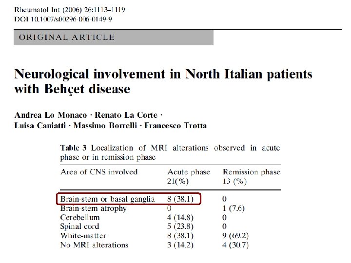
Neuroimaging & Neuro-Behçet Distribuzione delle lesioni RM Giunzione meso-diencefalica 46% Ponte/bulbo 40% Ipotalamo/talamo 23% Gangli della base/ cap. interna 18% Midollo cervicale 5% Cervelletto 5% Telencefalo 8% 65 paz. con neuro-Behçet e RMN cerebrale anormale Koçer N et al. AJNR 1999
Predominat brainstem involvement The brainstem-thalamic-basal ganglia lesions, in the proper clinical context, can strongly support the diagnosis of acute/subacute parenchymal NBD, and on occasions can raise this possibility even when the systemic features of BD are scarce large pontomesencephalic lesion in a 41 -year-old woman with a locked-in syndrome 1 week after the acute onset (personal observation)
Region predominantly involved in ANBD, CNBD, NPSLE, MS MRI ANBD CNBD MS NPSLE Basal ganglia and/or brain stem 100 36 0 14 Sc. hemisphere white matter 0 36 100 29 Cerebral cortex No parenchimal lesions 0 0 0 27 0 0 29 29 ANBD = acute neuro BD CNBD = chronic neuro BD NPSLE = NP Systemic Lupus Erythematosus MS = multiple sclerosis Coban et al. Neuroradiology 1999
Acute Neuro-Behçet Agosto: cefalea intensa, emiparesi sin. Settembre: ampie lacune mnesiche. Novembre: ipersonnia diurnia, iperfagia, apatia. RM: vasta lesione fronto-temporale dx sottocorticale interessante anche la regione dei nuclei della base con compressione del corno temporale e frontale del III ventricolo. (osservazione personale)
Non-parenchymal neuro. Behçet’s disease Right-sided transverse venous sinus thrombosis in a patient with MR venogram
Neuro-Sjogren The real burden of CNS involvement still represents a controversial issue Range = 0 - 68 % ? ? ? Govoni M et al. CNS Drugs 2001 More recent data : year % n. pz Anaya et al 2002 2. 5 120 Garcia-Carrasco 2002 1 400 Delalande et al. 2004 68 82 Jamilloux et al 2003 9. 7 420 Ramos-Casals 2010 2 the In Sjogren’s syndrome 1010 PNS is predominantly Morreale et al 2014 67. 5 involved 120 Moreira et al 2015 15 Range = 7 - 35 % 93
25/424 patients - 5. 8 % NP involvement Diffuse manifestations Focal or multifocal pictures MS-like disorders Isolated optic neuritis No. pts. % 10* 9 5 1 40 36 20 4 Outcome S I W 4 1 1 3 7 2 1 3 * FUP not available in 3 pts. S = stable; I = improved; W = worsened § Age at first CNS-NP event, mean (yrs) 46. 7 (13. 6) § Disease duration at first CNS-NP event, mean (yrs) 3. 4 (4. 6)
Aspetti clinici • Quadri NP polimorfi • Tutto il nevrasse può essere colpito • Esordio spesso insidioso (4 a - 5 a decade) • Modalità di decorso variabile (intermittente/remittente o lentamente progressivo) • Possibile manifestazione di esordio (*) nel 52 % dei casi della casistica di Ferrara 2010 nel 62 % casistica di Lafitte 2001
Clinical spectrum of CNS involvement • Focali – – – Deficit focali (mono/multipli) Afasia/disartria Convulsioni parziali (rare) S. tronco cerebrale S. cerebellare Sindromi extrapiramidali • Diffuse – – – encefalopatia Meningite asettica Meningoencefalite Disfunzioni cognitive Ansia, Depressione Cefalea • Mielopatia – – – Mielite trasversa acuta Mielite cronica progressiva S. Brown-Sequard Vescica neurogena M. del motoneurone • Altri – Neuropatia ottica – Quadri SM-like
Aspetti particolari • Quadri psichiatrici di tipo prev. neurotico – aumentati livelli di ostilità, ideazione paranoide, disforia, ansia, somatizzazione e compulsività, aspetti isteroidi, depressione • Frequenti disturbi del sonno (SFM), talora narcolessia • Disturbi cognitivi subdoli, raramente gravi (demenza) • Rari epilessia, psicosi e disturbi del movimento – – – Malinow et al Ann Int Med 1985 Drosos et al Clin Exp Rheumatol 1989 Tishler et al Clin Exp Rheumatol 1997 Valtysdottir et al J Rheumatol 2000 Mitsuo et al, Mod Rheumatol 2001
Risonanza Magnetica Overlap Sjogren & SM Atrofia cerebrale, dilatazione ventricoli; frequente riscontro Quadri mielitici (60 -80 %) di WMH sottocorticali e peri-ventricolari nei pz. con S. Sjogren (con o senza int. NP) Prevalenza del Sjogren nella SM 2. 3 - 3. 3 % Prevalenza della SM nel Sjogren up to 30 % casistica di Ferrara 20 % Ferrara dei quadri neurologici alterazioni RM nel 91 % dei pz. con int. SNC
Sjogren • • DD tra S. di Sjögren e SM Quadri multifocali con decorso remittente e ricorrente Quadri di neuropatia ottica e mielopatia acuta F : 24 yr Rilievo di BOC nel LCR Aspetti RM simili alla SM – Alexander et al, Ann Int Med 1986 – Barnet et al, Neurology 1995 – Collard et al, Neurology 1997 SM • • • S. sicca (soggettiva) in 1/3 dei casi F. Raynaud, piastrinopenia ANA nel 3 - 27 % dei casi Ac Ro/SSA nel 2 -15 % dei casi ACL nel 6 -8 % dei casi – Tourbah et al, Arch Neurol 1998 – Ijdo et al, Lupus 1999 – De Andres et al, Acta N Scand 2001 2005 2003 2007
Neuro-Sjögren vs SM • Neuropatia periferica • Vasculite cutanea • Presenza di Ac. anti-Ro/SSA e anti-La/SSB • Tumefazione delle gh. parotidi • Sialoadenite focale (FS 1) • HLA-B 8, DR 3, DRw 52 vs DR 2 • Minore quantità di BOC nel LCS
Summary • CNS is a potential and not rare target for damage in rheumatic diseases • Different pathogenetic pathways working in systemic rheumatic diseases can lead to NP involvement • Thinking of an underlying rheumatic disease when approaching a new onset NP picture - especially in young patients – is warmly advised