LeggCalve Perthes Disease FIRM 1 GRANDROUND PRESENTERS DR


")


 calcification lateral to")


- Slides: 44
Legg-Calve. Perthes Disease FIRM 1 GRANDROUND PRESENTERS: DR. MAINA/DR. ONDARI FACILITATOR: DR. T. MOGIRE 01/08/2013
Georg Perthes (1869 -1927)
First described by Karel Maydl
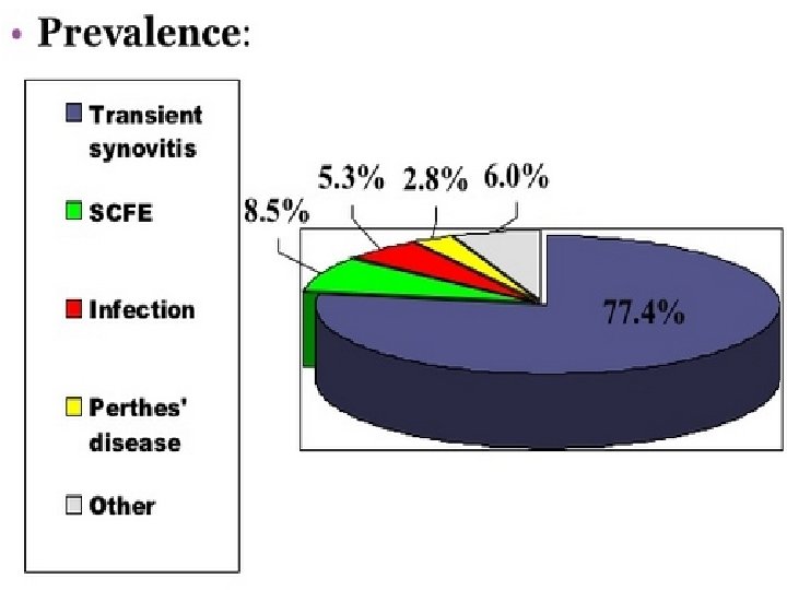
Epidemiology Incidence 1 -4/10, 000 Age 4 - 10 years; average 7 yrs As early as 2 yrs as late as teens Boys : girls 4: 1 Bilateral 10 -12% No evidence of inheritance Common in Caucasians; rare in black races
Etiology Idiopathic Past theories Infection, inflammation, trauma, congenital Most theories involve vascular compromise
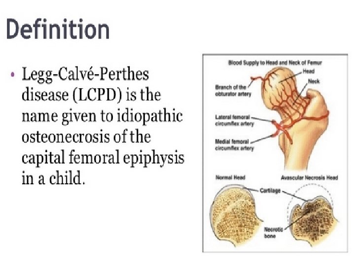
Pathophysiology Rapid growth occurs in relation to devt of blood supply Interruption of blood supply results in necrosis, removal of necrotic tissue, and its replacement with new bone. Bone replacement may be so complete and perfect that completely normal bone may result The adequacy of bone replacement depends on
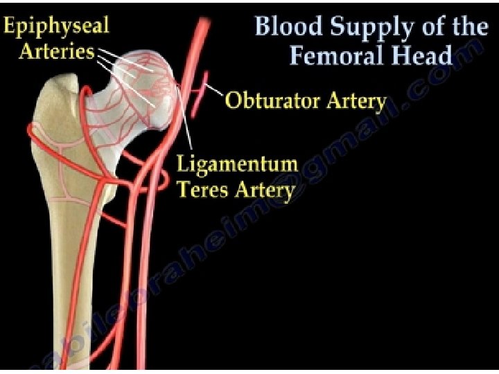
Sources of blood supply Up to 4 years Metaphyseal vessels Retinacular vessels Ligamentum teres – scanty 4 to 7 years Metaphyseal vessels ceases Above 7 years Vessels in ligamentum teres have developed
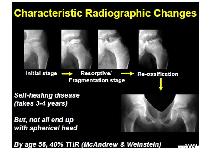
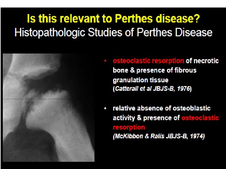
Pathology Goes through stages which may last 3 to 4 years Stage 1 Ischaemia and bone death, cartilage thickens Stage 2 Revascularization and repair Dead marrow replaced by granulation tissue Bone revascularized and new bone laid down Dead bone resorbed, replaced by fibrous tissue, fragmentation Stage 3 Distortion and remodelling Restoration of femoral archtecture or collapse
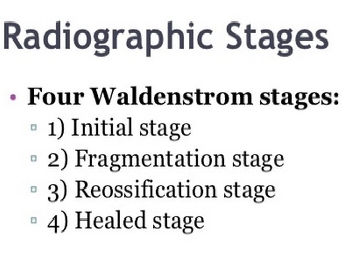
Classification Waldenstrom classification Catterall classification Salter and thompson classification Herring classification
Caterall classification Based on amt of involvement of femoral epiphysis Group I <1/2 of head involved , Group II Up to half of head. Some collapse of central portion Group III >1/2 of head involved with sclerosis, fragmentation and collapse of head Group IV Entire epiphysis involved
Caterall “head-at-risk” signs Associated with poor results lateral subluxation (most important) calcification lateral to the epiphysis Gage's sign: V shaped defect laterally
Caterall “head-at-risk” sign metaphyseal cysts
Gage's sign
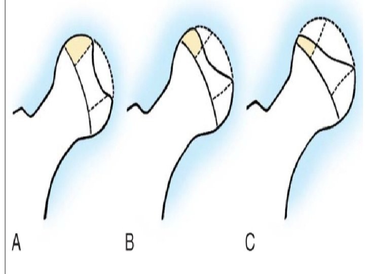
Salter and thompson classification Describes extent of subchondal fracture in the superolateral portion of femoral head Type A - <50% of femoral head Type B - >50% of femoral head can be observed radiographically earlier and more readily tan caterall classification Can be applied early in course of dz to determine management
Herring classificatin/lateral pillar Based on degree of collapse of lateral pillar during fragmentation stage Goup A No collapse, no progressive flattening Group B <50% collapse Group C >50% collapse Ritterbusch 1993 Has the highest predictive value and interobserver reliability
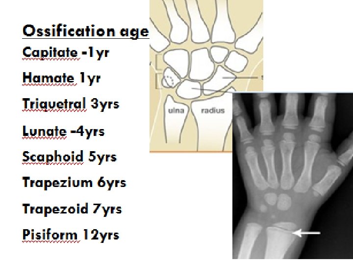
Bilateral involvement More severe dz than unilateral Boys and girls equally affected Independent event Bone age delayed in perthes disease
Examination Short stature Delayed bone age Early Decreased ROM Antalgic gait Late Decreased ROM of motion from acetabular impingement Disuse atrophy of thigh muscles Leg lenght descrepancy Trendelenburg gait
Investigations Blood tests haemogram, ESR, CRP Imaging Plain X-rays Hip U/S Bone scintigrpahy MRI Dynamic arthrography Assess spherity of femoral head Hinge abduction Bilateral perthes Skeleta survey as part of work-up
Song et al MRI findings on widened medial joint space Initial stage Overgrowth of cartilage Fragmentation stage Overgrown cartilage with widened true medial joint space Healing stage Widened true medial joint space
Treatment Goals of tratment Maintain femoral head spherity – containment Avoid severe degenerative arthritis Guided by Age Severity Limitation in ROM
Treatment cont. Initial Mx determined by sympts severity Analgesia Modification of activities Bedrest and short period of traction Wheelchair/crutch walking discouraged
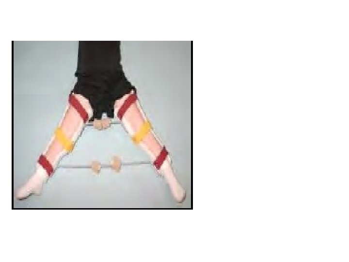
Treatment: Two main choices Conservative Pain control Gentle exercises Regular re-assessment Avoid sport and strenous activities Containment Hold hips widely abducted in cast/brace >1 yr Operation Varus osteotomy of femur Innominate osteotomy of pelvis Both
Herring Guidelines to treatment Children <6 years Symptomatic treatment Children >6 years; bone age more imp than chronological age Bone age at or <6 yrs Lateral pillar A or B/ caterall I and II Lateral pillar C/ Caterall III and IV Bone over 6 years Herring A and B/Caterall I and II Abduction brace or osteotomy Herring C/Caterall III and IV Symptomatic treatment Outcome unaffected by treatment Children 9 yrs and older Except in very mild cases, operative containment is the
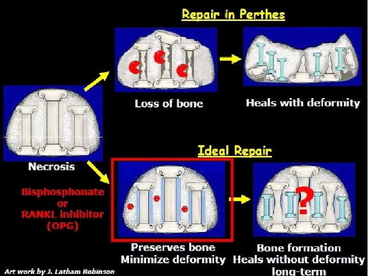
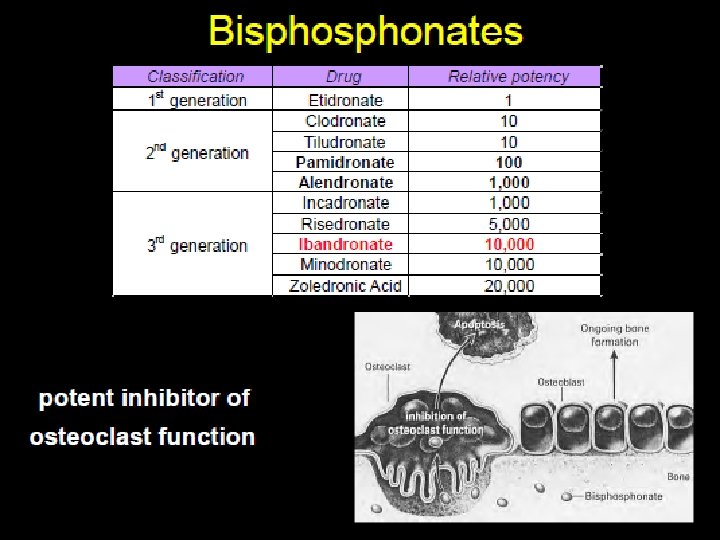
oseoclast-osteoblat interactio
Prognostic features Age Gender Herrings lateral pillar classification Salter and thompson grade B worse prognosis Caterral classification grade Caterral “head-at-risk” signs Girls have worse prognosis Classification grade <6 yrs; good regardless of treatment 6 -9 years; not always satisfactory with containment >10 yrs; questionable benefit from containment, poor prognosis The five signs carry worse prognosis Others Body weight, decreased ROM