Introduction to Womens Health Physical Therapy Tara Sullivan





- Slides: 71
Introduction to “Women’s Health” Physical Therapy Tara Sullivan, PT, DPT, PRPC
Personal Bio Doctorate in Physical Therapy A. T. Still University Master of Science in Human Movement A. T. Still University Bachelor of Science in Exercise and Sport Science Oregon State University Florida College of Natural Health Massage Therapy Certificate
Specialty Training Herman & Wallace, Pelvic Rehabilitation Institute Pelvic Floor 1 Pelvic Floor 2 a Pelvic Floor 2 b Pelvic Floor 3 Pregnancy and Postpartum Movement Systems Approach: Lumbar, Hip, & SIJ Women’s Health Summit Pediatric Incontinence Pudendal Neuralgia Sexual Medicine Post Prostatectomy Pelvic Rehabilitation Practitioner Certification (PRPC) Nationally recognized Pelvic Rehab Specialist/Expert
And I’m a Mom……
Lawson— 15 months
Almost 2. 5 yo #ham
Objectives Identify medical diagnosis in patient’s past medical history that may be a sign of pelvic floor dysfunction Identify when a patient may benefit from pelvic floor therapy based on presentation and results of current treatment Understand the function of pelvic floor muscles Understand the relationship between the pelvic floor, bladder and bowel Understand relationship between pelvic floor and hip musculature
“You do WHAT? !? !”
What is “Women’s Health”? Different Titles: Urogynecological Physical Therapist Pelvic Floor PT Women’s Health Therapist Pelvic Disorders Specialist Pelvic Rehab Therapist Urotherapist
What do we treat? Bowel Bladder Sexual Health Chronic Pelvic Pain *As it relates to the Pelvic Floor
Pelvic Floor Dysfunction is used to describe the difficulty of the muscles of the pelvic floor to tighten and relax normally. (National Center of Pelvic Pain) Other names for Pelvic Floor Dysfunction: Chronic pelvic pain syndrome Levator ani syndrome Prostatitis or chronic prostatitis Coccydynia (tailbone pain) Myofascial pain syndrome
PFD Pelvic Floor Dysfunction is an umbrella term used to encompass several different bowel, bladder, and sexual disorders, and chronic pain syndromes, that affect or are caused by the inability of the pfm to contract, relax, and bulge effectively. Are the pelvic floor muscles the Primary reason for the Patient’s dysfunction or a Secondary reflexive muscle response?
Common S&S of PFD associated w/ Pain Pelvic and/or lower abdomen pain (Bilateral or Ipsilateral) Sitting pain Pain during/after sex (with penetration and/or orgasm) Genital/groin/perineal pain Pain or relief during or after a bowel movement Pains increases with stress and activity Pain during or after urination (Dysuria) Bladder pain
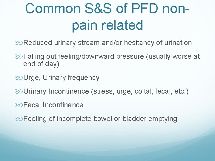
Common S&S of PFD nonpain related Reduced urinary stream and/or hesitancy of urination Falling out feeling/downward pressure (usually worse at end of day) Urge, Urinary frequency Urinary Incontinence (stress, urge, coital, fecal, etc. ) Fecal Incontinence Feeling of incomplete bowel or bladder emptying
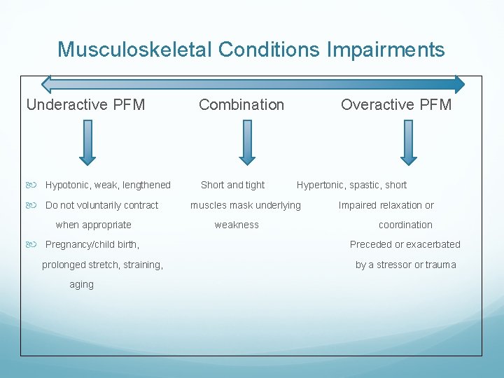
Musculoskeletal Conditions Impairments Underactive PFM Combination Hypotonic, weak, lengthened Short and tight Do not voluntarily contract when appropriate Pregnancy/child birth, prolonged stretch, straining, aging Overactive PFM Hypertonic, spastic, short muscles mask underlying weakness Impaired relaxation or coordination Preceded or exacerbated by a stressor or trauma
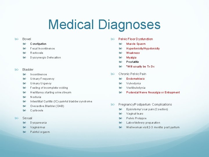
Medical Diagnoses Bowel Constipation Fecal Incontinence Rectocele Dyssynergic Defecation Bladder Incontinence Urinary Frequency Urinary Urgency Feeling of incomplete voiding Hestitancy starting urine stream Nocturia Interstitial Cystitis (IC)-painful bladder syndrome Overactive Bladder (OAB) Cystocele Sexual Dyspareunia Vaginismus Painful orgasm Pelvic Floor Dysfunction Muscle Spasm Hypertonicity/Hypotonicity Weakness Myalgia Prostatitis *Will usually be Tx Dx Chronic Pelvic Pain Endometriosis Vulvodynia Vestibulodynia Pudendal Nerve Neuralgia or Entrapment Pregnancy/Postpartum Complications Episiotomy/ scar pain (Csection) Vaginal tears Pelvic Prolapse Labor/delivery preparation Well woman visit 2 -3 months post partum
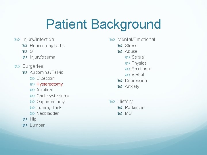
Patient Background Injury/Infection Reoccurring UTI’s STI Injury/trauma Surgeries Abdominal/Pelvic C-section Hysterectomy Ablation Cholecystectomy Oopherectomy Tummy Tuck Neobladder Hip Lumbar Mental/Emotional Stress Abuse Sexual Physical Emotional Verbal Depression Anxiety History Parkinson MS
General Overview Pelvic Floor Function The 5 S’s Sphincter control Sexual Function Support Stability Sump Pump Pelvic Floor/Bladder/Bowel coordination Pelvic Floor Strength/Endurance
Pelvic Floor Function Sphincter Control Opens/Closes openings of the urethra, vagina and rectum Dysfunction: Urinary and Fecal incontinence
Pelvic Floor Function Support Of organs against gravity and intra-abdominal pressure, tone for the vaginal and rectal walls Dysfunction: Pelvic Prolapse
Pelvic Floor Function Sexual health Orgasm, Blood flow, Mobility Dysfunction: dyspareunia, painful orgasm, vaginismus
Pelvic Floor Function Stability: Assists in stability of the SIJ, pubic symphysis, sacrococcygeal, lumbopelvic and hip joint (Lee) Critical in enabling effective load transfer from the lower extremity to the pelvis and spine (Sapsford, Hodges) “SUI as evidence of Failed Load Transfer through the Pelvis” by Diane Lee Dysfunction: Coccydynia, SIJ/hip pain
Pelvic Floor Function Sump Pump: A venous and lymphatic pump for the pelvis (Mitchell 1999) Further data/more recent research is needed assessing this postulation Dysfunction: Pelvic Congestion
Pelvic Floor/Bladder Coordination Bradley’s Loop 1: Voluntary Control of Micturition Without it we have no desire or awareness of voiding Bradley’s Loop 2: Maintains Duration of Detrusor Contraction Without it the ability to empty is lost, leads to stop and go urination and increased post-void residual Bradley’s Loop 3: Coordinates Detrusor with Sphincter Relaxation Without it we have uninhibited sphincter relaxation Reciprocal relationship of bladder to pelvic floor/external sphincter (quick flicks) Bradley’s Loop 4: Control of sphincters Keeps sphincters closed during bladder filling
Pelvic Floor/Bowel Puborectalis must relax in order for the stool to ‘come around the corner’ Feet on a stool (increases puborectal angle) Diaphragm breathing Bowel retraining schedule Dietary changes based on stool consistency GIKIDS. ORG “The Poo in You”
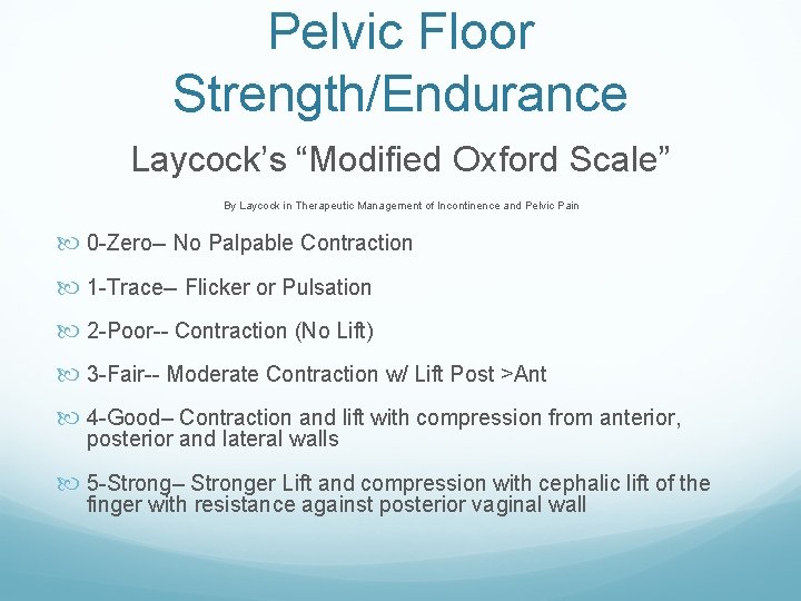
Pelvic Floor Strength/Endurance Laycock’s “Modified Oxford Scale” By Laycock in Therapeutic Management of Incontinence and Pelvic Pain 0 -Zero-- No Palpable Contraction 1 -Trace-- Flicker or Pulsation 2 -Poor-- Contraction (No Lift) 3 -Fair-- Moderate Contraction w/ Lift Post >Ant 4 -Good– Contraction and lift with compression from anterior, posterior and lateral walls 5 -Strong– Stronger Lift and compression with cephalic lift of the finger with resistance against posterior vaginal wall
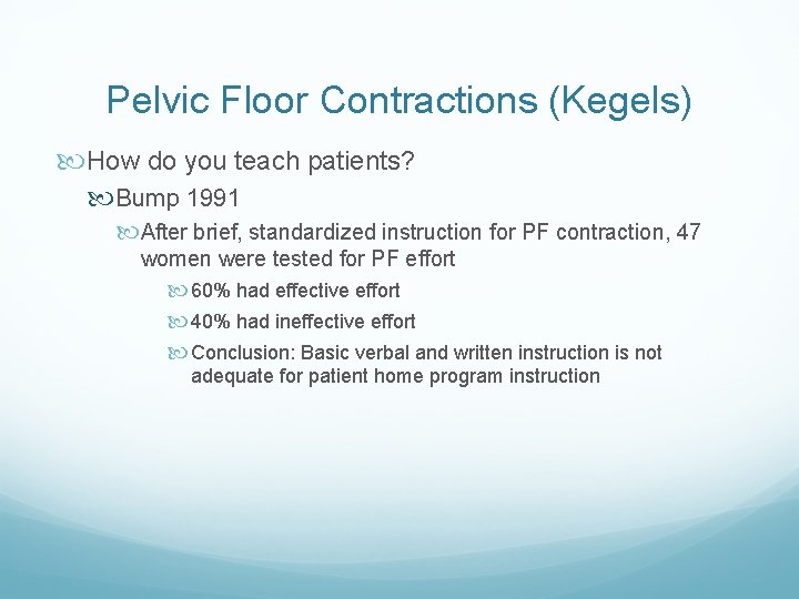
Pelvic Floor Contractions (Kegels) How do you teach patients? Bump 1991 After brief, standardized instruction for PF contraction, 47 women were tested for PF effort 60% had effective effort 40% had ineffective effort Conclusion: Basic verbal and written instruction is not adequate for patient home program instruction
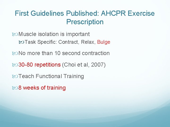
First Guidelines Published: AHCPR Exercise Prescription Muscle isolation is important Task Specific: Contract, Relax, Bulge No more than 10 second contraction 30 -80 repetitions (Choi et al, 2007) Teach Functional Training 8 weeks of training
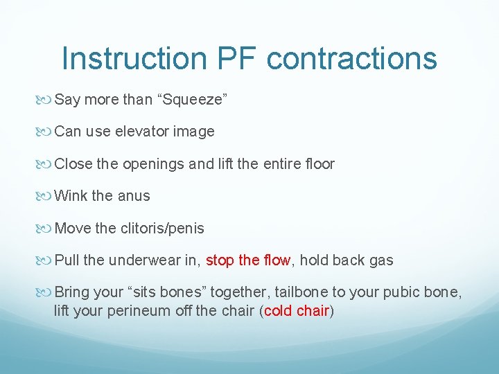
Instruction PF contractions Say more than “Squeeze” Can use elevator image Close the openings and lift the entire floor Wink the anus Move the clitoris/penis Pull the underwear in, stop the flow, hold back gas Bring your “sits bones” together, tailbone to your pubic bone, lift your perineum off the chair (cold chair)
To Kegel or To Not Kegel No Kegels with a Shortened/Tight Pelvic Floor and Pain presentation
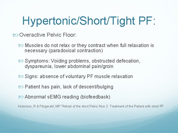
Hypertonic/Short/Tight PF: Overactive Pelvic Floor: Muscles do not relax or they contract when full relaxation is necessary (paradoxical contraction) Symptoms: Voiding problems, obstructed defecation, dyspareunia, lower abdominal pain/groin Signs: absence of voluntary PF muscle relaxation Patient has pain, lack of descent/bulging Abnormal s. EMG reading (biofeedback) Kotorinos, R & Fitzgerald, MP “Rehab of the short Pelvic floor 2: Treatment of the Patient with short PF
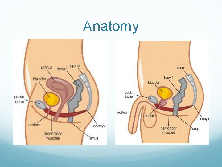
Anatomy
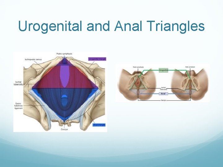
Urogenital and Anal Triangles
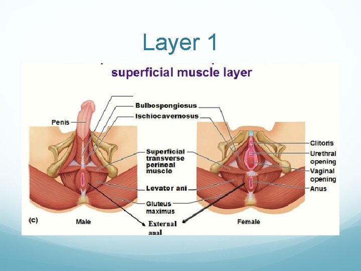
Layer 1
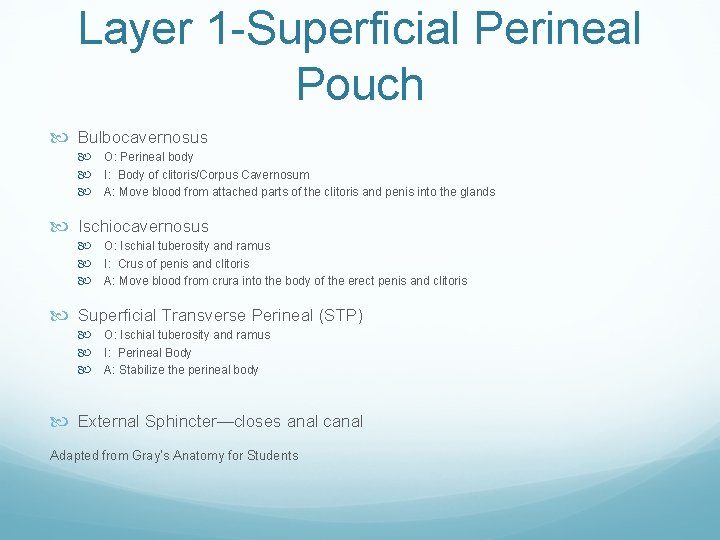
Layer 1 -Superficial Perineal Pouch Bulbocavernosus O: Perineal body I: Body of clitoris/Corpus Cavernosum A: Move blood from attached parts of the clitoris and penis into the glands Ischiocavernosus O: Ischial tuberosity and ramus I: Crus of penis and clitoris A: Move blood from crura into the body of the erect penis and clitoris Superficial Transverse Perineal (STP) O: Ischial tuberosity and ramus I: Perineal Body A: Stabilize the perineal body External Sphincter—closes anal canal Adapted from Gray’s Anatomy for Students
Layer 2
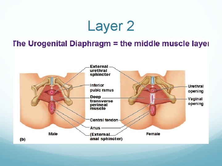
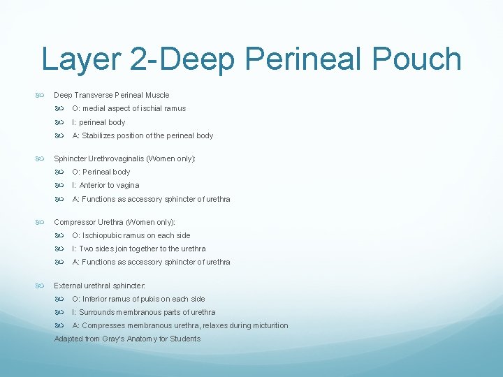
Layer 2 -Deep Perineal Pouch Deep Transverse Perineal Muscle O: medial aspect of ischial ramus I: perineal body A: Stabilizes position of the perineal body Sphincter Urethrovaginalis (Women only): O: Perineal body I: Anterior to vagina A: Functions as accessory sphincter of urethra Compressor Urethra (Women only): O: Ischiopubic ramus on each side I: Two sides join together to the urethra A: Functions as accessory sphincter of urethra External urethral sphincter: O: Inferior ramus of pubis on each side I: Surrounds membranous parts of urethra A: Compresses membranous urethra, relaxes during micturition Adapted from Gray’s Anatomy for Students
Layer 3
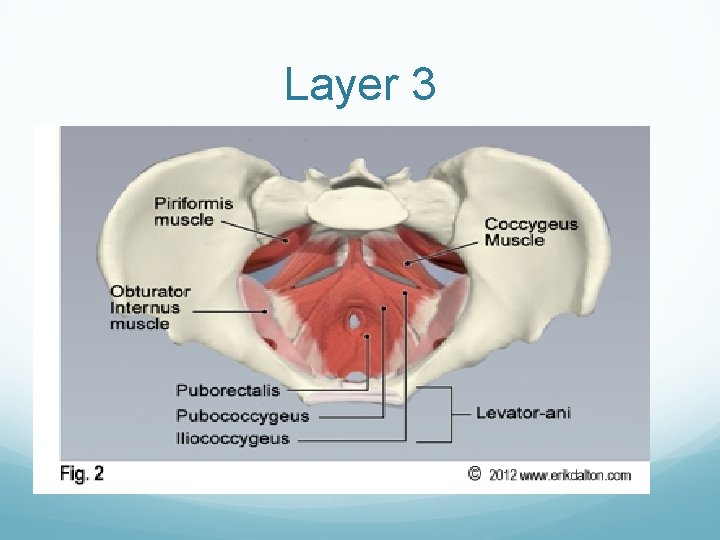
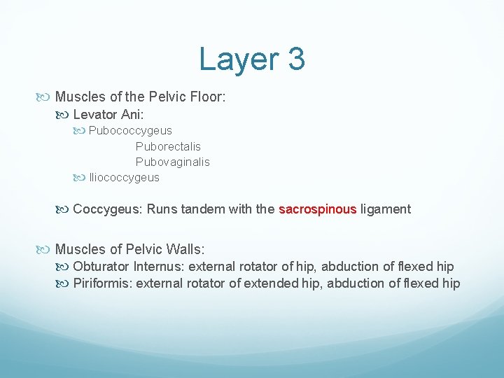
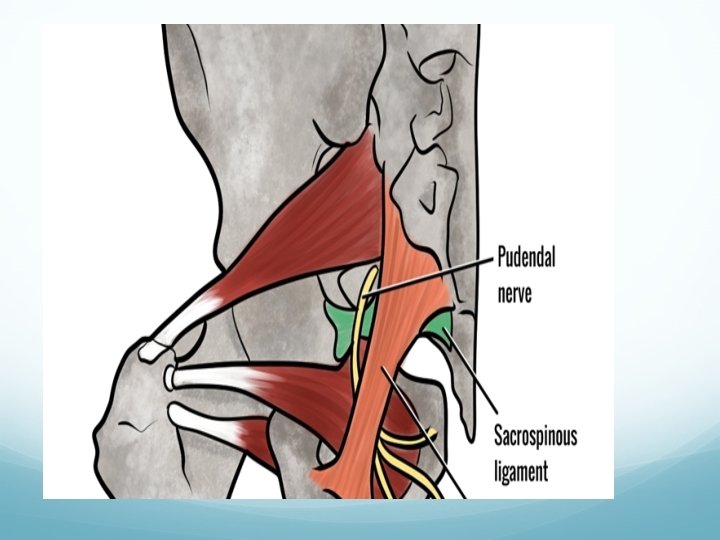
Layer 3 Muscles of the Pelvic Floor: Levator Ani: Pubococcygeus Puborectalis Pubovaginalis Iliococcygeus Coccygeus: Runs tandem with the sacrospinous ligament Muscles of Pelvic Walls: Obturator Internus: external rotator of hip, abduction of flexed hip Piriformis: external rotator of extended hip, abduction of flexed hip
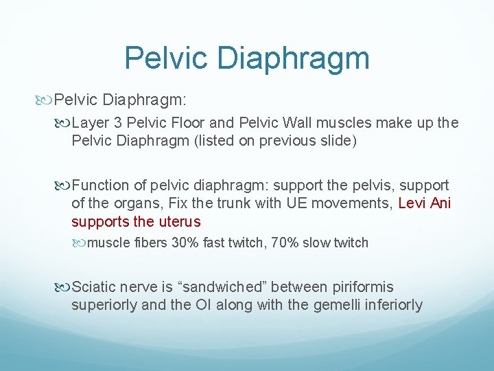
Pelvic Diaphragm: Layer 3 Pelvic Floor and Pelvic Wall muscles make up the Pelvic Diaphragm (listed on previous slide) Function of pelvic diaphragm: support the pelvis, support of the organs, Fix the trunk with UE movements, Levi Ani supports the uterus muscle fibers 30% fast twitch, 70% slow twitch Sciatic nerve is “sandwiched” between piriformis superiorly and the OI along with the gemelli inferiorly
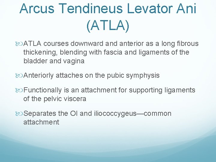
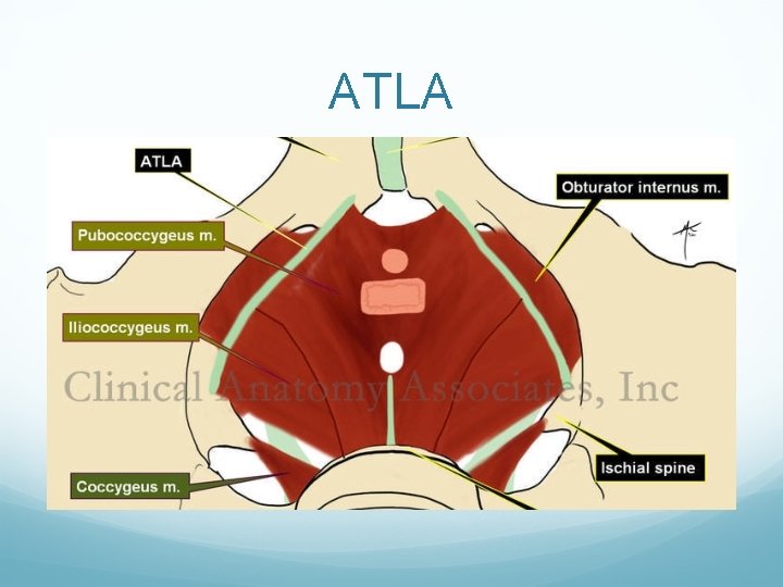
Arcus Tendineus Levator Ani (ATLA) ATLA courses downward anterior as a long fibrous thickening, blending with fascia and ligaments of the bladder and vagina Anteriorly attaches on the pubic symphysis Functionally is an attachment for supporting ligaments of the pelvic viscera Separates the OI and iliococcygeus—common attachment
ATLA
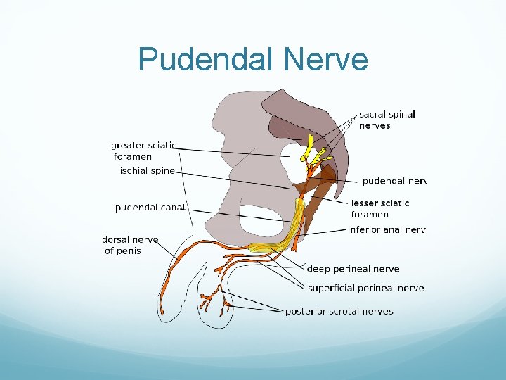
Pudendal Nerve
Pudendal Nerve Summary The pudendal nerve has 3 main branches: Inferior rectal or hemorrhoidal Perineal Dorsal nerve of clitoris or penis L 4 -S 4 (derived mainly from sacral branches) “S 2, 3, 4 keep poop and pee off the floor”
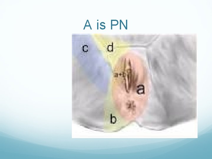
Pudendal Entrapment (PNE) vs Pudendal Neuralgia (PN) Entrapment and Neuralgia are not the same! PN refers to pain along the distribution of the PN (clitoris/penis, anus, perineum)—PN does not mean nerve is trapped, can be irritated from inflammation, tight/spastic muscles, or mechanical compression (prolonged sitting, poor sitting mechanics). WHERE is your pain? Not all sitting pain is the same. PNE will have symptoms of PN but nerve is ‘trapped or damaged’, pain is unrelenting and may not modify with positions, may be increased with sitting. PN and PFD not the same but have overlapping S&S d/t tight/spastic muscles, treatments are similar. Less B&B issues w/ PN vs PFD.
A is PN
Orthopedic Considerations SIJ Hypertonicity of the PF affects SIJ stability Levator ani causes counternutation (extension) of sacrum stiffening the SIJ (forward flexing the coccyx) Multifidi—nutation of the sacrum (flexion) and segmental spinal control (extension of lumbar spine) SIJ stability: PF contributes to lowering of vertical shear forces, increasing SIJ compression hence increasing SIJ stability. Teach Pelvic brace: Tr. A, M, IIO, and PFM Kathe Wallace, PT, BCB-PMD. Women’s Health Summit. 2014. 1 -32. Pel. 2008 Spitznagle, Theresa “Tracy” M. PT, DPT, MHS, WCS. 2013. Movement Systems Approach to Musculoskeletal Pelvic Pain. a. The Pelvic Girdle. Diane Lee 2000, pg 53.
Orthopedic Considerations Hip 64 -72% of patient’s with hip dysfunction also experience PF dysfunction Hip ER’s are lengthened due to increased hip adduction and IR; this causes PF pain and can cause physical stress on the pudendal nerves Pain patterns: posterior lateral hip pain, sciatica, lateral thigh pain, pain in WB or with prolonged sitting, pudendal neuralgia, dyspareunia, urgency/frequency.
Orthopedic Considerations When Hip ER’s, especially the piriformis and OI are shortened they cause compressive forces on the multiple branches of the pudendal nerves and/or can cause a “sciatic nerve sandwich” Pain Pattern: OI trigger points mimic piriformis syndrome and refers pain to the coccygeal region with spillover into the posterior thigh. Sx reported are fullness in the rectum, pain referred to ipsilateral thigh or coccyx.
The OI Connection Integral player in pelvic floor function and rehabilitation. PFM are very small and thin, yet able to produce enough force in isolation to counteract changes in intraabdominal pressure during less rigorous activities (standing, abdominal crunch). However, when the intraabdominal pressure exceeds the maximum force generated by the PFM’s in isolation, implies other parallel stuctures (OI) is recruited/required to assist PFM to maintain PF function including continence.
Journal of Women’s Health Physical Therapy Study 18 -35, same strength of PFM and OI at start of study measured by MMT Control group and Exercise group Each group maintained same level of fitness/exercise prior to study except: The exercise group did Monster Walks, Clam, and Isometric Hip ER against wall (3 sets of 10, 3 x/week for 12 weeks) Strengthening the deep rotators of the hip that surround the PFM, such as OI, improved PFM strength in healthy, young women. This could be particularly beneficial in patients who have difficulty performing the traditional Kegel exercise
Your Patients Consider your patient population: Fit, athlete, strong MMT----may need pfm in isolation Sedentary, inactive---benefit from hip/core strengthening Elderly population---cognitively cannot perform Kegels, may benefit from hip/core/adduction OVERFLOW may be enough, others may need to be referred out to PF PT for specific training Remember to review their medical history and ask the personal questions (incontinence, dyspareunia, etc. )
Orthopedic Considerations Adductor muscles Attachments at the abdominal fascia that can be limited with any kind of abdominal scar adhesions and fascial connections to layer 1 Trigger points refer pain to the urogenital region (layer 2) and increases urinary frequency and urgency. Shortened adductors cause anterior rotation of pelvis, counternutation (extension) of the sacrum, compression/shearing of symphysis pubis Pelvic Floor Function, Dysfunction and Treatment- Advanced PF 3. Herman & Wallace Inc. Pelvic Rehabilitation Institute. 2010 -2013 Travel and Simons. Myofascial Pain and Dysfunction. 1992.
Orthopedic Considerations Postural changes in pregnancy Greater than 50% of pregnant women who had LBP also had PFD with increased activity in PF muscles 92% of post-partum women who are incontinent at 12 weeks will be at 5 years unless treated 41 -83% of post-partum women 2 -3 months are identified to have sexual dysfunction LBP: PF 1: Pelvic Floor Muscle Disorders. Section on Women’s Health 2011. Incontinent/sexual dysfunction: Vicierup et al. 2000.
Post-Partum Hormones tested at 1 year Ask if they are leaking w/ activity or urge, bladder habits, bowel habits (constipation? ) Nursing posture Holding baby, stepping over baby gate, etc. Ask the questions no one else will ask---pain with intercourse? Baby blues? Brain fog?
Orthopedic Considerations Athletes and SUI/UUI Both types of incontinence rate high among competitive female athletes ages 18 -25, enough to alter activities around voiding habits. 52% of female athletes experience urine loss during sport or activities. If the athlete has incontinence their PF is weak, abdominal exercises and increasing intra-abdominal pressure will compress their pelvic organs down with each contraction. This repetitive compression will lengthen their PF ultimately making them weaker and exacerbating their problem PF 1: Pelvic Floor Muscles Disorders, Section on Women’s Health 2011 Dockter M. Koistand AM, Martin KA, Schiwal LJ. Prevalence of Urinary Incontinence: A Comparative Study of Collegiate Female Athletes and Non-Athletic Controls. JWHPT. 2007; 31: 12 -17 Nygaard IE, et al. Urinary Incontinence in Elite, Nulliparous Athletes. Obstet Gynecol. 1994; 84: 183 -187
How PT applies-Treatment Manual therapy Pelvic alignment, muscle balance Therapeutic Exercise Diet modifications, behavior modifications, postural adjustments, proper body mechanics Biofeedback Tactile or Visual feedback Electrical Stimulation To strengthen weak pelvic floor muscles (0 or 1)
Manual Therapy Techniques that correct pelvic asymmetries, reduce muscle spasm, decrease pain, and increase tissue mobility are central to the treatment of pelvic pain. Can be use post-surgical to ‘break down’ scar tissue/reducing adhesions Decrease pain Improve circulation Stretch tight muscles and free up connective tissue Facilitate weak muscles Improve visceral and muscle mobility
Therapeutic Exercises Many painful conditions of the pelvis have their origin at the pelvic floor and lumbosacral muscles. The function of the pelvic floor may be compromised by surgery, pregnancy and/or childbirth, disease or musculoskeletal injury. Specialized exercises are used to develop the strength and coordination of the pelvic floor. Dilators Core stabilization exercises Self-correction techniques Diaphragm Breathing!
Biofeedback Muscle pain and dysfunction can be caused by chronic muscles spasm of the lumbo-pelvic region. Surface electromyography (s. EMG) is used to detect painfully contracted muscles. Once revealed, the biofeedback systems help train patients to voluntarily relax painfully contracted muscles Can use mirror or any tactile or visual device
Electrical Stimulation Intravaginal electrical stimulation is utilized to facilitate pelvic floor contractions and help patients regain motor control. The electrical stimulation recruits the dormant musculature, developing elemental function that, with the use of biofeedback and cueing from therapist, can eventually be voluntarily controlled.
Patient Education MOST IMPORTANT TREATMENT!!
In Conclusion Most Common presentations of Pelvic Floor Dysfunctions: Hip, Low Back, Groin, or Pelvic pain Unspecified Abdominal Pain Coccydynia Dyspareunia (Pain during/after intercourse) Vulvadynia (or any vaginal pain) Endometriosis (painful periods) Pelvic Floor spasms (vaginal or rectal) Prostatitis (non-bacterial)
Conclusion Most Common presentations of Pelvic Floor Dysfunctions Continued: Bladder Pain/Interstitial Cystitis Dysuria (pain/burning w/ urination) Reoccurring Bladder/UTI Infections Urinary Urge/Frequency Nocturia (Nighttime voids) Feeling of Incomplete Emptying Hesitancy starting urine stream Urinary and Fecal Incontinence (Urge and Stress)
Conclusion Treatment for Pelvic Floor Disorders Include: Strengthening weak muscles, including the use of biofeedback and electrical stimulation Improving flexibility of short and tight muscles and connective tissue Improving timing and coordination of pelvic floor with bladder Core stabilization of the deep abdominal and trunk muscles Correction of joint restrictions in the spine, pelvis, and ribcage Self-care techniques along with deep relaxation techniques
Conclusion Recognize when to refer patients to Pelvic Floor therapy! Based on their history, presentation, and/or response to your treatment
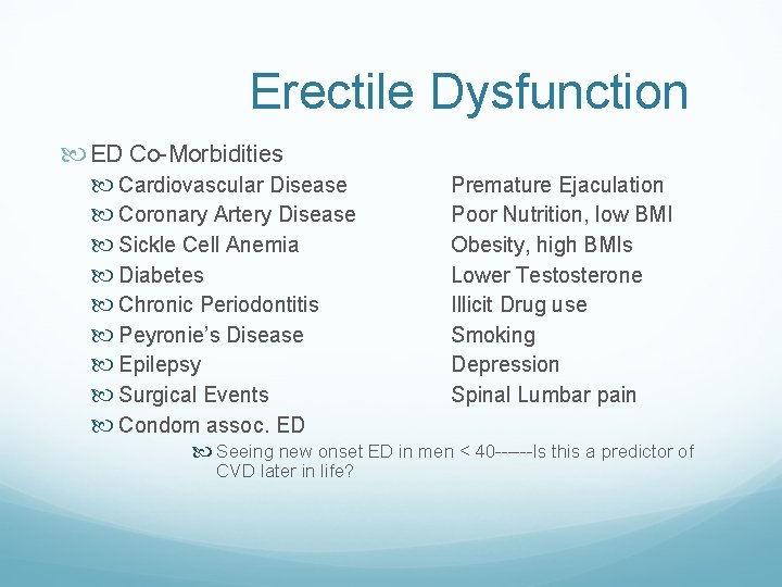
Erectile Dysfunction ED Co-Morbidities Cardiovascular Disease Coronary Artery Disease Sickle Cell Anemia Diabetes Chronic Periodontitis Peyronie’s Disease Epilepsy Surgical Events Condom assoc. ED Premature Ejaculation Poor Nutrition, low BMI Obesity, high BMIs Lower Testosterone Illicit Drug use Smoking Depression Spinal Lumbar pain Seeing new onset ED in men < 40 ------Is this a predictor of CVD later in life?
ED continued Morning erection? ? Mental component
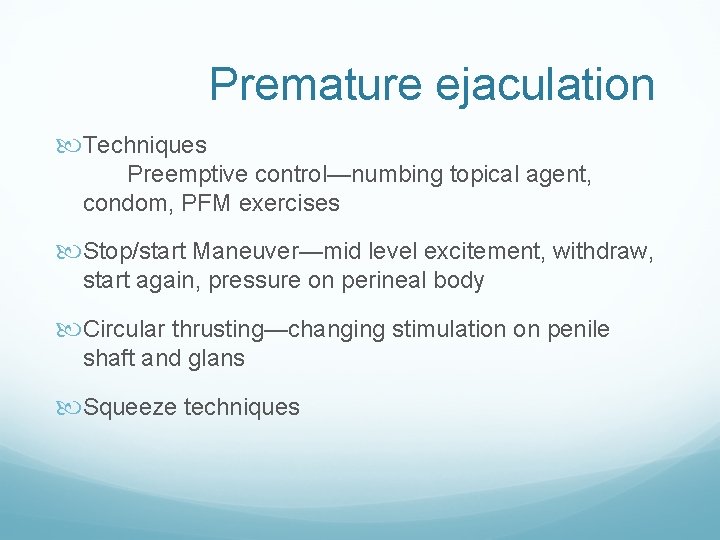
Premature ejaculation Techniques Preemptive control—numbing topical agent, condom, PFM exercises Stop/start Maneuver—mid level excitement, withdraw, start again, pressure on perineal body Circular thrusting—changing stimulation on penile shaft and glans Squeeze techniques
Sounds like……. . Pelvic Floor Dysfunction!!!