ICHTHYOLOGY UNIT II Skeletal System Axial and Appendicular


 Shark; B) Bony fish Dorsal (medial)")


- Slides: 22
ICHTHYOLOGY UNIT II Skeletal System: Axial and Appendicular Skeleton
Fish skeletal system • • • There are two different skeletal types: the exoskeleton, which is the stable outer shell of an organism, and the endoskeleton, which forms the support structure inside the body. The skeleton of the fish is made of either cartilage (cartilaginous fishes) or bone (bony fishes). The main features of the fish, the fins, are bony fin rays and, except for the caudal fin, have no direct connection with the spine. They are supported only by the muscles. The ribs attach to the spine. Bones are rigid organs that form part of the endoskeleton of vertebrates. They function to move, support, and protect the various organs of the body, produce red and white blood cells and store minerals. Bone tissue is a type of dense connective tissue. Bones come in a variety of shapes and have a complex internal and external structure. They are lightweight, yet strong and hard, in addition to fulfilling their many other functions. The axial skeleton consists of the skull and the vertebral column. The appendicular skeleton supports the fins in fish and associated girdles and median fin support
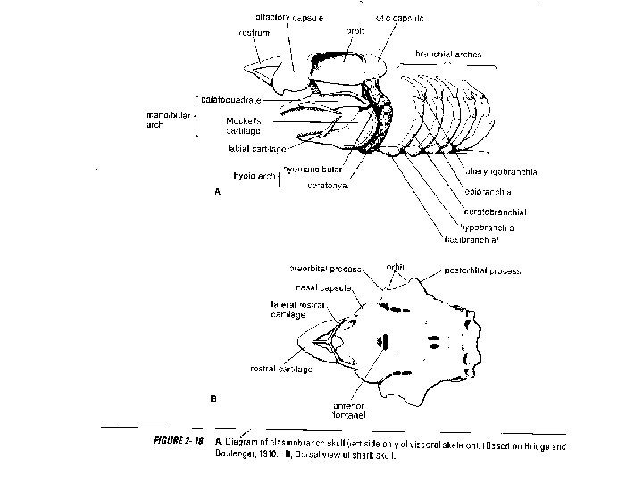
Fish Skulls • Neurocranium – Chondocranium – Dermatocranium • Splanchiocranium – Gills – Jaws
Basic fish anatomy
Jaw Structure • Premaxilla • Maxilla • Supramaxilla • Palatine • Dentary
Gill Coverings • • • Preopercle Opercle Subopercle Interopercle Branchiostegals
Vertebral column in fishes • vertebral column, in which the notochord (a stiff rod of uniform composition) found in all chordates has been replaced by a segmented series of stiffer elements (vertebrae) separated by mobile joints (intervertebral discs, derived embryonically and evolutionarily from the notochord). • However, a few fish have secondarily lost this anatomy, retaining the notochord into adulthood, such as the sturgeon. [9]
Fish vertebrae • • • In fish both sides of the centrum are concave, the space between is filled with a ball of cartilaginous substance that holds them a little apart allowing them to flex a bit. (There is an exception to this rule however, Garfish (Lepisostidae) have interlocking vertebrae much like those of reptiles. In other words the centra of the vertebrae are convex on the anterior or front face and concave on the posterior or behind face allowing them to fit into each other). The vertebrae that connect the skull to the spine are called the Atlas and the Axis, as in all vertebrates. In the picture above we can see representative vertebrae from three fish and two sharks, a Sturgeon, a Cod and a Salmon a White Shark and an Angel Shark.
Vertebral Column
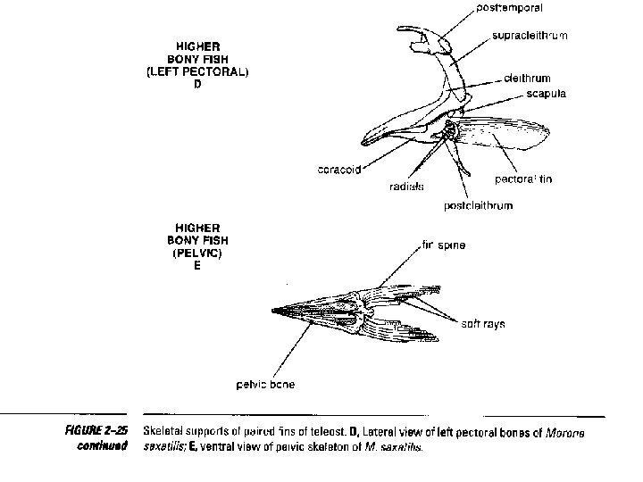
Appendicular skeleton in Fishes • Pectoral and pelvic girdles are primitively absent in the hag fishes and lampreys. Sharks have a coracoscapular cartilage that hangs more or less freely inside the body wall and has no attachment to the vertebral column. In rays, the pectoral girdle is attached to the fused anterior section of the vertebral column (synarchial condition) and also, byway of the propterygium of the pectoral girdle and antorbital cartilage, to the nasal capsules of the skull.
Paired Fins
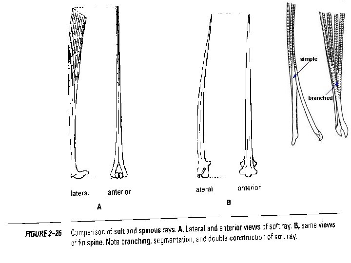
Spines vs. Rays Soft Finned Rays Soft, unpointed Segmented Branched Bilateral (lt. and rt. halves) Spines Hard, pointed tissue Unsgemented Unbranched Solid
Fins
Median fins • • The median or unpaired fins consist of the dorsal, and adipose fins along the dorsal and ventral profiles of the fish. In jawless fishes, cartilaginous rods support the median fins. In Chondrichthyes, the median fins are supported by ceratotrichia, horny fin rays composed of elastin and supportedby dermal cells. Below the ceratotrichia are three layers of radials – rodlike cartilages that support the fin rays and extend inward toward the vertebral column. In bony fishes, the ceratotrichia are replaced duringontogeny by lepidotrichia, bony supporting elements that are derived from scales. Ceratotrichia are present in lungfishes and larval actinopterygians. Primitive actinopterygianssuch as the Bowfin (Amia) still have three radials supporting each median fin ray, but these are reduced to two and then one in advanced teleost`s. The remaining elements then known as an interneural bone if it is under the dorsal fins or interhaemal bone if it is above the anal fin.
Fig 1 Skeletal support of Median fin A) Shark; B) Bony fish Dorsal (medial) Fins
Caudal Fins • Caudal fins are highly variable in shape but essentially serve the same function; that being forward motion, although some do it better than others and some provide additional benefits.
Tail in fishes • • • When the notochord extends straight to the tip of the tail and the caudal fin is divided equally then the tail is protocercal, as in embryonic fishes, adult cyclostomes, Heterocercal tail has the vertebral column bent upwards in the posterior region so that the caudal fin is divided into a narrow dorsal lobe and a broader ventral lobe. It is typical of elasmobranchs (Scoliodon) and some lower bony fishes. Diphycercal tail has lost the terminal upturned part of the vertebral column, and the vertebral column does not reach the end. Externally the tail has become symmetrical secondarily having two equal lobes of the caudal fin. It is found in Dipnoi, Polypterus and Latimeria. In homocercal tail, the terminal part of the vertebral column is bent upwards, but the dorsal lobe of the caudal fin is lost during development and the ventral lobe divides into two equal parts, so that the caudal fin appears symmetrical externally. This tail is common in bony fishes. Hypocercal tail has a larger lower lobe of the caudal fin into which the vertebral column is bent downwards. It is found in some primitive fishes
T