Essentials of Anatomy Physiology 4 th Edition Martini
- Slides: 66
Essentials of Anatomy & Physiology, 4 th Edition Martini / Bartholomew 8 The Nervous System Power. Point® Lecture Outlines prepared by Alan Magid, Duke University Slides 1 to 145 Copyright © 2007 Pearson Education, Inc. , publishing as Benjamin Cummings
The Nervous System What Two Organ Systems Control All the Other Organ Systems? • Nervous system characteristics • Rapid response • Brief duration • Endocrine system characteristics • Slower response • Long duration Copyright © 2007 Pearson Education, Inc. , publishing as Benjamin Cummings
The Nervous System What are the Two Anatomical Divisions? • Central nervous system (CNS) • • • Brain Spinal cord Peripheral nervous system (PNS) • • • All the neural tissue outside CNS Afferent division (sensory input) Efferent division (motor output) • Somatic nervous system • Autonomic nervous system Copyright © 2007 Pearson Education, Inc. , publishing as Benjamin Cummings
Neural Tissue Organization What is the Anatomic Organization of CNS Neurons? • White matter—Bundles of axons (tracts) that share origins, destinations, and functions Copyright © 2007 Pearson Education, Inc. , publishing as Benjamin Cummings
Neural Tissue Organization What is the Anatomic Organization of PNS Neurons? • Ganglia—Groupings of neuron cell bodies • Nerve—Bundle of axons supported by connective tissue • Spinal nerves • To/from spinal cord • Cranial nerves • To/from brain Copyright © 2007 Pearson Education, Inc. , publishing as Benjamin Cummings
Neural Tissue Organization What are the Pathways in the CNS? • Ascending pathways Carry information from sensory receptors to processing centers in the brain • Descending pathways Carry commands from specialized CNS centers to skeletal muscles Copyright © 2007 Pearson Education, Inc. , publishing as Benjamin Cummings
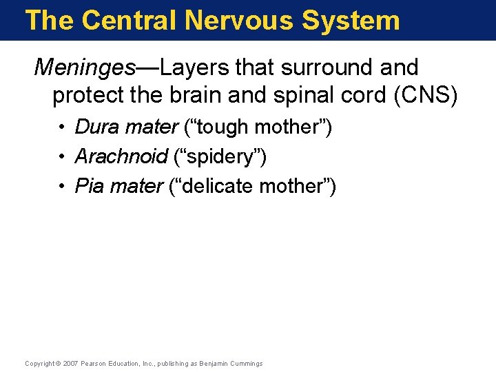
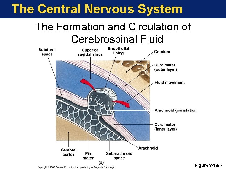
The Central Nervous System Meninges—Layers that surround and protect the brain and spinal cord (CNS) • Dura mater (“tough mother”) • Arachnoid (“spidery”) • Pia mater (“delicate mother”) Copyright © 2007 Pearson Education, Inc. , publishing as Benjamin Cummings
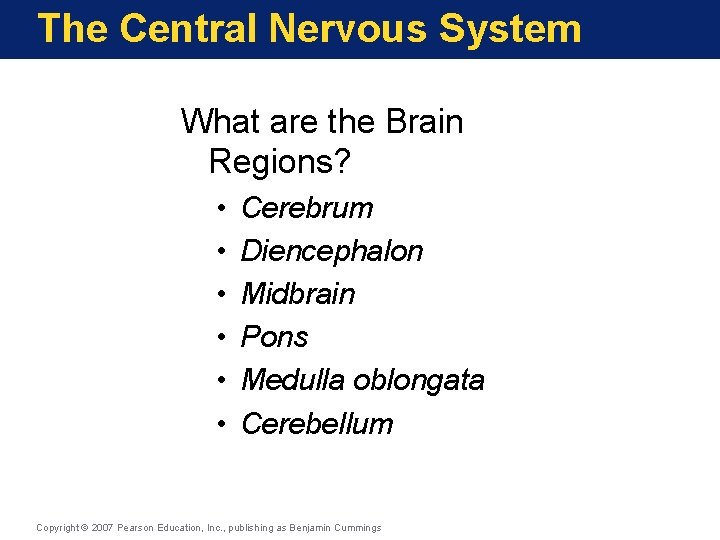
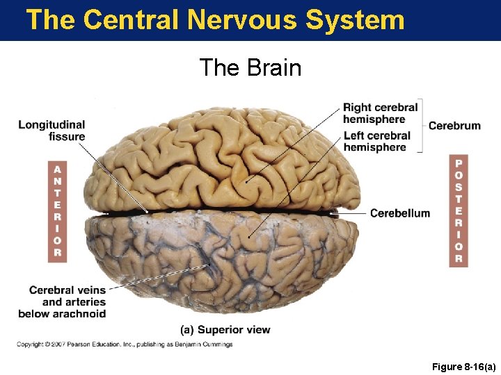
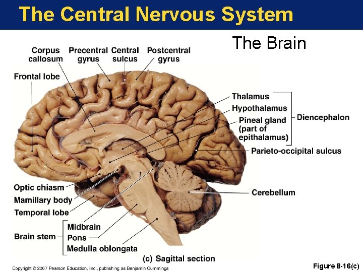
The Central Nervous System What are the Brain Regions? • • • Cerebrum Diencephalon Midbrain Pons Medulla oblongata Cerebellum Copyright © 2007 Pearson Education, Inc. , publishing as Benjamin Cummings
The Central Nervous System The Brain Figure 8 -16(a)
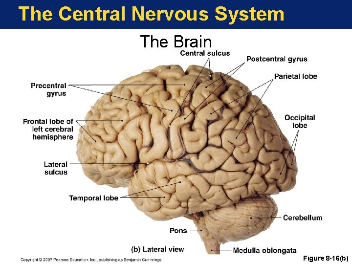
The Central Nervous System The Brain Figure 8 -16(b)
The Central Nervous System The Brain Figure 8 -16(c)
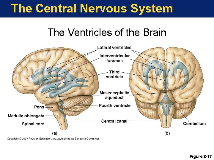
The Central Nervous System Brain—The four hollow chambers in the center of the brain filled with cerebrospinal fluid (CSF) Copyright © 2007 Pearson Education, Inc. , publishing as Benjamin Cummings
The Central Nervous System The Ventricles of the Brain Figure 8 -17
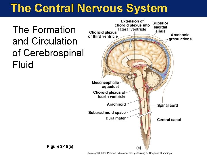
The Central Nervous System The Formation and Circulation of Cerebrospinal Fluid Figure 8 -18(a)
The Central Nervous System The Formation and Circulation of Cerebrospinal Fluid Figure 8 -18(b)
The Central Nervous System What are the Functions of the Cerebrum? • • Conscious thought Intellectual activity Memory Origin of complex patterns of movement Copyright © 2007 Pearson Education, Inc. , publishing as Benjamin Cummings
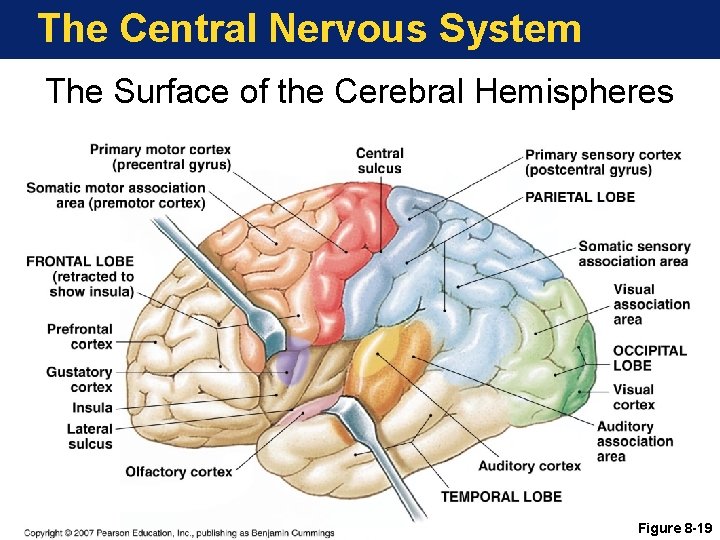
The Central Nervous System What are the Functions of the Cerebral Cortex? • Primary motor cortex (precentral gyrus) • Directs voluntary movement • Primary sensory cortex (postcentral gyrus) • Receives somatic sensation (touch, pain, pressure, temperature) • Association areas • Interpret sensation • Coordinate movement Copyright © 2007 Pearson Education, Inc. , publishing as Benjamin Cummings
The Central Nervous System The Surface of the Cerebral Hemispheres Figure 8 -19
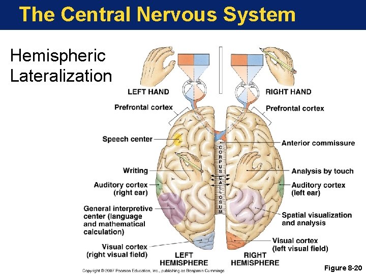
The Central Nervous System Hemispheric Lateralization Figure 8 -20
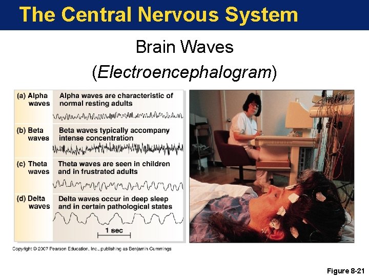
The Central Nervous System Brain Waves (Electroencephalogram) Figure 8 -21
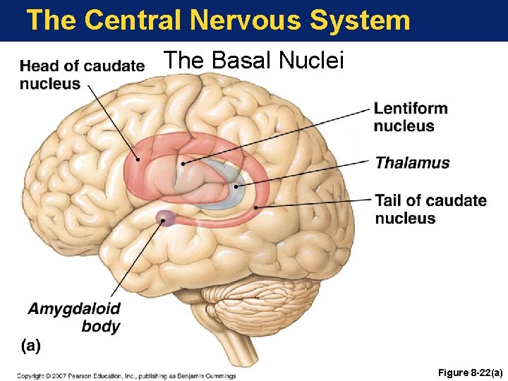
The Central Nervous System The Basal Nuclei Figure 8 -22(a)
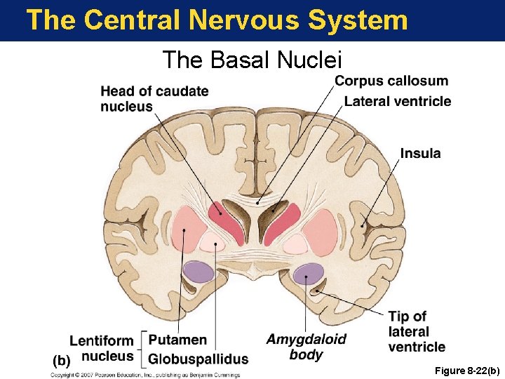
The Central Nervous System The Basal Nuclei Figure 8 -22(b)
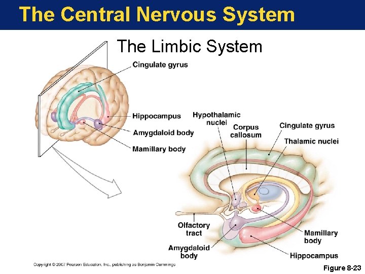
The Central Nervous System What are the Functions of the Limbic System? • Establish emotions and related drives • Control reflexes associated with eating • Store and retrieve long-term memories Copyright © 2007 Pearson Education, Inc. , publishing as Benjamin Cummings
The Central Nervous System The Limbic System Figure 8 -23
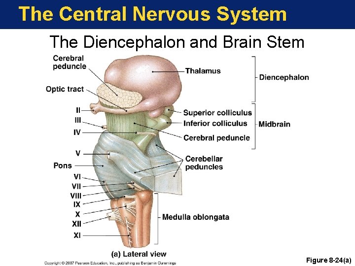
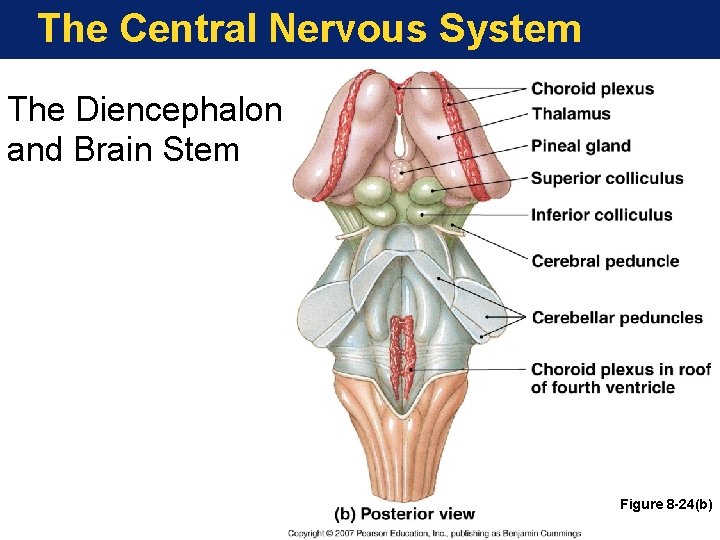
The Central Nervous System What is the Diencephalon? • Switching and relay center • Components include: • Epithalamus • Thalamus • Hypothalamus Copyright © 2007 Pearson Education, Inc. , publishing as Benjamin Cummings
The Central Nervous System The Diencephalon and Brain Stem Figure 8 -24(a)
The Central Nervous System The Diencephalon and Brain Stem Figure 8 -24(b)
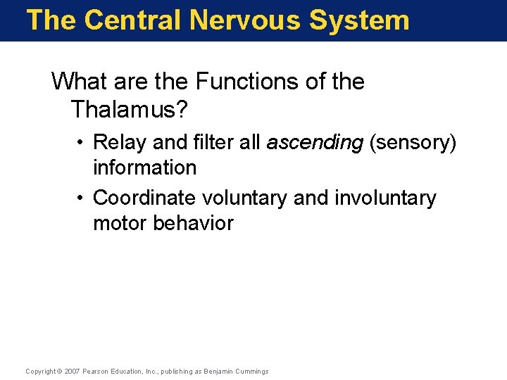
The Central Nervous System What are the Functions of the Thalamus? • Relay and filter all ascending (sensory) information • Coordinate voluntary and involuntary motor behavior Copyright © 2007 Pearson Education, Inc. , publishing as Benjamin Cummings
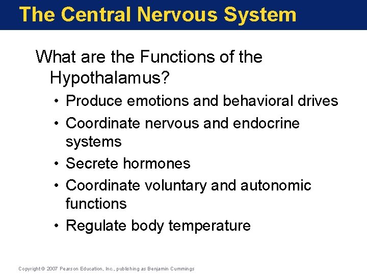
The Central Nervous System What are the Functions of the Hypothalamus? • Produce emotions and behavioral drives • Coordinate nervous and endocrine systems • Secrete hormones • Coordinate voluntary and autonomic functions • Regulate body temperature Copyright © 2007 Pearson Education, Inc. , publishing as Benjamin Cummings
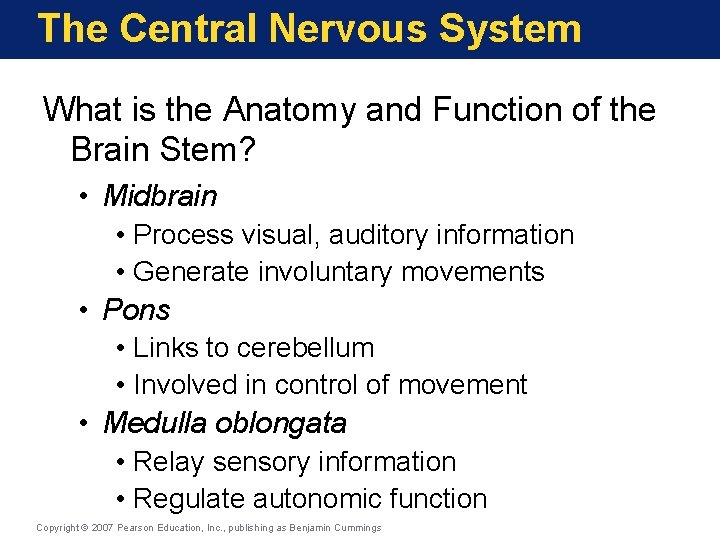
The Central Nervous System What is the Anatomy and Function of the Brain Stem? • Midbrain • Process visual, auditory information • Generate involuntary movements • Pons • Links to cerebellum • Involved in control of movement • Medulla oblongata • Relay sensory information • Regulate autonomic function Copyright © 2007 Pearson Education, Inc. , publishing as Benjamin Cummings
The Central Nervous System What is the Anatomy and Function of the Cerebellum? • Oversees postural muscles • Stores patterns of movement • Fine tunes most movements Copyright © 2007 Pearson Education, Inc. , publishing as Benjamin Cummings
The Central Nervous System What are the Functions of the Medulla Oblongata? • Relays ascending information to cerebral cortex • Controls crucial organ systems by reflex • Cardiovascular centers • Respiratory rhythmicity centers Copyright © 2007 Pearson Education, Inc. , publishing as Benjamin Cummings
The Central Nervous System Key Note The brain, a large mass of neural tissue, contains internal passageways and chambers filled with CSF. The six major regions of the brain have specific functions. As you ascend from the medulla oblongata to the cerebrum, those functions become more complex and variable. Conscious thought and intelligence are provided by the cerebral cortex. Copyright © 2007 Pearson Education, Inc. , publishing as Benjamin Cummings
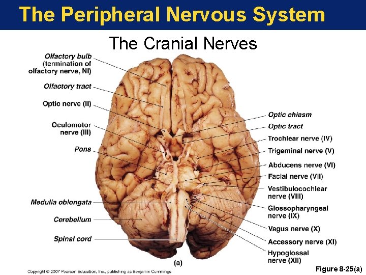
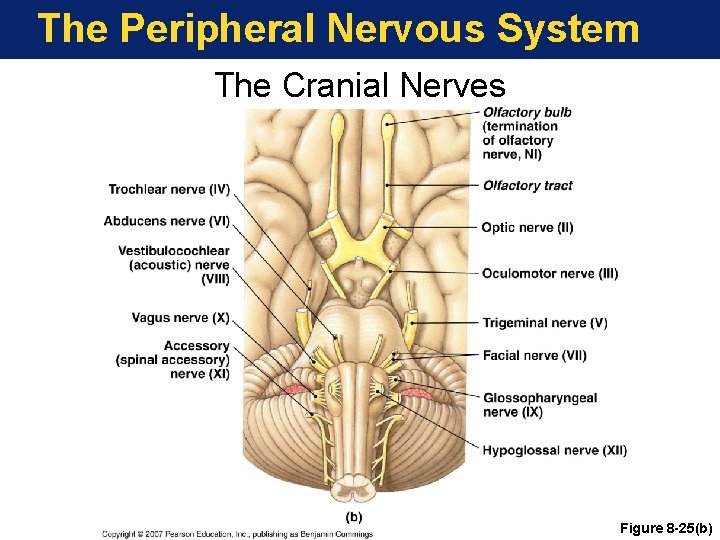
The Peripheral Nervous System What are the Twelve Pairs Of Cranial Nerves? • Olfactory (CN I) • Sense of smell • Optic (CN II) • Sense of vision • Oculomotor (CN III) • Eye movement Copyright © 2007 Pearson Education, Inc. , publishing as Benjamin Cummings
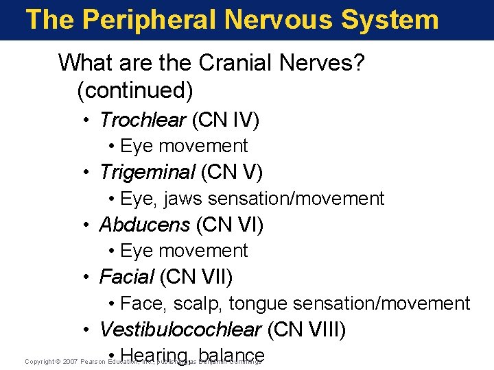
The Peripheral Nervous System What are the Cranial Nerves? (continued) • Trochlear (CN IV) • Eye movement • Trigeminal (CN V) • Eye, jaws sensation/movement • Abducens (CN VI) • Eye movement • Facial (CN VII) • Face, scalp, tongue sensation/movement • Vestibulocochlear (CN VIII) • Hearing, balance Copyright © 2007 Pearson Education, Inc. , publishing as Benjamin Cummings
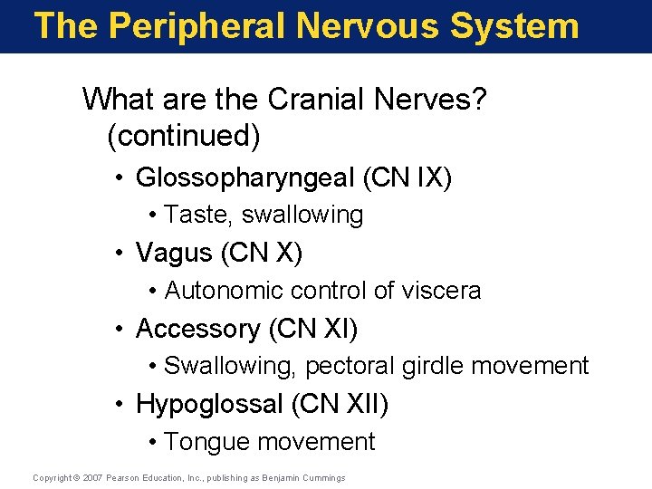
The Peripheral Nervous System What are the Cranial Nerves? (continued) • Glossopharyngeal (CN IX) • Taste, swallowing • Vagus (CN X) • Autonomic control of viscera • Accessory (CN XI) • Swallowing, pectoral girdle movement • Hypoglossal (CN XII) • Tongue movement Copyright © 2007 Pearson Education, Inc. , publishing as Benjamin Cummings
The Peripheral Nervous System The Cranial Nerves Figure 8 -25(a)
The Peripheral Nervous System The Cranial Nerves Figure 8 -25(b)
The Peripheral Nervous System Key Note The 12 pairs of cranial nerves are responsible for the special senses of smell, sight, and hearing/balance, and control movement of the eye, jaw, face, tongue, and muscles of the neck, back, and shoulders. They also provide sensation from the face, neck, and upper chest and autonomic innervation to thoracic and abdominopelvic organs. Copyright © 2007 Pearson Education, Inc. , publishing as Benjamin Cummings
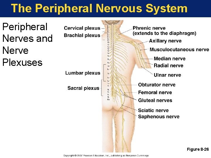
The Peripheral Nervous System Nerve Plexus—A complex, interwoven network of nerves Copyright © 2007 Pearson Education, Inc. , publishing as Benjamin Cummings
The Peripheral Nervous System Peripheral Nerves and Nerve Plexuses Figure 8 -26
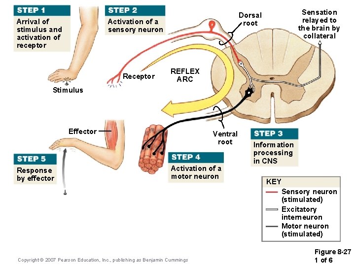
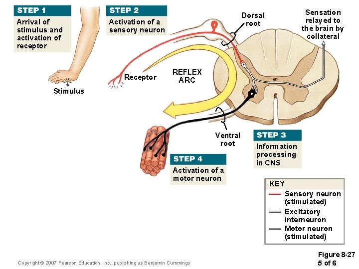
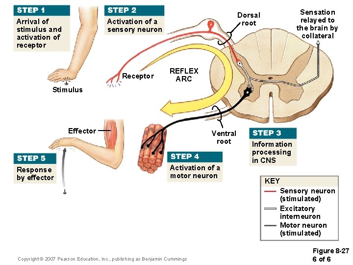
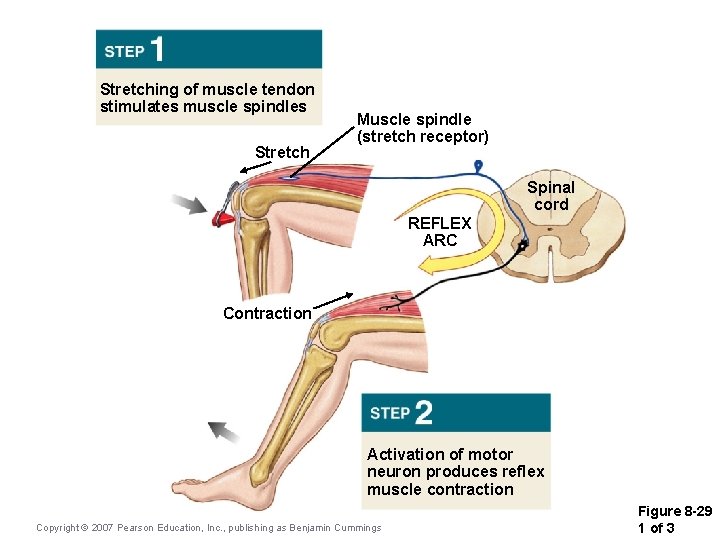
The Peripheral Nervous System Reflex—An automatic involuntary motor response to a specific stimulus Copyright © 2007 Pearson Education, Inc. , publishing as Benjamin Cummings
Arrival of stimulus and activation of receptor Activation of a sensory neuron Receptor Sensation relayed to the brain by collateral Dorsal root REFLEX ARC Stimulus Effector Response by effector Ventral root Activation of a motor neuron Copyright © 2007 Pearson Education, Inc. , publishing as Benjamin Cummings Information processing in CNS KEY Sensory neuron (stimulated) Excitatory interneuron Motor neuron (stimulated) Figure 8 -27 1 of 6
Arrival of stimulus and activation of receptor Stimulus Copyright © 2007 Pearson Education, Inc. , publishing as Benjamin Cummings Figure 8 -27 2 of 6
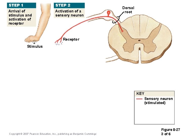
Arrival of stimulus and activation of receptor Activation of a sensory neuron Dorsal root Receptor Stimulus KEY Sensory neuron (stimulated) Copyright © 2007 Pearson Education, Inc. , publishing as Benjamin Cummings Figure 8 -27 3 of 6
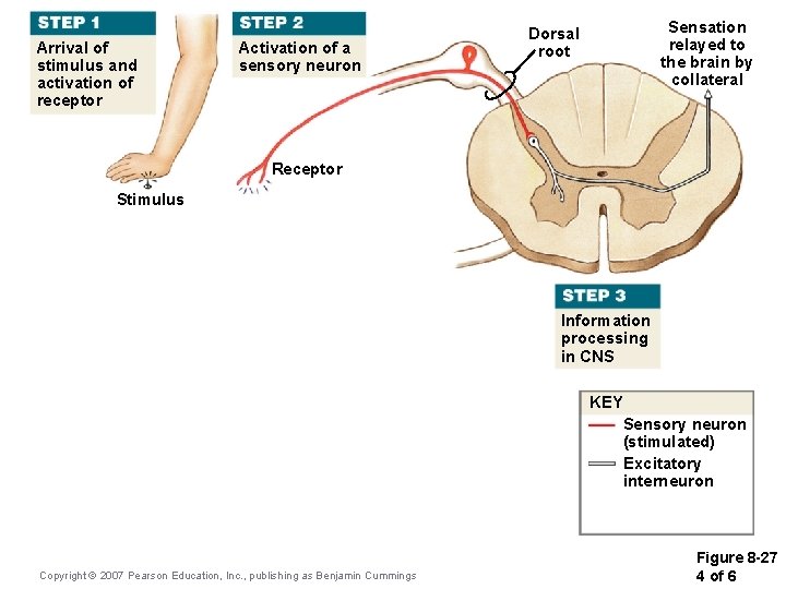
Arrival of stimulus and activation of receptor Activation of a sensory neuron Sensation relayed to the brain by collateral Dorsal root Receptor Stimulus Information processing in CNS KEY Sensory neuron (stimulated) Excitatory interneuron Copyright © 2007 Pearson Education, Inc. , publishing as Benjamin Cummings Figure 8 -27 4 of 6
Arrival of stimulus and activation of receptor Activation of a sensory neuron Receptor Sensation relayed to the brain by collateral Dorsal root REFLEX ARC Stimulus Ventral root Activation of a motor neuron Copyright © 2007 Pearson Education, Inc. , publishing as Benjamin Cummings Information processing in CNS KEY Sensory neuron (stimulated) Excitatory interneuron Motor neuron (stimulated) Figure 8 -27 5 of 6
Arrival of stimulus and activation of receptor Activation of a sensory neuron Receptor Sensation relayed to the brain by collateral Dorsal root REFLEX ARC Stimulus Effector Response by effector Ventral root Activation of a motor neuron Copyright © 2007 Pearson Education, Inc. , publishing as Benjamin Cummings Information processing in CNS KEY Sensory neuron (stimulated) Excitatory interneuron Motor neuron (stimulated) Figure 8 -27 6 of 6
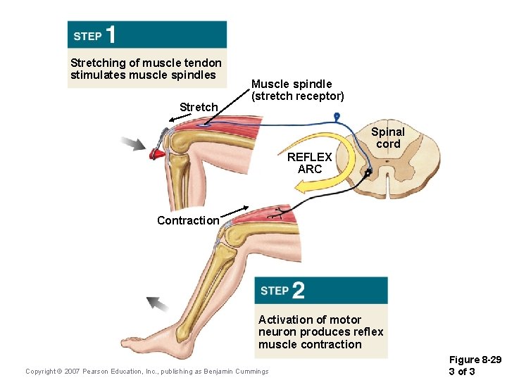
Stretching of muscle tendon stimulates muscle spindles Stretch Muscle spindle (stretch receptor) Spinal cord REFLEX ARC Contraction Activation of motor neuron produces reflex muscle contraction Copyright © 2007 Pearson Education, Inc. , publishing as Benjamin Cummings Figure 8 -29 1 of 3
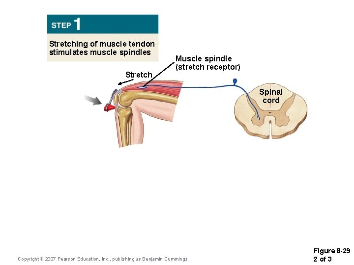
Stretching of muscle tendon stimulates muscle spindles Stretch Muscle spindle (stretch receptor) Spinal cord Copyright © 2007 Pearson Education, Inc. , publishing as Benjamin Cummings Figure 8 -29 2 of 3
Stretching of muscle tendon stimulates muscle spindles Stretch Muscle spindle (stretch receptor) Spinal cord REFLEX ARC Contraction Activation of motor neuron produces reflex muscle contraction Copyright © 2007 Pearson Education, Inc. , publishing as Benjamin Cummings Figure 8 -29 3 of 3
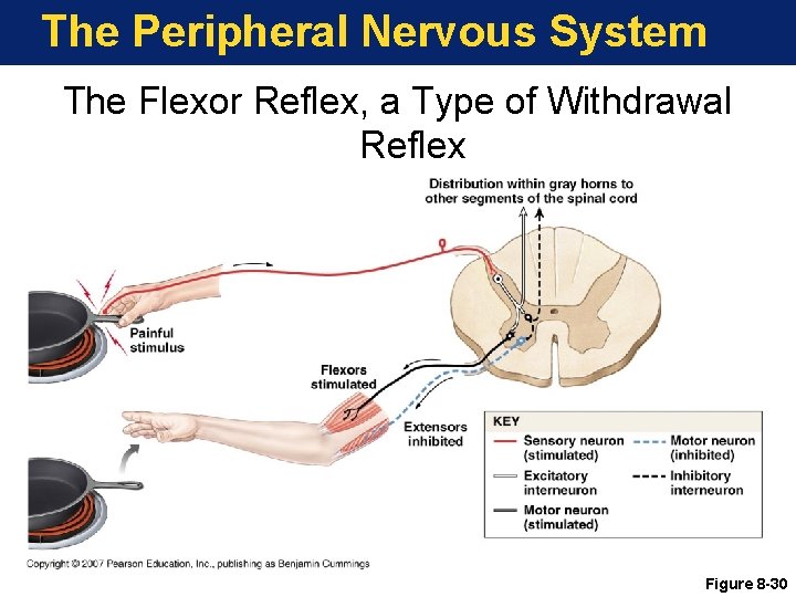
The Peripheral Nervous System The Flexor Reflex, a Type of Withdrawal Reflex Figure 8 -30
The Peripheral Nervous System Key Note Reflexes are rapid, automatic responses to stimuli that “buy time” for the planning and execution of more complex responses that are often consciously directed. Copyright © 2007 Pearson Education, Inc. , publishing as Benjamin Cummings
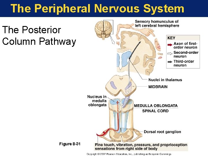
The Peripheral Nervous System The Posterior Column Pathway Figure 8 -31
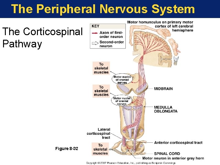
The Peripheral Nervous System The Corticospinal Pathway Figure 8 -32
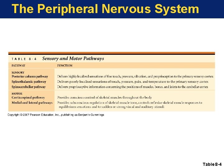
The Peripheral Nervous System Table 8 -4
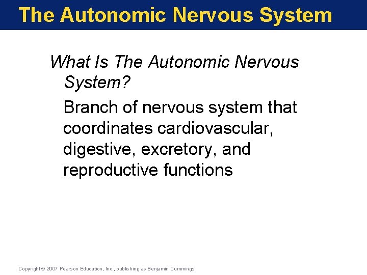
The Autonomic Nervous System What Is The Autonomic Nervous System? Branch of nervous system that coordinates cardiovascular, digestive, excretory, and reproductive functions Copyright © 2007 Pearson Education, Inc. , publishing as Benjamin Cummings
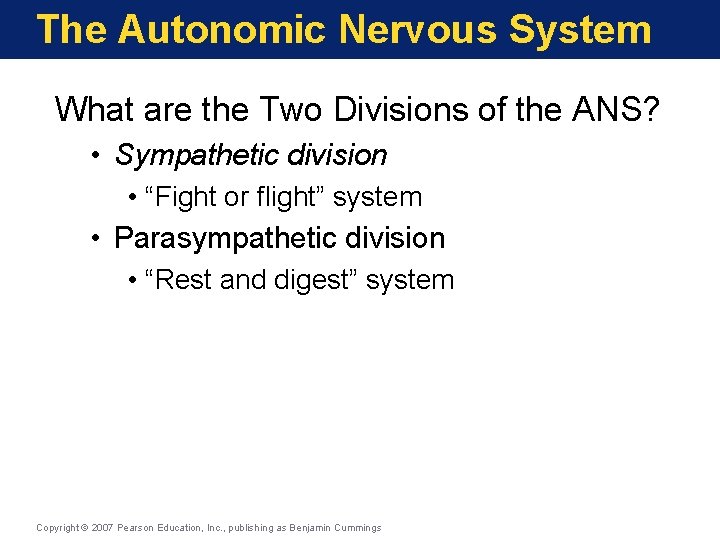
The Autonomic Nervous System What are the Two Divisions of the ANS? • Sympathetic division • “Fight or flight” system • Parasympathetic division • “Rest and digest” system Copyright © 2007 Pearson Education, Inc. , publishing as Benjamin Cummings
The Autonomic Nervous System Key Note The two divisions of the ANS operate largely without our awareness. The sympathetic division increases alertness, metabolic rate, and muscular abilities; the parasympathetic division reduces metabolic rate and promotes visceral activities such as digestion. Copyright © 2007 Pearson Education, Inc. , publishing as Benjamin Cummings
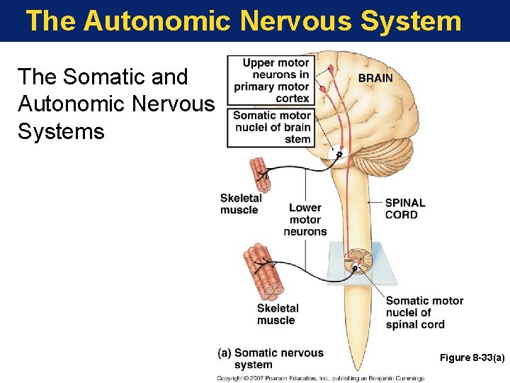
The Autonomic Nervous System The Somatic and Autonomic Nervous Systems Figure 8 -33(a)
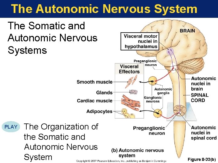
The Autonomic Nervous System The Somatic and Autonomic Nervous Systems PLAY The Organization of the Somatic and Autonomic Nervous System Figure 8 -33(b)
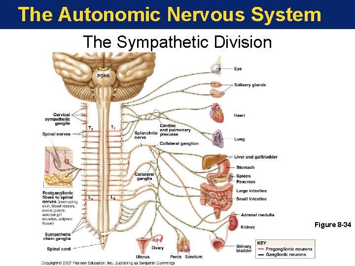
The Autonomic Nervous System The Sympathetic Division Figure 8 -34
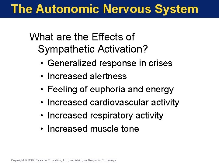
The Autonomic Nervous System What are the Effects of Sympathetic Activation? • • • Generalized response in crises Increased alertness Feeling of euphoria and energy Increased cardiovascular activity Increased respiratory activity Increased muscle tone Copyright © 2007 Pearson Education, Inc. , publishing as Benjamin Cummings
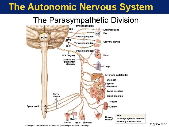
The Autonomic Nervous System The Parasympathetic Division Figure 8 -35
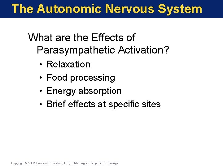
The Autonomic Nervous System What are the Effects of Parasympathetic Activation? • • Relaxation Food processing Energy absorption Brief effects at specific sites Copyright © 2007 Pearson Education, Inc. , publishing as Benjamin Cummings
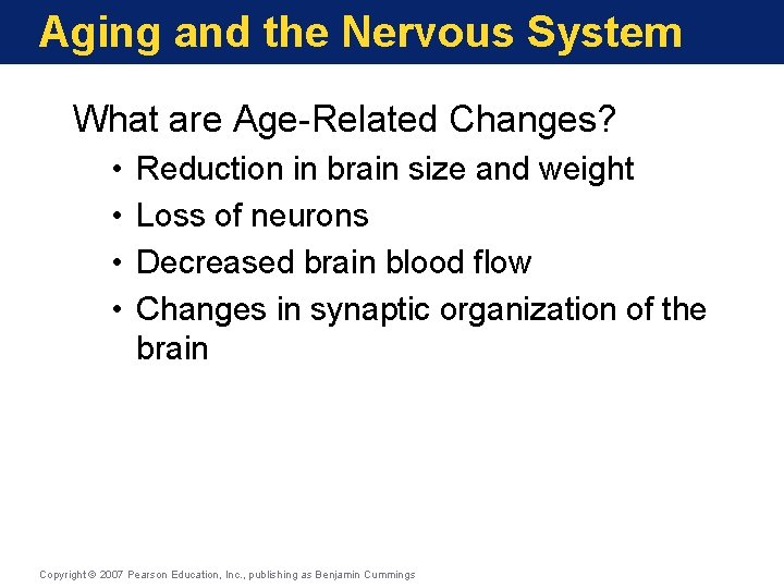
Aging and the Nervous System What are Age-Related Changes? • • Reduction in brain size and weight Loss of neurons Decreased brain blood flow Changes in synaptic organization of the brain Copyright © 2007 Pearson Education, Inc. , publishing as Benjamin Cummings