Esophageal Disorders Dr Nadia zawi Department of Gastroenterology

- Slides: 42
Esophageal Disorders Dr. Nadia zawi Department of Gastroenterology BMC
Objectives • To understand the anatomy and the physiology of the oesophagus and their relation ship to disease. • The clinical features , investigations , and treatment of different types of diseases.
• Oesophageal diseases due to : - • • Motility. Reflux. Infectious. Neoplastic. • Oesophageal varices ( liver cirrhosis).
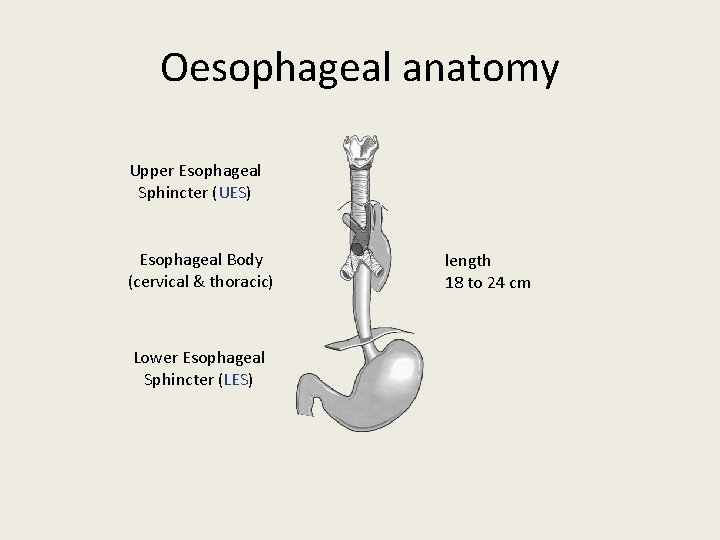
Oesophageal anatomy Upper Esophageal Sphincter (UES) Esophageal Body (cervical & thoracic) Lower Esophageal Sphincter (LES) length 18 to 24 cm
physiology • The upper esophageal sphincter (UES) is tonically closed and contracts during inspiration, preventing air from entering into the gastrointestinal tract, and reflux. It relaxes during swallow, belching and vomiting. • The lower esophageal sphincter (LES) maintains a steady baseline tone (~20 mm Hg) to prevent gastric contents from entering the esophagus. The LES also contracts during periods of increased intra-abdominal pressure, preventing reflux due to the pressure build up in the abdomen
• . The esophagus has 2 muscle layers: the inner circular layer and the outer longitudinal layer. • The longitudinal muscle shortens the esophagus, while the circular muscle forms lumen-occluding ring contractions. The combination of these localized contractions is responsible for peristalsis. • The proximal esophagus contains striated muscle. • the distal esophagus smooth muscle.
I - Motility Disorders (classification) • upper esophageal – UES disorders – neuromuscular disorders • esophageal body – achalasia – diffuse esophageal spasm – nutcracker esophagus – nonspecific esophageal dysmotility • LES – achalasia – hypertensive LES • primary disorders – achalasia – diffuse esophageal spasm – nutcracker esophagus – nonspecific esophageal dysmotility • secondary disorders – severe esophagitis – scleroderma – diabetes – Parkinson’s – stroke
Achalasia • idiopathic disorder of muscle of oesophagus due to loss of ganglionic cells in the mesenteric plexus. • loss of peristalsis. • increased resting tone of lower sphincter. • loss of normal relaxation with swallowing. • muscular spasm.
epidemiology *1 -2 per 200, 000 population *usually presents between ages 25 to 60 *male=female *Caucasians > others *average symptom duration at diagnosis: 2 -5 years
Clinical Presentation • clinical presentation – solid dysphagia 90 -100% (75% also with dysphagia to liquids) – post-prandial regurgitation 60 -90% – chest pain 33 -50% – weight loss – nocturnal cough and recurrent aspiration
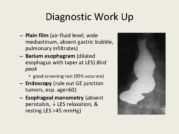
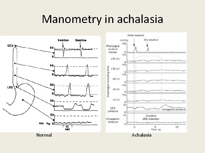
Diagnostic Work Up – Plain film (air-fluid level, wide mediastinum, absent gastric bubble, pulmonary infiltrates) – Barium esophagram (dilated esophagus with taper at LES) Bird peak • good screening test (95% accurate) – Endoscopy (rule out GE junction tumors, esp. age>60) – Esophageal manometry (absent peristalsis, LES relaxation, & resting LES >45 mm. Hg)
Manometry in achalasia Normal Achalasia
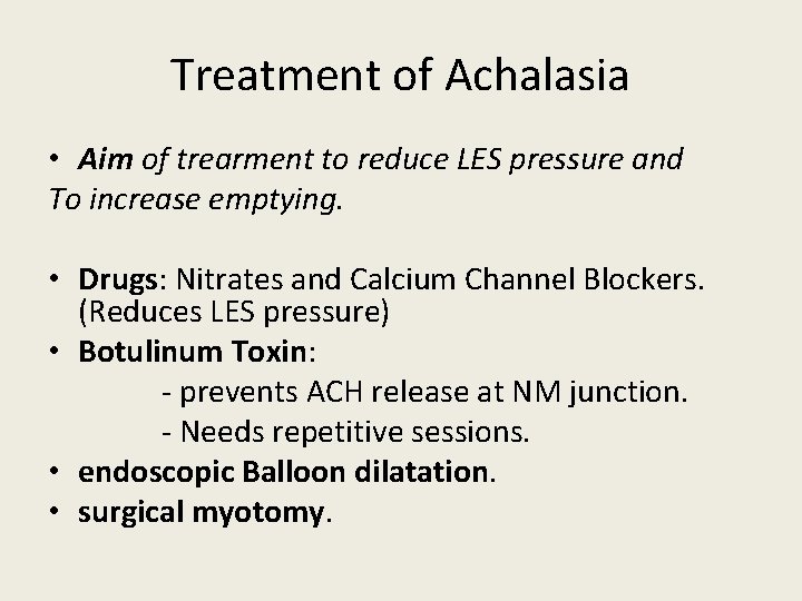
Treatment of Achalasia • Aim of trearment to reduce LES pressure and To increase emptying. • Drugs: Nitrates and Calcium Channel Blockers. (Reduces LES pressure) • Botulinum Toxin: - prevents ACH release at NM junction. - Needs repetitive sessions. • endoscopic Balloon dilatation. • surgical myotomy.
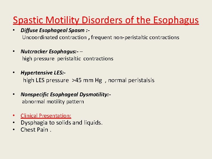
Spastic Motility Disorders of the Esophagus • Diffuse Esophageal Spasm : Uncoordinated contraction , frequent non-peristaltic contractions • Nutcracker Esophagus: - – high pressure peristaltic contractions • Hypertensive LES: - high LES pressure >45 mm Hg , normal peristalsis • Nonspecific Esophageal Dysmotility: abnormal motility pattern • Clinical Presentation: • Dysphagia to solids and liquids. • Chest Pain.
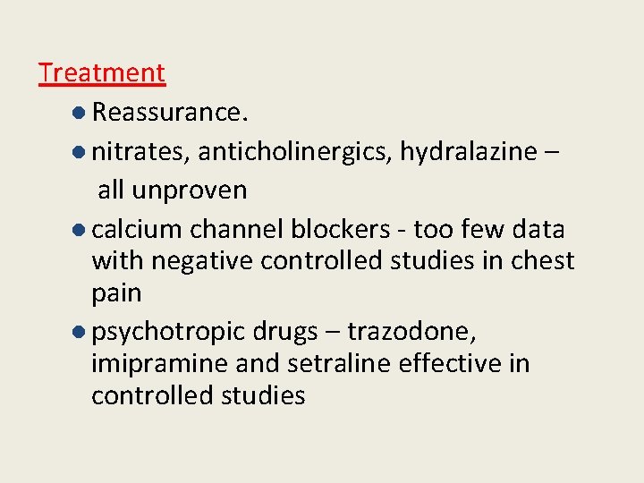
Treatment l Reassurance. l nitrates, anticholinergics, hydralazine – all unproven l calcium channel blockers - too few data with negative controlled studies in chest pain l psychotropic drugs – trazodone, imipramine and setraline effective in controlled studies
II- Disorders due to reflux GERD • Abnormal retrograde movement of stomach contents to oesophagus(Hydrochloric acid, pepsin)
• Normally, reflux prevented by: -Lower oesophageal sphincter -Anatomic structure (acute angle with stomach, RT crus of diaphragm) -Oesophageal peristaltic clearance -Swallowed saliva -Gravity • Reflux more likely to occur when: -Decreased tone of sphincter -Sliding hiatal hernia -Decreased oesophageal clearance -Decreased saliva production -When lying down
Symptoms: • heartburn, • acid regurgitation, • water brash, • dysphagia, • A typical symptoms -asthma. - chest pain. - laryngitis. - cough.
• Diagnosis: Diagnosis of GERD remains doutful. – 50 -60% of patients have heartburn or acid regurgitation symptoms. PH study is used : - if the diagnosis of GERD is in doubt - to determine whether the chest pain or other atypical symptoms correlate with acidreflux events. - esophageal p. H studies: 24 hours monitoring of PH: reduction in ph less than 4.
endoscopy: very low incidence of endoscopic findings. microscopic evidence of oesophagitis indications of endoscopy: *Alarm symptoms *Empiric therapy failure *Preoperative evaluation(HH). *Detection of Barrett’s esophagus “PPI Test” may be best and most cost-effective approach.
• Treatment: • Lifestyle modifications – avoid coffee, fatty foods, smoking; lose weight, raise head of bed, eliminate late night meals. • Acid suppressin via PPI’s. • Surgery (Laparoscopic Nissen Fundoplication).
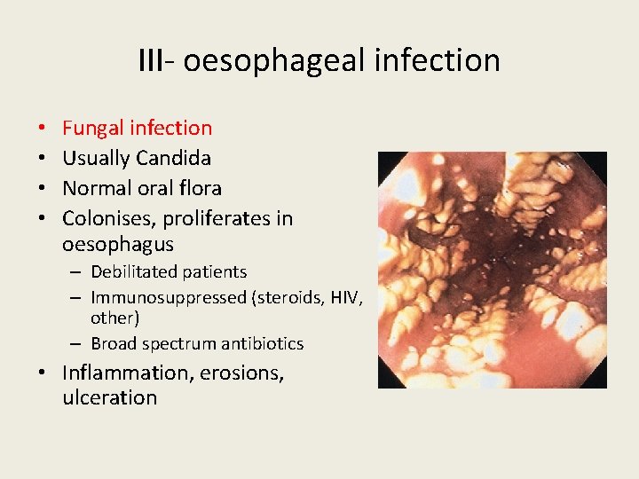
III- oesophageal infection • • Fungal infection Usually Candida Normal oral flora Colonises, proliferates in oesophagus – Debilitated patients – Immunosuppressed (steroids, HIV, other) – Broad spectrum antibiotics • Inflammation, erosions, ulceration
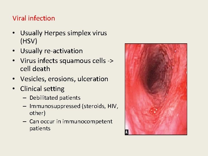
Viral infection • Usually Herpes simplex virus (HSV) • Usually re-activation • Virus infects squamous cells -> cell death • Vesicles, erosions, ulceration • Clinical setting – Debilitated patients – Immunosuppressed (steroids, HIV, other) – Can occur in immunocompetent patients
IV- oesophageal tumours • Malignant > common than benign. • Unfortunately, esophageal cancer discovered late & overall 5 y ear prognosis is bad < 10. • Even for potentally resectable cancer eso, 5 y survival is < 30%
Beniegn tumours • The gastrointestinal stromal tumour (GIST, another name for leimymoma), usually asymptomatic but may cause bleeding or dysphagia • Uncommon, include: • fibrovascular polyps, leiomyomas, papillomas, lipomas, neurofibromas, granular cell tumors. • When large, can cause dysphagia or chest pain from obstruction or stretch. • Usually discovered incidentally. • Found most often in distal third of esophagus. • Usually solitary, but may be multiple (3%).
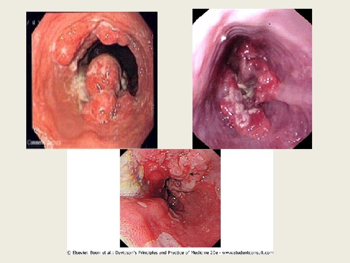
Carcinoma esophagus • common in men than women 3 -4: 1 • Should be considered in the cases that presenting with dysphagia. • Histopathological types: • adenocarcinoma : the lower third of the oesophagus from Barrett's oesophagus or from the cardia of the stomach. • squamous cancers: (Upper oesophagus) • Small-cell cancer is a rare : (any part of the oesophagus.
• Risk factors: • • • Alcohol. Tobacco smoking. SCC of the head & neck. Achalasia. Papilloma virus infection. Plummer-Vinson syndrome Tylosis (familial hyperkeratosis of palms & soles). Celiac disease. Radiation exposure. Post-cricoid web
• SYMPTOMS • is progressive dysphagia over a several-month period until only liquids can be taken. • Unexplained persistent chest pain • weight loss • Hematemesis • Coughing after drinking fluid • Hoarseness from involvement of the recurrent laryngeal nerve by tumor are unusual symptoms.
• Signs: • • Paller. clubbing ( benign & malignant tumors). Vricho’s node in left supracalvicular region. Early diagnosis affords the only chance for cure.
Diagnosis: • upper GI endoscopy with cytology & biopsy. • A barium swallow demonstrates the site& length of the stricture but adds little useful information. • Thoracic & abdominal CT to stage the tumour& define operability. • most sensitive modality is EUS(endoscopic USS) to define the TNM stage. • Blood: • 1 -Complete blood picture: iron deficiency anemia. • 2 - Occult blood in stool • 3 - Tumor markers: CEA - CA 15 -3
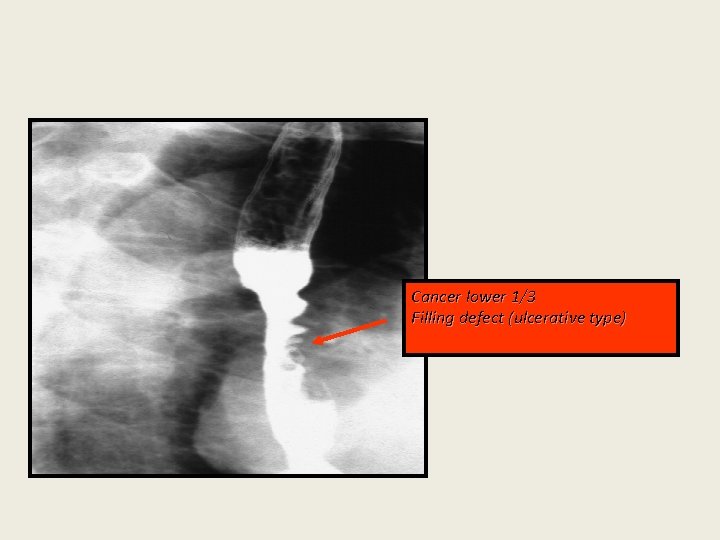
Cancer lower 1/3 Filling defect (ulcerative type)
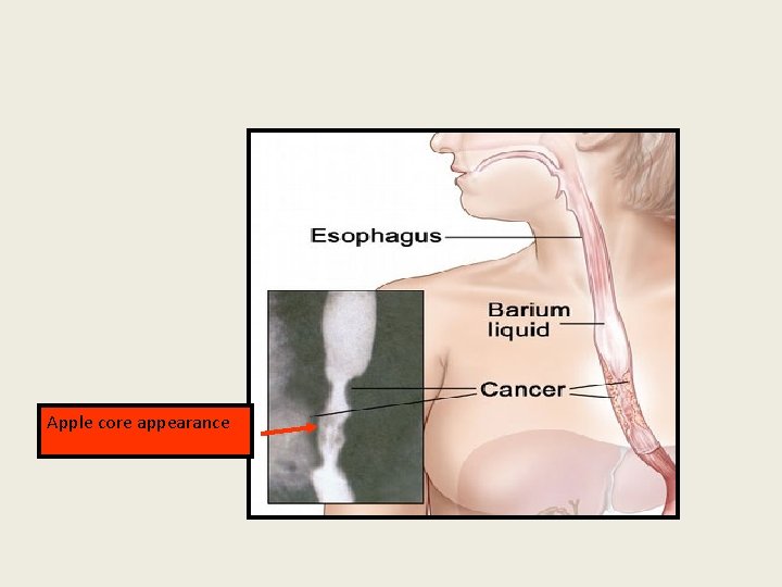
Apple core appearance
• Treatment I- operable: surgery followed by chemotherapy. II- inoperable: palliative procedure. Critera for inoperability: • Unfit patient. • Distant metastasis. • Unresectable tumor. • Infiltration of important structures such as aorta, trachea. Despite modern treatment, the overall 5 -year survival of oesophageal cancer is 6 -9%.
• Although SCC are radiosensitive, radiotherapy alone is associated with a 5 -year survival of only 5%. • 70% have extensive disease at presentation; in these, treatment is palliative & based upon relief of dysphagia &pain. • Endoscopically directed tumour ablation using laser therapy or insertion of stents is the major method of improving swallowing. • Palliative radiotherapy • may induce shrinkage of both SCC/ adeno carcinomas but symptomatic response may be slow. • Quality of life can be improved by nutritional support /appropriate analgesia.
Summary • Oesophageal disorders ranges from motility disorders to malignant tumours. • Dysphagia is the most commom presenting symptoms. • Oesophageal manometry helps in differentiating different types of motility disorder • Early detection of tumours improve the outcome.
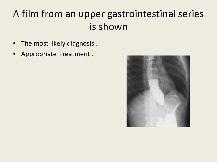
Case 1 • A 45 -year-old man has a 2 -year history of dysphagia that was initially induced only by solid foods but has progressed over the past year to include both solids and liquids. He also describes intermittent chest pain, Retrosternal burning with occasional regurgitation of undigested food, and a 4. 5 -kg weight loss over the past 2 years.
• Physical examination is normal. Upper endoscopy shows a small amount of retained food in the distal esophagus but is otherwise normal. An esophageal motility study demonstrates a hypertensive lower esophageal sphincter that does not relax fully with wet swallows.
A film from an upper gastrointestinal series is shown • The most likely diagnosis. • Appropriate treatment.
Case 2 • A 67 -year-old woman has an 8 - to 10 -month history of intermittent dysphagia for both solid foods and liquids. The dysphagia initially developed over the course of 10 to 14 days. She reports no abdominal symptoms but has lost 13. 5 kg during this time. • An upper gastrointestinal barium radiograph shows a smooth narrowing in the distal esophagus. An esophageal motility study is consistent with achalasia.
• The most likely diagnosis. • the most appropriate next step in managing this patient?