DYSPHAGIA Dr S Jaya Sandeep INTRODUCTION Dysphagia refers


: For direct examination of esophageal mucosa and biopsy. � Associated")


- Slides: 22
DYSPHAGIA Dr S Jaya Sandeep
INTRODUCTION Dysphagia refers to difficulty in swallowing which results from interference or obstruction to the food passage. It can be divided into two types: Oropharyngeal (difficulty in transferring food bolus from oropharynx to upper esophagus) Esophageal (difficulty in transporting bolus through the body of esophagus).
ODYNOPHAGIA : painful swallow. is more marked in ulcerative and inflammatory lesions of food passages-oral cavity, oropharynx and oesophagus. Patient with dysphagia may complaint of : Foreign body sensation Throat discomfort Feel of hold up Absolute difficulty in swallowing
STAGES OF DEGLUTITION Mastication Oral phase Pharynge al phase Salivation Movement of tongue, soft palate Closure of oral, nasopharyngeal isthmus and larynx. Opening of cricopharynx Esophage al phase Involuntary propulsion of bolus
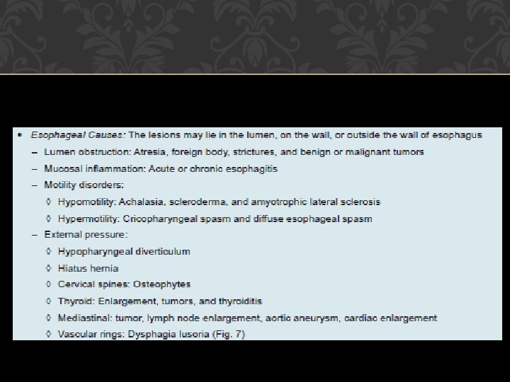
CAUSES
EVALUATION History �Sudden onset: Foreign body or impaction of food on a stricture or malignancy, and neurological lesions. Gradually progressive: Malignancy (short history with weight loss) and peptic strictures (long history with heartburn). Chronic, continuous and not progressive: Stricture. �Intermittent : Spasms or spasmodic episodes over an organic lesion. �Liquids/solid s: More with liquids (paralytic lesions), more with solids, and progressing to liquids (malignancy). �
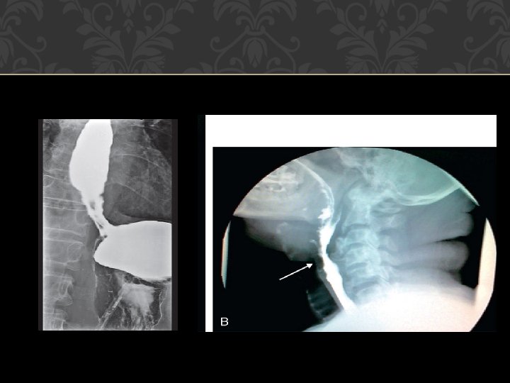
Intolerance to acid food and fruit juices: Ulcerative lesions. Associated complaints: Regurgitation and heartburn: Hiatus hernia. Regurgitation of undigested food in lying down and coughing in night: Hypopharyngeal diverticulum. Nasal regurgitation: Palatal paralysis. Aspiration into lungs: Laryngeal paralysis.
Physical Examination The physical examination should include oral cavity, oropharynx, hypopharynx, larynx, neck, chest and nervous system including cranial nerves. The lesions of oral cavity and pharynx can be seen during physical examination whereas esophageal causes require investigations such as barium swallow and esophagoscopy.
Investigations Upper endoscopy, barium swallow, manometry and p. H recording are the mainstay of dysphagia evaluation. � Hemogram: Iron deficiency anemia in Plummer-Vinson syndrome. � X-ray chest: For advanced esophageal lesions, cardiovascular, pulmonary and mediastinal diseases or pneumomediastinum
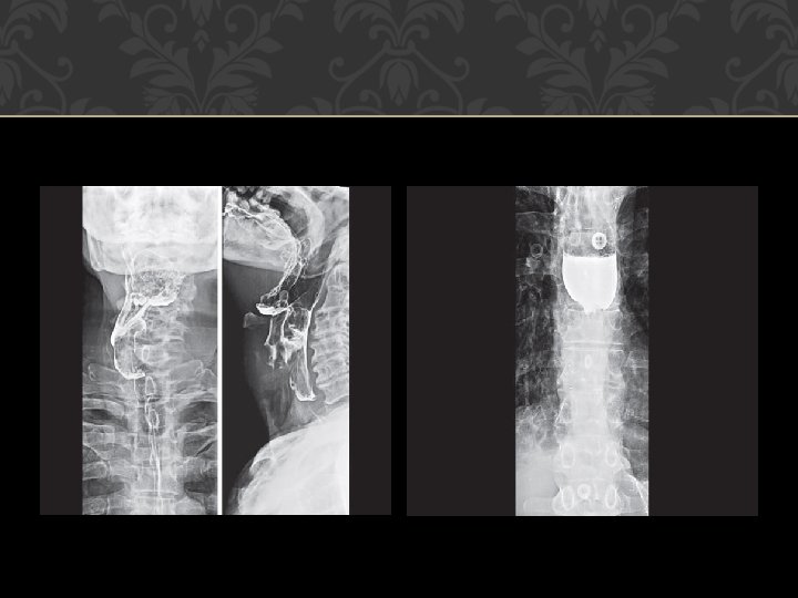
X-ray neck lateral and frontal projections: For cervical osteophytes, lesions of post-cricoid or retropharyngeal region. a. Useful in children. b. Shows radiopaque foreign bodies. c. Saying “e” during exposure brings the tongue anterior, and shows oropharynx better. d. Blowing through closed mouth distends hypopharynx.
Computed radiography and digital radiography: In this filmless technique, image is captured on an array of digital elements, and is read directly into a computer. Advantages: Soft tissue differences: It can emphasize subtle soft tissue differences even if the image is suboptimally exposed. Invert image: Radiodense elements appear whiter than radiolucent elements on traditional films but computer technology can invert the image and some aspects of anatomy, and pathology can be visualized better.
Barium swallow: For malignancy, cardiac achalasia, strictures, diverticulum, hiatus hernia or esophageal spasms. � Cineradiography: For motility disorders of esophageal wall or sphincters. �Ultrasound : Transesophageal echosonography can evaluate the depth of malignant ulcer, which helps in staging the disease.
Manometry and p. H studies: For motility disorders, gastroesophageal reflux and esophageal spasm (spontaneous or acid induced). A pressure transducer along with a p. H electrode and an open-tipped catheter are introduced into the esophagus. They measure the pressures of esophageal wall and sphincters. Gastroesophageal reflux disease is measured by p. H electrode, which also measures the effectiveness of esophagus to clear the acid load.
Esophagoscopy (flexible or rigid): For direct examination of esophageal mucosa and biopsy. � Associated investigations: They vary from case to case such as bronchoscopy for bronchial carcinoma, cardiac catheterization for vascular anomalies, and thyroid scan for malignant thyroid.
Oral Candidiasis FB esophagus
Schatzki’s ring Esophageal web
Parapharyngeal mass Achalasia cardia
NORMAL BARIUM SWALLOW
THANK YOU