Dura mater and dural venous sinuses The Meninges


 Dura mater Endosteal layer Meningeal layer They are")


 for")


 is a large venous space")


- Slides: 39
Dura mater and dural venous sinuses
The Meninges
Cranial Meninges - 3 layer protective membrane 1. Dura Mater - Composed of two layers: a) Periosteal – outer layer, attaches to bone. b) Meningeal – inner layer, closer to brain. Two layers fused, except to enclose the dural sinuses 2. Arachnoid Layer - ‘spider’ web like. 3. Pia Mater - delicate, follows convolutions.
Superior sagittal sinus (Dural venous sinus) Dura mater Endosteal layer Meningeal layer They are closely united except along certain lines; they are separated to form venous sinuses Subdural space Coronal section of the upper part of the head
Folds of the Dura Mater 1. Falx cerebri. 2. Tentorium cerebelli. 3. Falx cerebelli. 4. Diaphragma sellae.
Falx cerebri 1. large sickle-shaped fold of dura mater occupying median longitudinal fissure between two cerebral hemispheres. 2. narrow anterior end is attached to crista galli and broad posterior end on to upper surface of tentorium cerebelli along the median plane. 3. Its convex upper margin is attached to lips of sagittal sulcus of skull vault and its lower concave margin is free and lies just above corpus callosum.
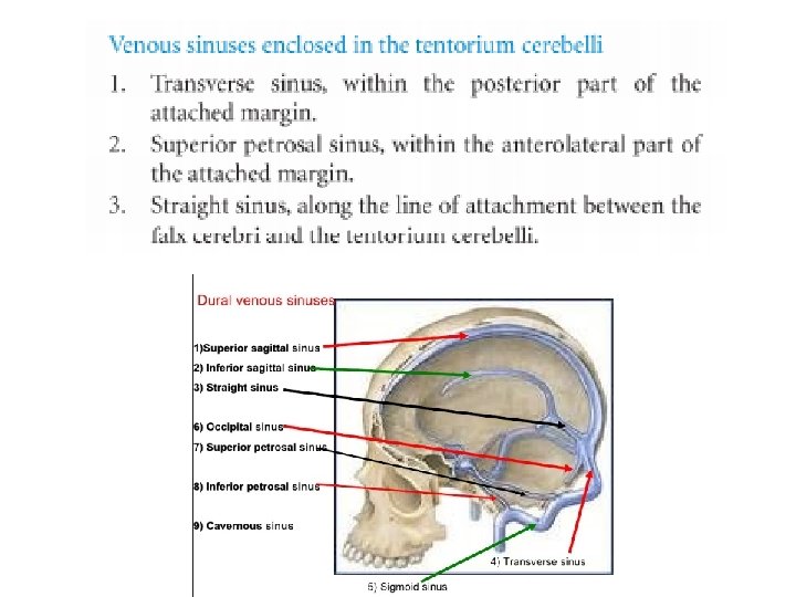
Venous sinuses enclosed in the falx cerebri 1. Superior sagittal sinus is enclosed within convex upper border. 2. Inferior sagittal sinus is enclosed within lower concave margin. 3. Straight sinus lies along line of attachment of falx cerebri with tentorium cerebelli
Tentorium cerebelli It is a tent-shaped fold of the dura mater forming roof of posterior cranial fossa. It separates the cerebellum from occipital lobes of the cerebrum. It has two margins and two surfaces.
Margins 1. The inner free margin is Ushaped and encloses tentorial notch (incisure) for the passage of midbrain. The anterior ends of the concave free margin are attached toanterior clinoid processes. 2. The outer attached margin is convex and attached on each side (from before backward) to the posterior clinoid process, the posteroinferior angle of the parietal bone, and the lips of transverse sulci on the occipital bone.
Falx cerebelli • Small sickle shaped fold in sagittal plane projecting forward in posterior cerebellar notch. • Extends from internal occipital protuberance toposterior margin of foramen magnum. • Has free concave anterior margin and convex attached posterior border. Venous sinus enclosed in the falx cerebri: Occipital sinus, along with its posterior attached part
Diaphragma sellae a small, circular, horizontal sheet of dura mater. forms a roof oversella turcica and covers the pituitary gland. The infundibulum (also known as pituitary stalk) passes into pituitary fossa through a central opening in diaphragma.
Dural Nerve Supply • Branches of the trigeminal, vagus, and first three cervical nerves and branches from the sympathetic system pass to the dura. • The dura is sensitive to stretching, which produces the sensation of headache.
1. In the anterior cranial fossa: by the anterior and posterior ethmoidal nerves (and receives some twigs from the maxillary nerve). 2. In the middle cranial fossa: by the meningeal branch of the maxillary nerve (in the anterior part) and the meningeal branch of the mandibular nerve (nervus spinosus) in the posterior part. 3. In the posterior cranial fossa: by the meningeal branches of the vagus and hypoglossal nerves. These are the C 1 and C 2 fibres carried by the cranial nerves. The dura mater around the foramen magnum is directly supplied by the C 2 and C 3 cervical nerves.
• Blood Supply of the Dura Mater Inner layer: The inner layer of the dura mater is more fibrous and requires very little blood to nourish it. Outer layer: The outer layer of the dura mater is richly vascular and provides nourishment to the adjacent bone. In the supratentorial compartment, it is supplied by the following arteries: 1. In the anterior cranial fossa: by the meningeal branches of the ophthalmic, anterior, and posterior ethmoidal arteries, and a branch of the middle meningeal artery. 2. In the middle cranial fossa: by the middle and accessory meningeal arteries and by the meningeal branches of the internal carotid and ascending pharyngeal arteries. 3. In the posterior cranial fossa: by the meningeal branches of the vertebral and occipital arteries
Superior cistern Chiasmatic cistern Interpeduncular cistern Pontine cistern Cerebellomedullary cistern Median sagittal section to show the subarachnoid cisterns & circulation of CSF
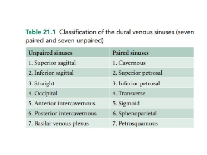
Cranial venous sinuses • The dural venous sinuses (also called dural sinuses, cerebral sinuses, or cranial sinuses) are venous channels found between layers of dura mater in brain. • They receive blood from internal and external veins of brain, receive cerebrospinal fluid (CSF) from the subarachnoid space, and ultimately empty into internal jugular vein.
Features 1. Lie between the layers of the dura mater. 2. Have no muscle in their walls. 3. Lined by endothelium only (muscular coat is absent). 4. Are devoid of values in their lumen. 5. Receive venous blood and CSF. 6. Receive valveless emissary veins which regulate blood flow and maintain the equilibrium of venous pressure within and outside the skull.
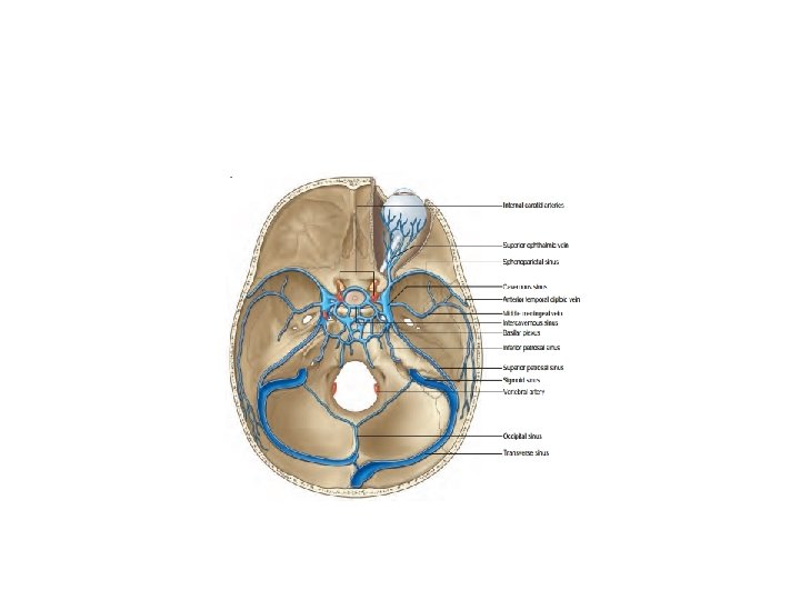
CAVERNOUS SINUS • (2 cm long, 1 cm wide) is a large venous space situated on either side of the body of the sphenoid and sella turcica in middle cranial fossa • The floor of the sinus is formed by the endosteal layer, while the lateral wall, roof, and medial wall by meningeal layer. •
• Medially, the roof is continuous with the diaphragm sellae. Posteriorly, the roof has a triangular depression between the attached margin (edge) of tentorium cerebelli to the posterior clinoid process and ridge raised by the free margin (edge) of tentorium cerebelli as it extends forward to gain attachment on the anterior clinoid process. • The oculomotor and trochlear nerves pierce this triangle to enter the cavernous sinus.
• Relations Superior 1. Optic chiasma. 2. Optic tract. 3. Internal carotid artery. 4. Anterior perforated substance. Inferior 1. Foramen lacerum. 2. Junction of body and greater wing of sphenoid Medial 1. Pituitary gland (hypophysis cerebri). 2. Sphenoid air sinus. Lateral 1. Temporal lobe (uncus) of the cerebral hemisphere. 2. Cavum trigeminale containing the trigeminal ganglion. Anterior 1. Superior orbital fissure. 2. Apex of the orbit. Posterior 1. Crus cerebri of midbrain. 2. Apex of petrous temporal bone
Structures Present in the Lateral Wall of the Sinus From above downward these are as follows: 1. Oculomotor nerve. 2. Trochlear nerve. 3. Ophthalmic nerve. 4. Maxillary nerve.
Structures Passing Through Cavernous Sinus 1. Internal carotid artery surrounded by sympathetic plexus of nerves. 2. Abducent nerve (it enters the sinus by passing below petrosphenoid ligament and accompaniesartery on its inferolateral aspect
Tributaries of the Cavernous Sinus The cavernous sinus receives blood from three sources: orbit, meninges, and brain. From orbit 1. Superior ophthalmic vein. 2. Inferior ophthalmic vein. 3. Central vein of retina (sometimes). From meninges 1. Sphenoparietal sinus. 2. Anterior (frontal) trunk of the middle meningeal vein. From brain 1. Superficial middle cerebral vein. 2. Inferior cerebral veins (only few)
Communications of Cavernous Sinus 1. Transverse sinus via superior petrosal sinus. 2. Internal jugular vein via inferior petrosal sinus. 3. Pterygoid venous plexus via emissary veins which pass through foramen ovale, foramen lacerum, and emissary sphenoidal foramen. 4. Facial vein via two routes: (a) Superior ophthalmic vein → angular vein → facial vein (b) Emissary veins → pterygoid venous plexus → deep facial vein → facial vein. 5. Opposite cavernous sinuses via anterior and posterior intercavernous sinuses. 6. Superior sagittal sinus via superficial middle cerebral vein and superior anastomotic vein. 7. Internal vertebral venous plexus, via basilar venous plexus.
Cavernous sinus thrombosis: • • • The septic thrombosis of cavernous sinus may be caused by its numerous communications. The commonest cause of thrombosis is the passage of septic emboli from the dangerous area of the face through facial vein—deep facial vein— pterygoid venous plexus—emissary vein. Cavernous sinus thrombosis gives rise to the following signs and symptoms: – Severe pain in the eye and forehead, due to involvement of the ophthalmic nerve. – Ophthalmoplegia (paralysis of ocular muscles) due to involvement of the 3 rd, 4 th, and 6 th cranial nerves. – Marked edema of eyelids with exophthalmos, due to congestion of orbital veins following obstruction of ophthalmic veins
Dural venous sinuses Superior sagittal sinus: Lies along superior margin of falx cerebri. receives in its coursesup. cerebral vein Tributaries 1. Superior cerebral veins. 2. Parietal emissary veins. 3. Small vein from the nasal cavity (if foramen cecum is patent). 4. Veins of the frontal air sinus.
Inferior sagittal sinus: Lies along inferior margin of falx cerebri. It ends by joining the great cerebral vein to form straight sinus. It drains the lower part of medial surface of each cerebral hemisphere.
Straight sinus: Lies at intersection of falx cerebri and tentorium cerebelli. continuation of the inferior sagittal sinus and terminates usually into left transverse sinus which at mastoid angle of the parietal bone becomes continuous with the left sigmoid sinus. Confluence of sinuses: Common confluence of superior sagittal sinus and straight sinus.
Venous sinuses
Transverse: Begins at confluence of sinuses. Extends along edges of tentorium cerebelli. Right receives blood from superior sagittal sinus. Left receives blood from straight sinus. receives sup. petrosal sinus, inf. cerebral n cerebellar veins n diploic veins
Sigmoid: Continuation of straight sinus. “S”-shaped. Ends at jugular foramen: Joins internal jugular vein. Its tributaries are: 1. Mastoid and condylar emissary veins. 2. Cerebellar veins. 3. Internal auditory vein.
Superior and inferior petrosal sinuses • Petrous part of temporal bone • Sup. petrosal sinus drains the cavernous sinus into transverse sinus • Inf. petrosal sinus drains the cavernous sinus into IJV
SUMMARY
The flowing of the blood in dural sinus Sup. sagittal sinus Inf. sagittal sinus Straight sinus Confluence of sinus Transverse sinus Sup. petrosal sinus Sigmoid sinus Cavernous sinus Inf. petrosal sinus Internal jugular vein
Name Drains to Inferior sagittal sinus Straight sinus Superior sagittal sinus Typically becomes right transverse sinus or confluence of sinuses Straight sinus Typically becomes left transverse sinus or confluence of sinuses Occipital sinus Confluence of sinuses Right and Left transverse sinuses Sphenoparietal sinuses Cavernous sinuses Superior and inferior petrosal sinuses Superior petrosal sinus Transverse sinuses Sigmoid sinus Inferior petrosal sinus Sigmoid sinuses Internal jugular vein