MENINGES OF THE CENTRAL NERVOUS SYSTEM OUTLINE MENINGES


 - membranes covering the brain and spinal cord.")


- Slides: 28
MENINGES OF THE CENTRAL NERVOUS SYSTEM
OUTLINE � MENINGES � VENOUS SINUSES
LEARNING OBJECTIVES � Understand the type of meninges � To have understanding of the dural sinuses
INTRODUCTION � Meninges (singular – meninx) - membranes covering the brain and spinal cord. � Meninges � Three – cranial and spinal meninges (3) layered membranes namely ○ Pia mater- Innermost ○ Arachnoid mater- Middle ○ Dura mater- Outermost Subarachnoid space subdural space N. B Cranial and spinal meninges are continuous at the foramen magnum.
CNS meninges
PIA MATER � This is a thin and delicate membrane � Adherent to the surface of the brain. � Transverse � It the grooves and fissures is also closely applied to the roots of the cranial nerves at their origin.
ARACHINOID MATER � Thin avascular layer � Spider like connected to pia � Enters only the longitudinal fissures. � From its inner surfaces arises thin trabeculae. � These extend down and cross the subarachnoid space to continue with the pia mater.
DURAL SPACES � Extradural space � Subarachinoid space /cistern
DURAL MATER � It is the thick, tough , outer covering of the brain. � it is made up of two layers � Outer periosteal layer which is attached to the skull � Inner meningeal layer – is in contact with arachinoid and continuous with the skull � The two layers are separated by the dural folds and sinuses.
Dural folds or Reflections or partitions. �Falx cerebri �Falx cerebelli �Tentorium cerebelli �Diaphragmatic sellae Do you Know other examples?
Falx cerebri � Separates the cerebral hemispheres Found within the longitudinal fissure. � Crescent shape or sickle � � Attachment � Anteroirly; crista galli of ethmoid and frontal crest of frontal bone. � � Tentorium cerebelli Found between and separates cerebellum and brain stem from the occipital lobes. � Attachment � � Posteriorly; occipital bone along the groove for the transverse sinuses � Laterally; superior border of petrous temporal � Anteriorly; anterior and posterior cliniod processes. � � Anterior /medial border are free This give passage to midbrian as tectorial notch. Posteriorly; tentorium cerebelli.
Falx cerebelli � It lies inferior to the tentorium cerebelli � � Sellae diaphragm � It is small and circular Situated posteriorly in the � Formed a roof over the posterior cranial fosse. pituitary fossa as a Attachment; covering on the gland. � It has opening for the Posteriorly; internal occipital crest infundibulum and � Superiorly; tentorium accompanying vessels cerebelli � Anterior ; free, separates the cerebellar hemispheres �
Arterial supplies � Anterior meningeal ; a branch of ethmoidal as in ant. Cranial fossa. � Middle and accessory- branch of maxillary in middle cranial fossa � Posterior meningeal aa-terminal branch of ascending pharyngeal artery in post. Cranial fossa. � Others – occipital and vertebral
Innervations � Floor and anterior part of falx cerebri; ethmiodal nn branch of ophthalmic nn. � Posterior part of falx cerebri and tentorium cerebelli- meningeal branch of ophthalmic nn � Middle cranial fossa – meningeal br of maxillary nn and mandibular nn � Posterior cranial fossa-meningeal br. Of 1 st , 2 nd and 3 rd cervical nerve
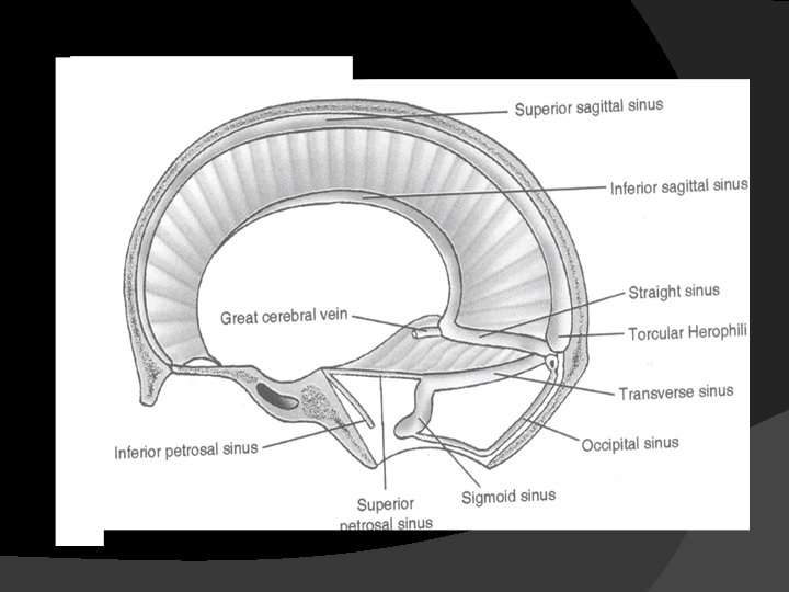
Dural or venous sinuses � Paired � Unpaired � Superior �Transverse �Carvanous �Greater petrosal �Lesser petrosal �Spenoparietal �Sigmiod �Basilar � Inferior sagital � Straight � Occipital � Intercavernous Do you Know other examples?
Sigmoid sinuses � Paired � It is continuous with the transverse sinus anteriorly. � It also formed the jugular bulb
Basilar sinus � Situated on the clivus
Superior sagittal sinus It occupies the longitudinal cerebral fissures. Begins: Ends: � Inferior sagittal sinus � Begins above the cribriform plate. � Ends in the straight sinus
� Transverse sinus Commence at internal occipital protuberance � Lies in the margin of tentorium cerebelli � Into sigmoid sinus � Empty into internal jugular vein �
Straight sinus or sinus rectus � Posterior end of falx cerebri � Middle of tentorium cerebelli � Recieves • Greater cerebral vein • Inferior sagittal vein • Empties into the confluence of sinuses
Cavernous sinus Content Lies on either sides of sphenoid bone � Joined by the intercavarnous sinuses Occulomotor nerve CN III � Troclear nerve CN IV Ophthalmic and Maxillary divisions of the Trigeminal nerve. Abducens nerve CN VI Internal carotid artery
Sphenoparietal sinus Drains into the cavernous sinus. � Receive tributaries from the superficial middle cerebral artery. � Posterior intercavarnous sinus It connects the left and right cavernous sinuses
� Superior petrosal sinus � Inferior petrosal sinus
SUMMARY NAME OF SINUS ORIGIN Superior sagittal sinus Foramen caecum where it Transverse sinus receives veins from the nasal cavity Junction of falx cerebri Transverse sinus and tentorium cerebelli Straight sinus Occipital sinus Confluence of sinuses Sphenoparietal sinuses foramen magnum by several small venous channels Internal occipital protruberance Small veins from sphenoid and parietal bones SINUS DRAIN TO Confluence of sinus Convergence of many sinuses Cavernous sinuses
SUMMARY Superior petrosal Cavernous sinus/sphenoid bone Transverse sinuses Internal occipital protruberance Inferior petrosal Inferior part of sinus cavernous sinus Cavernous sinuses Body of sphenoid bone Sigmoid sinuses Sigmoid sinus Superior Inferior sinuses and petrosal End of transverse Internal jugular vein sinus
Drainages of the sinuses
THE END THANK YOU ? QUESTIONS