
Divisions of the Nervous System Chapter 11 Meninges
- Slides: 55
Divisions of the Nervous System Chapter 11
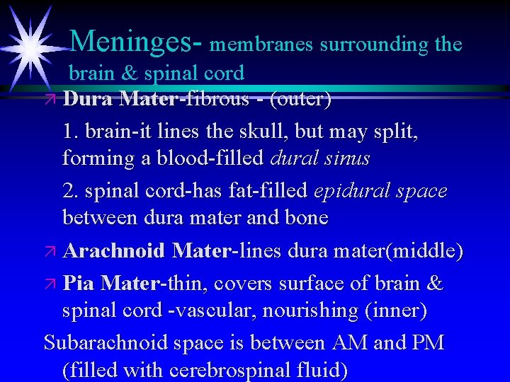
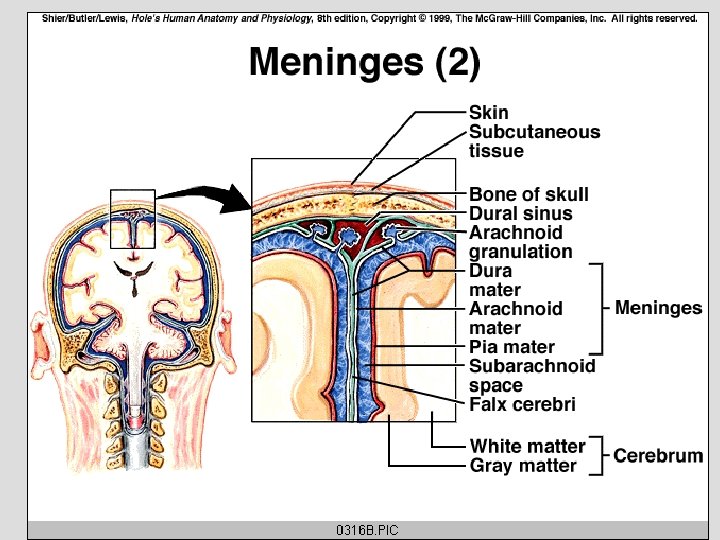
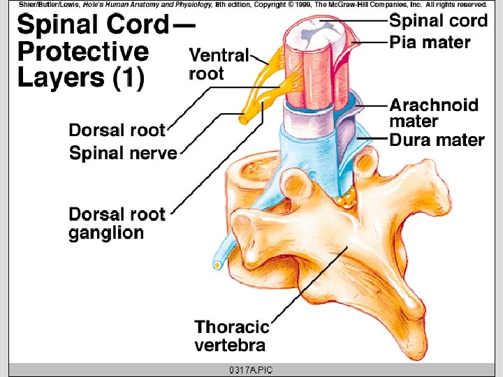
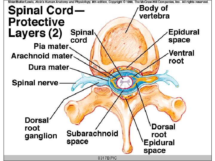
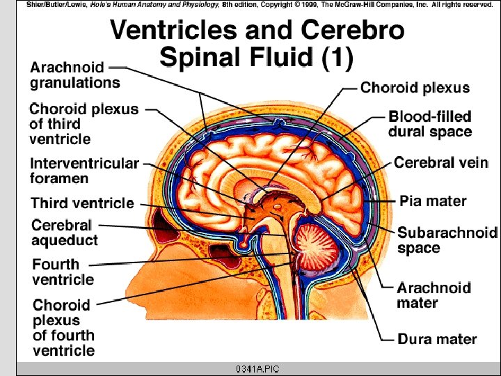
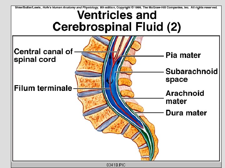
Meninges- membranes surrounding the brain & spinal cord ä Dura Mater-fibrous - (outer) 1. brain-it lines the skull, but may split, forming a blood-filled dural sinus 2. spinal cord-has fat-filled epidural space between dura mater and bone ä Arachnoid Mater-lines dura mater(middle) ä Pia Mater-thin, covers surface of brain & spinal cord -vascular, nourishing (inner) Subarachnoid space is between AM and PM (filled with cerebrospinal fluid)
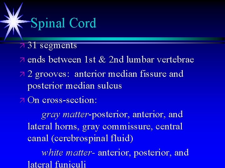
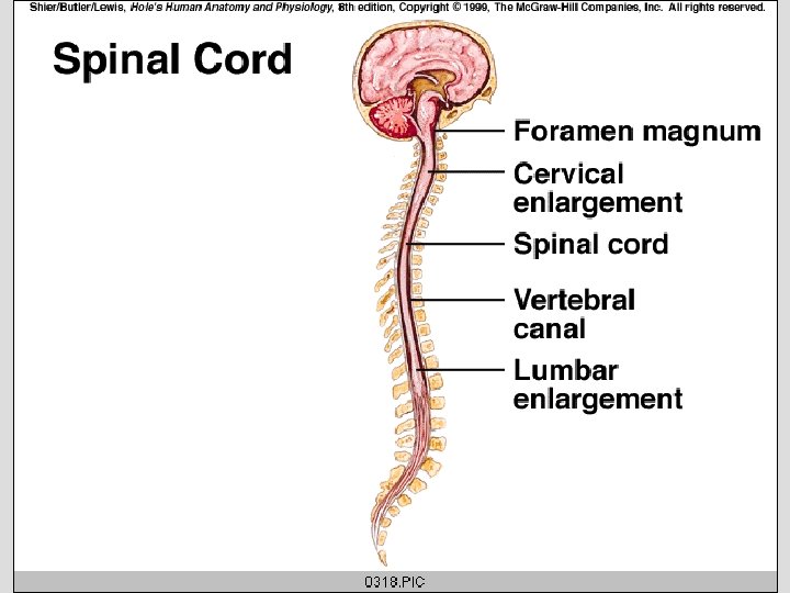
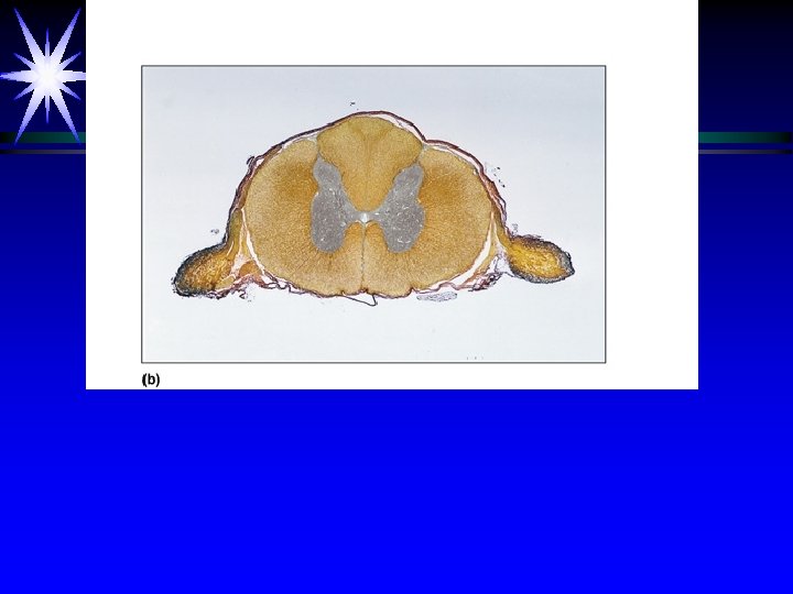
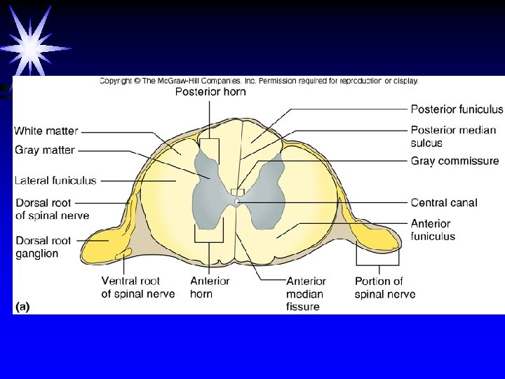
Spinal Cord ä 31 segments ä ends between 1 st & 2 nd lumbar vertebrae ä 2 grooves: anterior median fissure and posterior median sulcus ä On cross-section: gray matter-posterior, and lateral horns, gray commissure, central canal (cerebrospinal fluid) white matter- anterior, posterior, and lateral funiculi
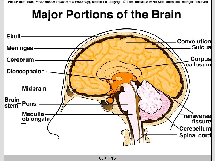
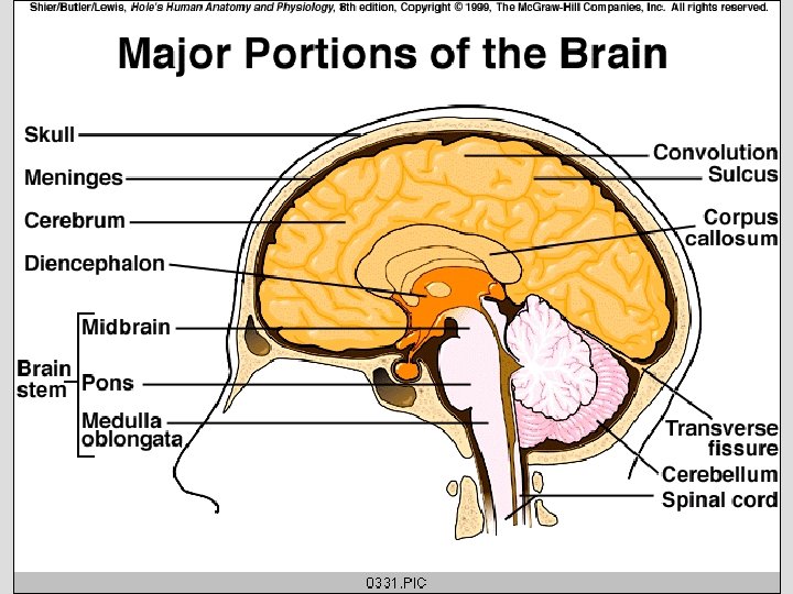
Brain Three Main Regions ä Cerebrum - largest ä Cerebellum ä Brainstem
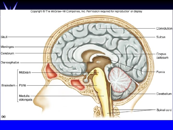
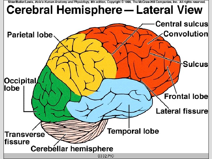
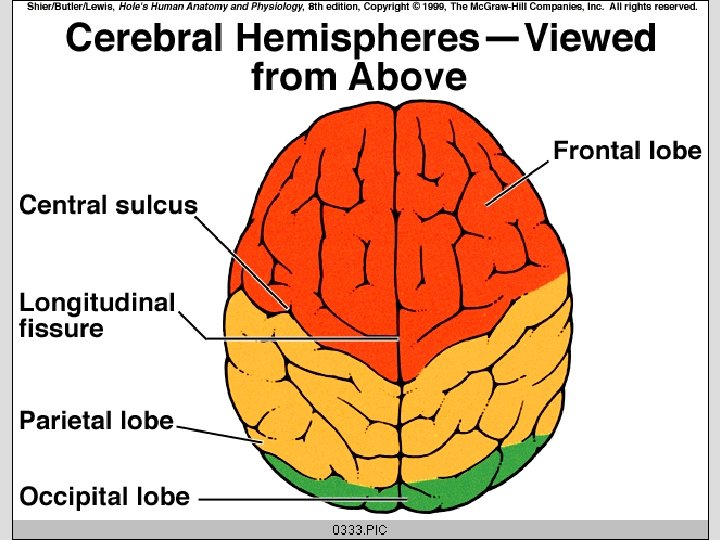
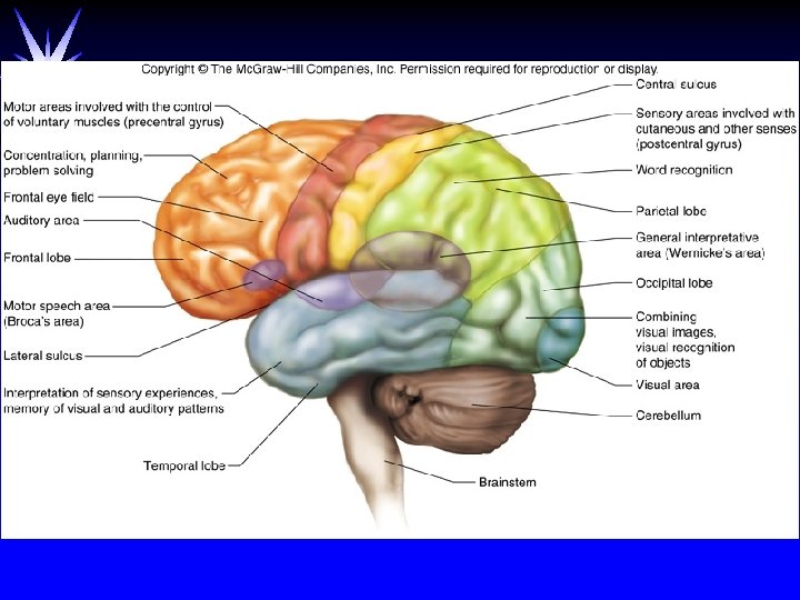
Cerebrum ä 2 cerebral hemispheres connected by band of white matter called the corpus callosum ä shallow grooves on surface are sulci(sulcus) ä deep grooves are fissures (longitudinal fissure, transverse fissure, lateral fissure) ä folds/convolutions between sulci are gyri(gyrus) ä Lobes: frontal, parietal, occipital, temporal ä Cerebral Cortex: outer, thin layer of gray matter
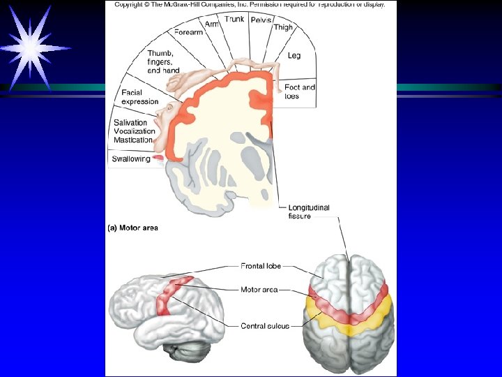
Figure 11. 15 a
Functions of Cerebrum
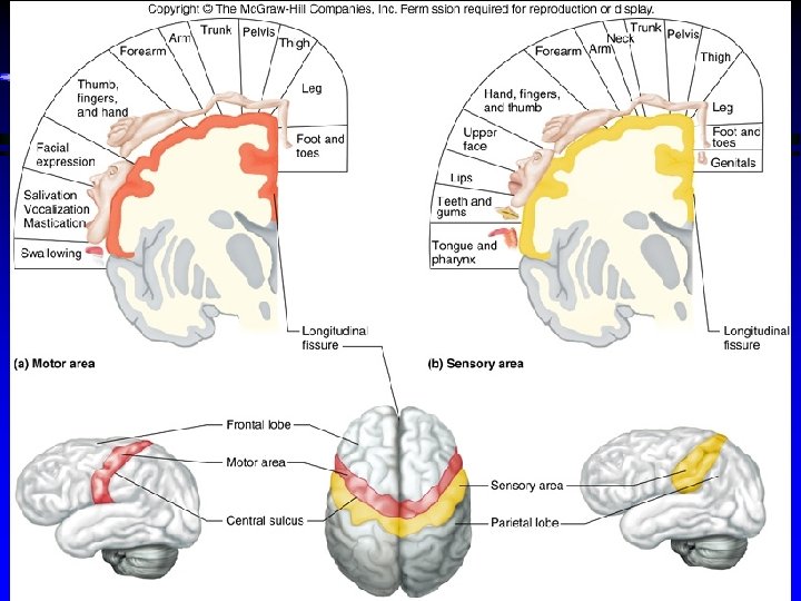
Functional Regions of Cerebral Cortex 1. Motor Regions a. Primary Motor Area-in precentral gyrus(directly in front of the central sulcus)controls voluntary muscle movements(left hemisphere controls right side, vice versa) b. Broca’s area-usually in left hemis. muscle actions associated with speech -regulates the patterns of breathing and vocalization needed for normal speech
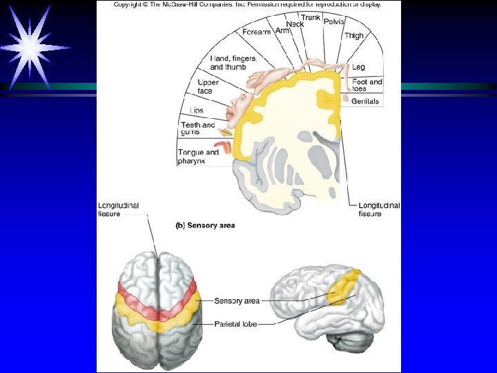
2. Sensory Regions a. Primary Sensory Area-in postcentral gyrus (behind the central sulcus) - receive somatic sensory info from receptors for touch, pressure, pain, vibration, taste, and temp. b. Hearing – responsible for monitoring sound; recognizes sounds such as spoken words
ä c. Visionä Recognizes images and puts them together ä Ex: when you see the symbols C, A, R this area of the brain recognizes that those are the letters that form the word car.
3. Association Areas-all lobes memory, reasoning, judgment, emotional expressions a. General Interpretive Area (Wernicke area)-word recognition and expression (written interpretation) - Plays an imp. Role in your personality
Figure 11. 17
http: //www. youtube. com/watch? v=HVGlfc. P 3 ATI http: //www. youtube. com/watch? v=yd 46 Hs 7 p. Tow&feature=relate d
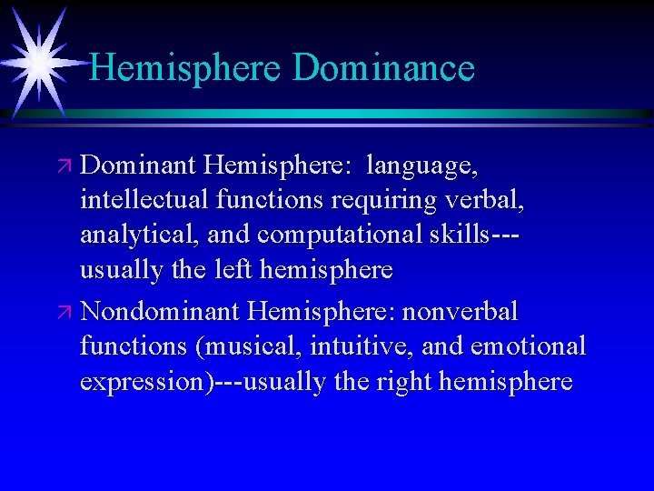
Hemisphere Dominance ä Dominant Hemisphere: language, intellectual functions requiring verbal, analytical, and computational skills--usually the left hemisphere ä Nondominant Hemisphere: nonverbal functions (musical, intuitive, and emotional expression)---usually the right hemisphere
Memory ä short-term: few seconds--as long as impulse travels ä recent: minutes or days--lost if not consciously recalled ä long-term: results from repeated recall or experience---promotes facilitation of neurons
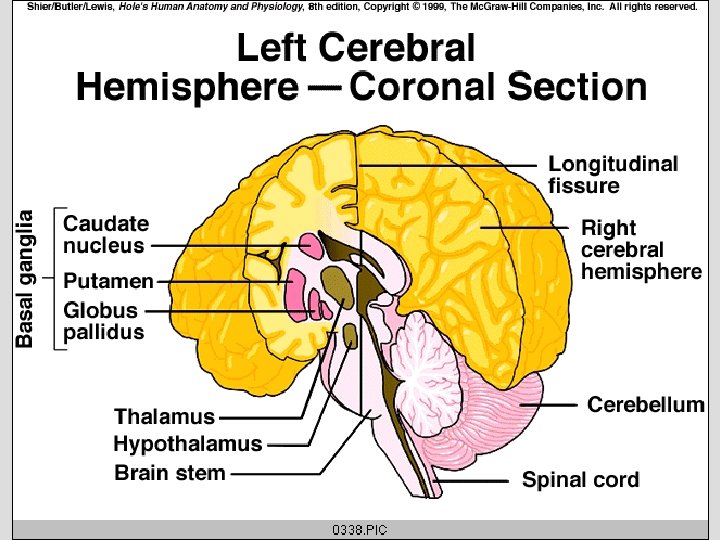
Basal Ganglia/Basal Nuclei ä Basal ganglia/nuclei are masses of gray matter deep within each cerebral hemisphere ä They serve as a relay for motor impulses from cortex and as a source of inhibitory neurotransmitters such as dopamine (lack of dopamine causes Parkinson’s disease) ä Inhibit muscular activities
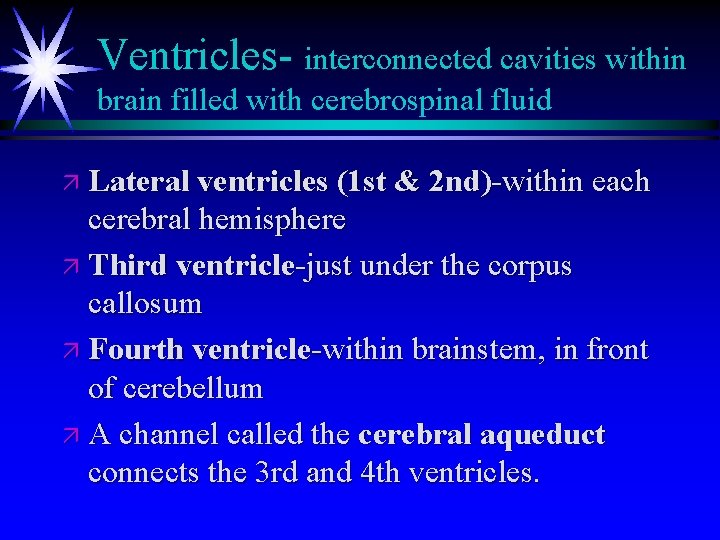
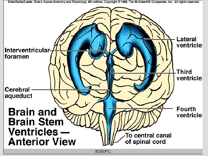
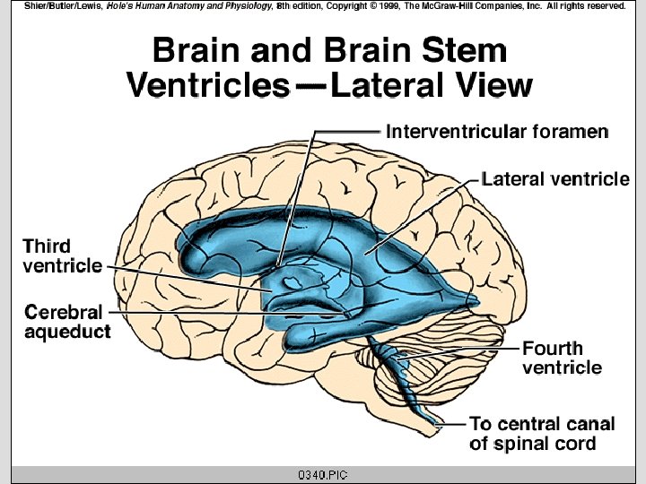
Ventricles- interconnected cavities within brain filled with cerebrospinal fluid ä Lateral ventricles (1 st & 2 nd)-within each cerebral hemisphere ä Third ventricle-just under the corpus callosum ä Fourth ventricle-within brainstem, in front of cerebellum ä A channel called the cerebral aqueduct connects the 3 rd and 4 th ventricles.
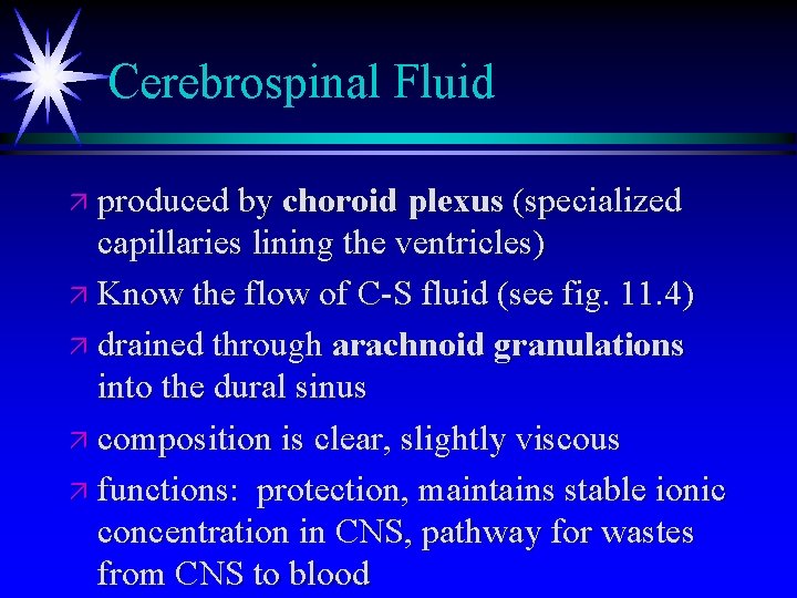
Cerebrospinal Fluid ä produced by choroid plexus (specialized capillaries lining the ventricles) ä Know the flow of C-S fluid (see fig. 11. 4) ä drained through arachnoid granulations into the dural sinus ä composition is clear, slightly viscous ä functions: protection, maintains stable ionic concentration in CNS, pathway for wastes from CNS to blood
Diencephalon- surrounds the 3 rd ventricle ä Thalamus-sides of dienceph. -relay station for incoming sensory impulses from other parts of the nervous system--general awareness of pain, touch, temp. , etc. ä Hypothalamus-anterior floor of dienceph. connects to pituitary; controls pituitary; body’s thermostat ä Mammillary bodies (2)-olfaction and swallowing reflexes ä Pineal gland-posterior projection
ä Limbic System-all structures in general region of diencephalon-Because they are connected to the cortex, they help in production of emotional feelings (fear, anger, pleasure, etc. )
Brainstem ä Midbrain-surrounds cerebral aqueduct; contains all descending voluntary motor tracts; center for visual and auditory reflexes; postural reflexes ä Pons-bulge on anterior surface of brainstem ä Medulla Oblongata-connects to spinal cord --contains all ascending and descending tracts---contains many vital control centers: cardiac, vasomotor, respiratory, vomiting, coughing, etc.
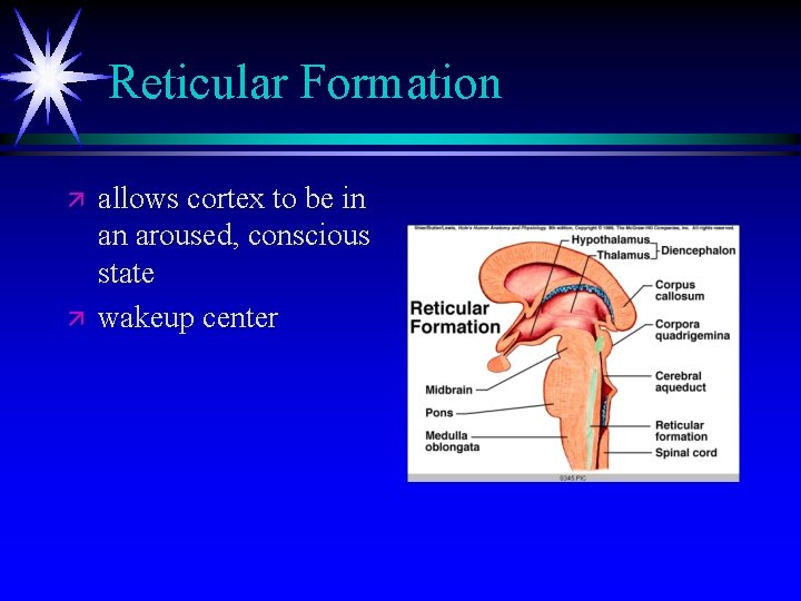
Reticular Formation ä ä allows cortex to be in an aroused, conscious state wakeup center
Cerebellum ä 2 hemispheres ä gray matter surrounds the white matter ä white matter is called the arbor vitae (tree of life) ä functions: 1. control of coordination of skeletal muscles 2. equilibrium (receives impulses from semicircular canals of inner ears)
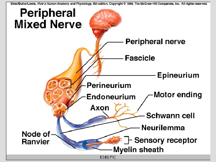
Peripheral Nervous System Each nerve is made of bundles of neuron fibers bound by C. T. An endoneurium surround each individual fiber within the nerve.
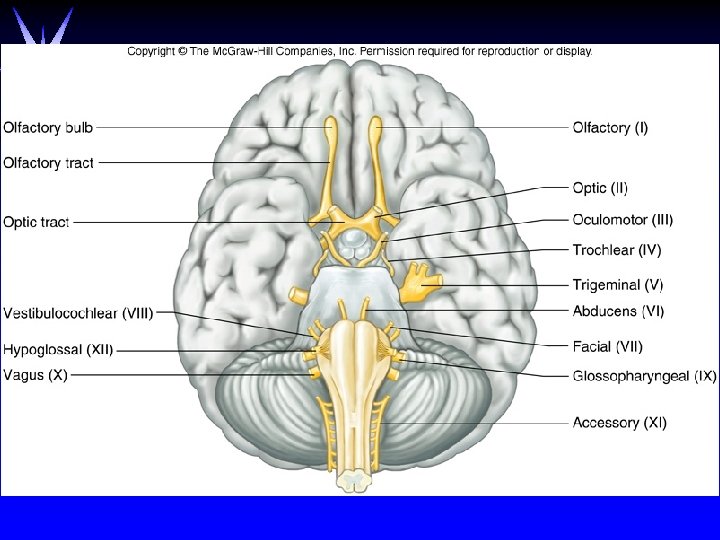
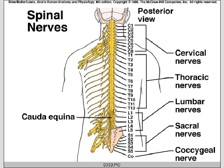
Types of Nerves ä ä Cranial Nerves 12 pairs some sensory, some motor, most mixed Name and number each (Table 11. 9) Old Otto Owens Told Tom About Finding Very Good Vacations Around Hawaii ä ä ä Spinal Nerves 31 pairs, all mixed cauda equina-group of descending spinal nerves below end of spinal cord but within vertebral column all form plexuses except some thoracic nerves-->become intercostal nerves
Figure 11. 25
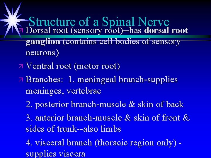
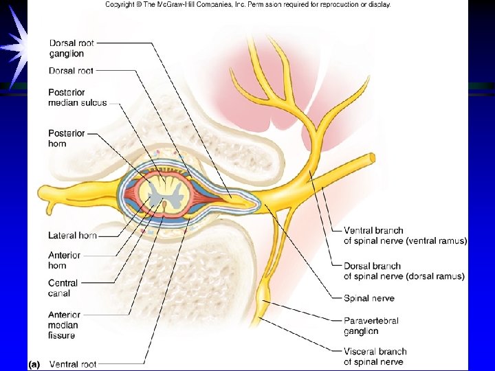
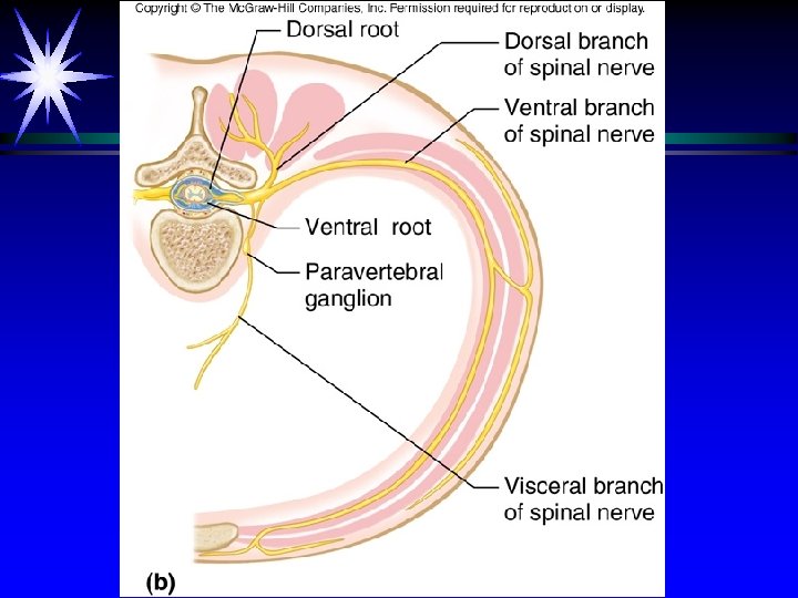
Structure of a Spinal Nerve ä Dorsal root (sensory root)--has dorsal root ganglion (contains cell bodies of sensory neurons) ä Ventral root (motor root) ä Branches: 1. meningeal branch-supplies meninges, vertebrae 2. posterior branch-muscle & skin of back 3. anterior branch-muscle & skin of front & sides of trunk--also limbs 4. visceral branch (thoracic region only) supplies viscera
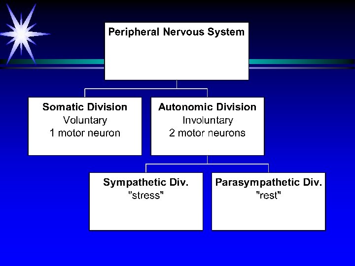
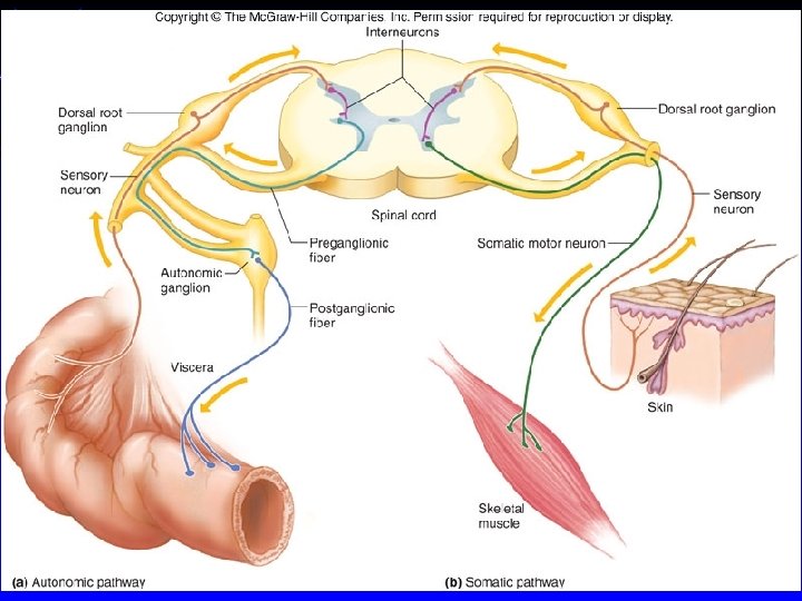
Somatic Division of PNS ä voluntary ä Motor impulses lead to skeletal muscles. ä Motor pathway has 1 motor neuron.
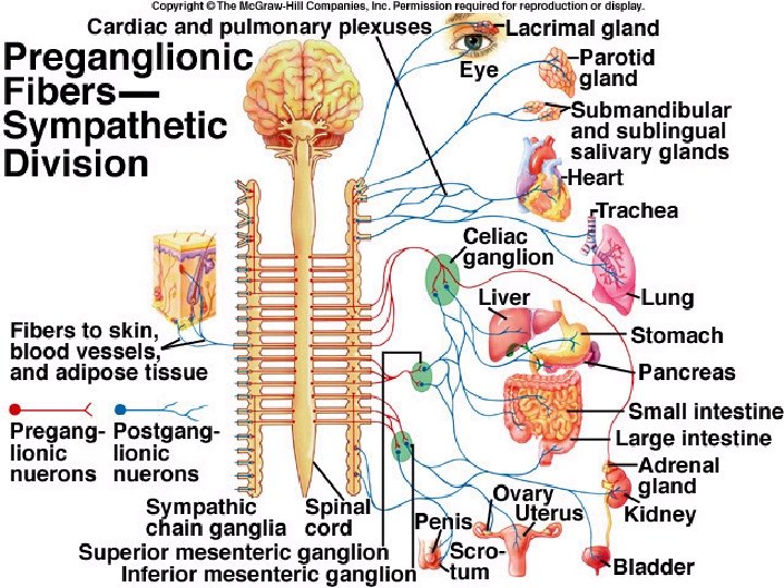
Autonomic Division of PNS ä involuntary ä Motor impulses lead to smooth muscles and glands. ä Motor pathway has 2 motor neurons: preganglionic fiber (myelinated) begins in CNS and ends at a PNS ganglion; postganglionic fiber (unmyelinated) begins at ganglion and ends at effector.
Comparison of the effects of sympathetic & parasympathetic divisions on each organ ä Every organ is supplied by both sympathetic and parasympathetic impulses. ä The effects are opposite. ä See Table 11. 10