Day 3 Meninges and Spinal Cord Meninges Covers


- Slides: 17

Day 3 Meninges and Spinal Cord

Meninges �Covers brain and Spinal cord � 3 layers Dura Arachnoid Pia

Dura Mater �Outer most layer �Tough, white fibrous connective tissue �Contains many blood vessels and nerves �Forms sheath around spinal cord �Epidural space Made of loose connective tissue and adipose tissue Provides a protective pad around spinal cord

Arachnoid Mater �Thin, net-like membrane �Lacks blood vessels �Found between dura and pia mater �Spreads over brain and spinal cord �Subarachnoid space Contains cerebrospinal fluid (CSF) ▪ Clear watery fluid

Pia Mater �Very thin �Contains many nerve and blood vessels that aid in nourishing the underlying cells of the brain and spinal cord �Attached to brain and spinal cord

Meninges Review �What are the meninges? �What are the layers of the meninges? �Where is cerebrospinal fluid located in the meninges?

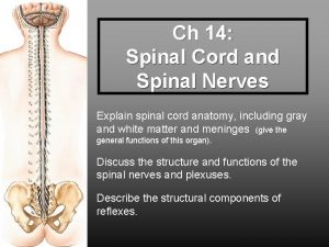
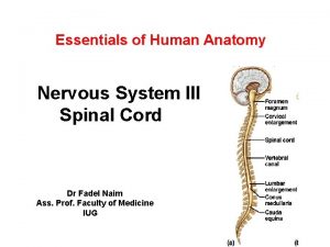
Spinal Cord �Slender nerve column that passes downward from the brain �Starts at the foramen magnum �Ends at the intervertebral disk that separates L 1 and L 2

Structure � 31 segments-each give rise to a pair of spinal nerves �Cervical Enlargement In neck, supplies nerves to upper limbs �Lumbar Enlargement In lower back, supplies nerves to lower limbs

Spinal Cord Structure Cont. �Two Grooves Anterior median fissure ▪ Deep Posterior median sulcus ▪ Shallow �Divide spinal cord into R and L halves

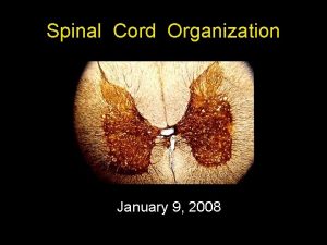
Gray or White? �Spinal Cord is made up of gray and white matter. Gray matter resembles a butterfly Gray matter makes up the horns ▪ Posterior, Anterior, and Lateral White is divided into funiculi by gray matter ▪ Anterior, Lateral, and Posterior funiculi


Structure Cont. �Nerve Pathways = Nerve Tracts �Central Canal Contains CSF (cerebral spinal fluid)

Function of the Spinal Cord � 2 major functions Conducting nerve impulses Center for spinal reflexes �Nerve Tracts provide a two-way communication system between brain and body parts outside the nervous system.

Tracts �Ascending Tracts: Carry sensory info to brain �Descending Tracts Conduct motor impulses form the brain to muscles and glands �Names of tracts are determined by origin and termination points Example: spinothalmic tract starts in the Sp. C. and ends in the thalamus of brain. �Nerve Fibers within A and D tracts are called AXONS


Spinal Functions Cont. �Spinal Reflexes Withdrawal and knee-jerk reflexes pass through spinal cord

Spinal Cord Review �Describe the structure of the spinal cord. �What are some general functions of the spinal cord? �How do you distinguish between the ascending and descending tracts of the spinal cord?
 Epineurium
 Spinal nerves labeled
 Median nerve innervates
 Exercise 15 spinal cord and spinal nerves
 Spinal cord covered by
 Spinal meninges and associated structures
 Spinal meninges
 Spinal meninges
 Day 1 day 2 day 3 day 4
 Foramen costo transversarium
 Spinal cord and brain
 Pns
Pns Poikilothermism and spinal cord injury
 Ans
Ans Spinal cord denticulate ligament
 Nervous sysytem
 Causes of spinal cord compression
 Spinal cord

