Development of the Spinal Cord Neurulation The process


 2) 3) 4) Cross section varies in different regions.")


- Slides: 26
Development of the Spinal Cord
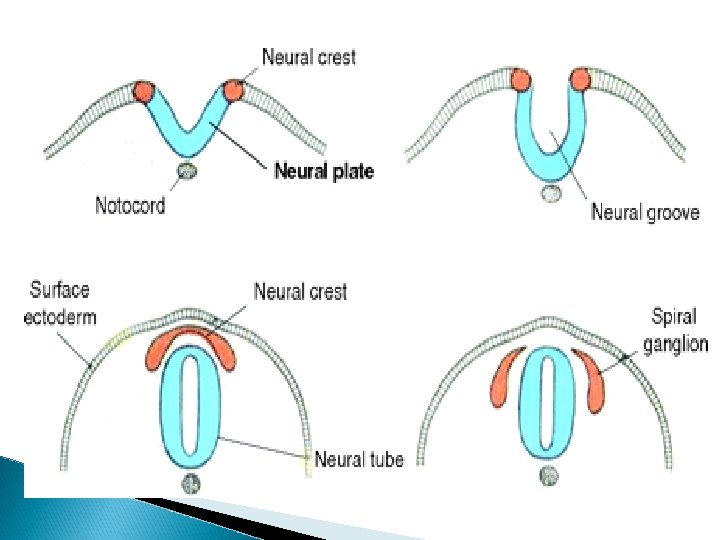
Neurulation • • The process involved in the formation of the neural tube which is the precursor of the CNS. The notochord induces the overlying ectoderm to form the neural plate. This becomes elevated at the sides forming the neural folds and groove (in the midline). The folds approach each other and fuse in the midline forming the neural tube with a cavity neural canal.
Spinal cord development • • The spinal cord develops from the neural tube caudal to the 4 th pair of somites. Neuroepithelial cells line the wall of the neural tube and give rise to the neurones and macroglial cells of the spinal cord. First, they produce neuroblasts which form the mantle layer – gray matter of spinal cord. The marginal layer contains fibres of neurones myelinated – white matter.
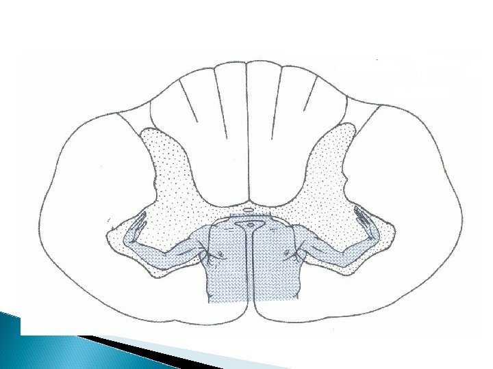
• • • Continuous addition of neuroblasts produces the ventral and dorsal thickenings and reduces the neural canal into a minute central canal. Ventral thickening – Basal plate (Ant. Horn cells) Dorsal thickening – Alar plate (Sensory horn) Sulcus limitans – boundary between them. Roof and floor plates have no neuroblasts, only nerve fibres crossing. Dorsal septum and ventral fissure formed by enlargement of alar and basal plates.
or septum
Spinal nerve • • • Neuroblasts → Neurons From T 1 – L 2 sympathetic part of ANS forms a small intermediate horn. Basal plate N break through the marginal layer and form Ventral Motor root of spinal nerve. Sensory horn N penetrate marginal layer and ascend forming association neurones. Neural crest cells form dorsal root ganglia, central process forms Dorsal Sensory root and peripheral process joins ventral root to form the trunk of the spinal nerve.
Pseudounipolar
Spinal Nerve Components
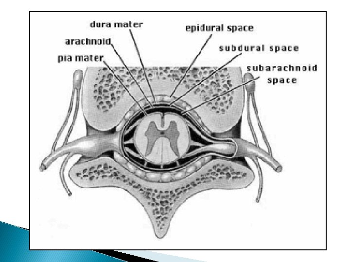
Development of spinal meninges Mesenchme surrounding the neural tube condenses to form the primordial membranes. Outer layer thickens – Dura mater Inner layer –Arachnoid & pia mater (lepto) Fluid filled spaces appear in the leptomeninges and coalesce to form subarachnoid space. Embryonic CSF begins to form in 5 th week of IUL and thus separates them.
“Ascent” of the spinal cord At 3 rd month of IUL, spinal cord length is equal to that of vertebral column. Differential growth causes disparity. In the newborn, spinal cord ends at L 2/3 while in adults it ends at L 1. Conus medullaris Cauda equina Filum terminale - Pia mater Lumbar punture, epidural anaesthesia
Cervical – 1 Upper thoracic - 2 Lower thoracic - 3
Internal Organization of the Spinal Cord
Introduction The spinal cord is the caudal extension of the central nervous system beginning at C 1 – L 1. Measures about 45 cm in length, 1. 5 cm in diameter enclosed within vertebral column. Divided into segments i. e. length of SC giving origin to the rootlets of one spinal nerve. Is made up of 31 segments: 8 cervical, 12 thoracic, 5 lumbar, 5 sacral & 1 coccygeal.
Cervical enlargement Lumbar enlargement
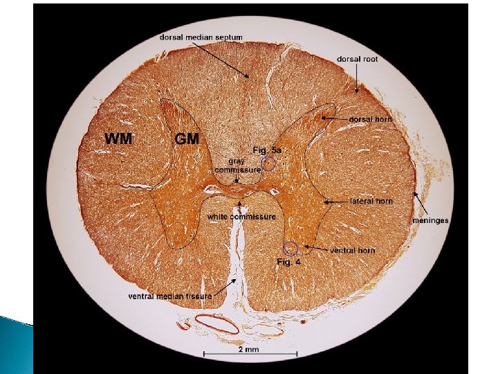
Central gray matter surrounded by white matter connected by commissures. • Separated by Dorsal septum & Ventral fissure. • Posterolateral sulcus –entrance of dorsal roots • Anterolateral sulcus – exit of ventral roots • Divides cord into: Posterior (Dorsal) funiculus (white matter) Lateral funiculus Anterior (Ventral) funiculus Gray matter divided into dorsal & ventral horns. Intermediolateral horn present in thoracic and lumbar segments. •
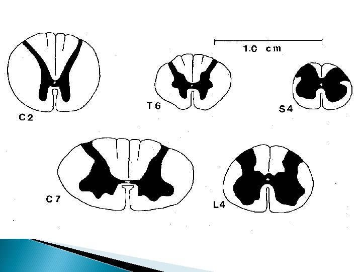
Spinal cord levels • 1) 2) 3) 4) Cross section varies in different regions. Area of white matter decreases caudally. Cervical and lumbar enlargements have increased gray matter (ventral horns). Intermediolateral horn and nucleus dorsalis of Clark found only b/w T 1 and L 2. Dorsal intermediate sulcus is present only in T 6 and above (Cuneate fasciculus)
Gray Matter Organization The organization of gray matter was previously presented as discreet nuclei within the dorsal, intermediate, intermediolateral and ventral horns. In 1952, Rexed found that cell clusters in the spinal cord are arranged into 10 zones or laminae.
Rexed Terminology versus Older Terminology
Rexed Terminology Lamina I - IV– Exteroceptive sensations • Laminae V & VI – Proprioceptive (position, movement. • Lamina VII – Acts as relay b/w midbrain and cerebellum • Lamina VIII – Modulates motor activity • Lamina IX – Main motor area (somatotopically organized) • Lamina X – Surrounds central canal (Onuf’s nucleus in Lam IX of S 1 – 4 anal, uret. sph. ) •
Spinal cord neurones There are 2 types of neurones in the spinal cord: Principal Neurones further divided into: 1)Projection (Tract) neurones – form tracts 2) Root neurones - form root of spinal nerve Interneurones e. g. Renshaw cell