DEPARTMENT OF RADIODIAGNOSIS Scalp swelling A rare presentation


 & shows")

")


![Mechanism of scalp metastasis[3] Abdominal visceral malignancy Venous communications to vertebral venous plexus Intracranial](https://slidetodoc.com/presentation_image_h2/9c1411cab9ad432f87713594ef5adeb0/image-26.jpg "Mechanism of scalp metastasis[3] Abdominal visceral malignancy Venous communications to vertebral venous plexus Intracranial")


- Slides: 31
DEPARTMENT OF RADIO-DIAGNOSIS Scalp swelling – A rare presentation of a metastasis Presenting Resident: Dr Nagi Reddy (JR-III) Guide: Dr Sanjay M Khaladkar Head of the Department : Dr R S Kuber
History 44 year old female patient, presented with – • Tender swelling over the right frontotemporal region. • Associated with pain and watering in the right eye. • No H/O trauma/fever.
USG- LOCAL SWELLING • • • An ill-defined heterogeneous solid lesion over the right temporo-frontal region measuring approx. 4 x 3 x 1 cm, with central and peripheral vascularity on color Doppler. However underlying soft tissue and bone involvement could not be clearly delineated. MRI was advised.
Radiograph skull- Waters view
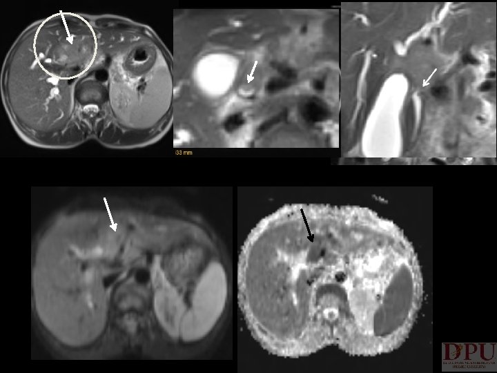
T 1 WI T 2 WI
Pre-con DWI Postcon
After 2 months Patient came with fresh complaints; • C/O yellowish discoloration of eyes since 1 week • Pain over the right hypochondriac region. • Newly developed swelling over the right arm.
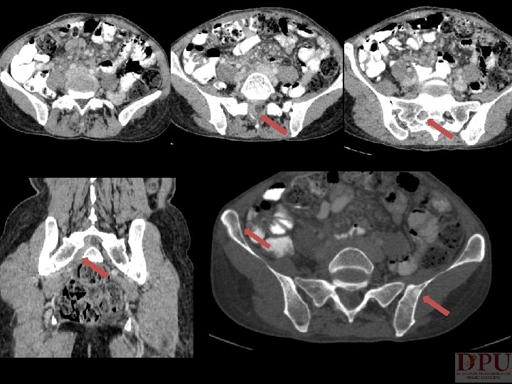
USG - ABDOPELVIS • Liver shows asymmetrically dilated Intrahepatic biliary radicals in both lobes, due to a nearly isoechoic ill-defined mass lesion at the bifurcation of common hepatic duct which is also marginally infiltrating into the cystic duct. This mass lesion measured around 6 x 5 x 4 cm compressing & displacing the portal vein which appears normal in caliber.
• Gall bladder is slightly over-distended (Volume is around 80 cc) & shows a significantly thickened wall, measuring around 7. 5 mm. Gall Bladder lumen shows mild sludge seen within. CBD collapsed & measuring around 1 mm in diameter.
Unenhanced Venous Portal Arterial Delayed
Maximum Intensity Projection (MIP)
Post biliary stenting
Biopsy from liver lesion Pathology - Poorly differentiated sclerosing cholangiocarcinoma.
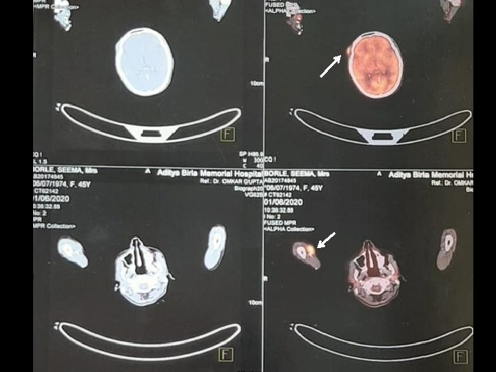
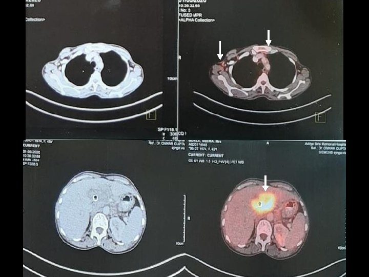
DIAGNOSIS Hilar cholangiocarcinoma with hepatic parenchymal infiltration and involving 1 st order RHD, LHD, proximal CBD and cystic duct. [ Bismuth classification - Type V ] Regional and distant nodal spread, skeletal, spinal and cutaneous metastasis -- being the rarity of this case. TNM staging - Stage IVB
TREATMENT • • • Extrahepatic biliary drainage and metallic biliary stent placement done with two stents. 4 Cycles of chemotherapy was given with FOLFOX. Kept on Capecitabine tablets.
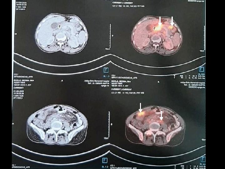
Follow up scan after 2 months Arterial Venous Delayed
DISCUSSION • • Cholangiocarcinomas are rare malignant epithelial tumors arising from the biliary tree and have a poor prognosis and high morbidity Types of Cholangiocarcinoma: o 1) Intrahepatic o 2) Hilar o 3) Extrahepatic
• • These are prone to metastasize to the lung, liver, peritoneum, and retroperitoneal lymph nodes, while cutaneous metastasis is uncommon. Cutaneous metastasis o 1) Seeding along o PTBD tract o 2) Distant metastasis o hematogeneous route
• There are few reports about cutaneous metastasis of cholangiocarcinoma, included following sites: – – Scalp - most common region for cholangiocarcinoma Axillary region Thigh Arm [1, 2]
Mechanism of scalp metastasis[3] Abdominal visceral malignancy Venous communications to vertebral venous plexus Intracranial venous sinuses Emissary veins Scalp veins
Approach to cutaneous metastasis • Cutaneous metastasis are solitary/multiple, usually <2 cm in size and comprises of 5% visceral malignancies and 2% of cutaneous malignancies. [4, 5] Few underlying malignant causes : • Melanoma (Overall most common malignancy) • Ca. Breast (MC visceral malignancy) • Ca. Lung • Ca. Ovary • Colorectal carcinoma • Oral squamous cell carcinoma • Ca. Prostate • Rarely cholangiocarcinoma.
Clinically or Radiologically suspicious cutaneous lesion Basic radiological survey Female CXR USG Breast/Mammo USG abdo-pelvis Male CXR USG abdo-pelvis TRUS (If indicated) Further cross sectional imaging can be done with CT/MRI/PET based on the findings obtained through above mentioned investigations
CONCLUSION • Whenever addressing scalp metastasis of unknown origin, we should keep the possibility of bile duct-derived carcinoma in mind.
REFERENCES 1. Varma K, Singh A, Misra V. Cutaneous Metastasis from Cholangiocarcinoma Presenting as Thigh Mass. Journal of clinical and diagnostic research: JCDR. 2016 Sep; 10(9): ED 23. 2. Baghmar S, Panda D, Arora A, Patidar Y, Yadav V, Rastogi A. Cutaneous metastasis from intrahepatic cholangiocarcinoma. Tropical Gastroenterology. 2017 Jun 22; 38(2): 125 -7. 3. Lu CI, Wong WR, Hong HS. Distant cutaneous metastases of cholangiocarcinoma: report of two cases of a previously unreported condition. Journal of the American Academy of Dermatology. 2004 Aug 1; 51(2): 108 -11. 4. Juan YH, Saboo SS, Tirumani SH et-al. Malignant skin and subcutaneous neoplasms in adults: multimodality imaging with CT, MRI, and 18 F-FDG PET/CT. AJR Am J Roentgenol. 2014; 202 (5): W 422 -38. doi: 10. 2214/AJR. 13. 11424 - Pubmed citation 5. Manohar K, Mittal BR, Bhattacharya A et-al. Asymptomatic Distant Subcutaneous Metastases Detected by (18)F-FDG-PET/CT in a Patient with Breast Carcinoma. World J Nucl Med. 2012; 11 (1): 24 -5. doi: 10. 4103/1450 -1147. 98742 - Free text at pubmed Pubmed citation
Thank you