CONGENITAL DIAPHRAGMATIC HERNIA CDH Dr RAJAT PIPLANI MS
 Dr. RAJAT PIPLANI MS, M. Ch. (MAMC), DNB, MNAMS, DHM")

")


 Anterior central tendon --Septum transversum (4")
Uncorrectable")

 –")
 • *Lung to head ratio (LHR) • <1")

 measurements to assess")


 • 1970’s – Early")

 / thoracoscopic repair • Abdominal/ thoracic •")

 In utero – Surgical repair - >70% mortality – Tracheal ligation")

 a. b. c. d. In CDH, the most common defect is Anterior morgagni")
 CDH is a/an a. Physiologic/ medical emergency b. Surgical emergency c. Both a")
 2 kg newborn child Day 1, with respiratory distress, Scaphoid abdomen, Bowel sounds")
 Antenatal Usg s/o Polyhydramnios and absent stomach bubble, most prabable diagnosis s/o a.")
 Day 4 Child with CDH on ventilator with high settings, ECHO s/o PDA,")
 Child with CDH on ventilator requires a. Aggressive ventilation with high PIP b.")
 CDH associated with better prognosis is a. Liver up b. Premature c. LHR")
 Antenatal review to pediatric surgery OPD, G 1 P 1, 30 wks pregnant")
 Surgery for CDH a. Reduction of contents and primary repair b. Primary repair")
 Recent advances in treatment of CDH antenatally a. ECMO b. HFOV c. FETO")

- Slides: 33
CONGENITAL DIAPHRAGMATIC HERNIA (CDH) Dr. RAJAT PIPLANI MS, M. Ch. (MAMC), DNB, MNAMS, DHM Hugh Greenwood Scholar (BAPS) Associate Professor Department of Pediatric Surgery, AIIMS Rishikesh
History 1679 – Riverius recorded the first CDH 1761 – Morgagni desribed types of CDH 1905 – Heidenhain repair CDH 1925 – Hedbolm suggested that CDH led to pulmonary hypoplasia and early operation improve survival • 1946 – Gross correct CDH < 24 hours of age • 1980 -1990 – delayed correction become widely accepted • •
Introduction • 1 in 2000 - 5000 births • 95. 8% posterolateral defects (Bochdalek) – 84. 4% left side – 13. 2% right side – 2. 4% bilateral • Morgagni and pars sternalis hernias rare • 10 -50% will have associated anomalies.
36 36
Embryology
ANATOMY Diaphragmatic development- begins 4 th week 1) Anterior central tendon --Septum transversum (4 wk)- forms the inferior portion of pericardial cavity and defines rudimentary pleuroperitoneal membrane. 2) Dorsolateral portion--Closure of pleuroperitoneal canals and formation of pleuroperitoneal membrane (6 -8 wk). 3) Dorsal crura– dorsal esophageal mesentery 4) Muscularisation of diaphragm- from thoracic intercostal muscle group and post hepatic mesenchyme. Failure of closure of pleuroperitoneal canal (9 -10 wk) Posteriolateral lumbocostal trigone (weak area)- by Bochdalek (1848)
Pathophysiology • Combination of events… A. Physical compression B. Pulmonary hypoplasia bilaterally (ipsilateral >contralateral)Uncorrectable A. Smaller bronchi B. Less branching C. Decreased alveolar SA C. Pulmonary hypertensionpotentially reversible A. B. Lack of pulmonary arterioles Increased thickness of muscle • Abnormally high vascular reactivity of vessels • Right to left shunt
Diagnosis • Prenatally – U/S – can diagnose 50%, 50% “normal”, – as early as 15 weeks » » Polyhydramnios Absent or intrathoracic stomach bubble Mediastinal or cardiac shift away from hernia Assess other organs – Amniocentesis for chromosomes, alpha fetoprotein (18 weeks) – Fetal MRI
Diagnosis • Postnatal – RD in the first 24 hours. scaphoid abdomen (majority) – After 24 h with vomiting, cough, cyanosis, gastric outlet obstruction, herniation of liver, spleen, bowel.
Prognostic Indicators • *Herniated organs (liver) • *Lung to head ratio (LHR) • <1 poor and <0. 85 severe, 0. 6 -1. 35 61% survive, >1. 35 100% survive • Associated anomalies • Birth weight and Apgar score ( the CDH study group) • Measurement of L/T ratio L/T normal ~ 0. 52, severe < 0. 26 • Others- Antenatal diagnosis<24 wks, polyhydramnios, position of stomach, right sided defect, Mcgoon’s index, Pulm. Artery Index, OI, MVI
Prenatal Diagnosis • Check for associated anomalies • Plan for delivery at tertiary perinatal centre +/ECMO • Options for parents – Prenatal therapies, termination, treatment after delivery
Investigations • Arterial blood gas – frequent arterial blood gas (ABG) measurements to assess for p. H, Pa. CO 2, and Pa. O 2. – Pa. O 2 may be higher from a preductal (right-hand) sampling site. • Chromosome studies – frequent association with chromosomal anomalies (trisomy 13, 18) – If dysmorphic features are observed on examination, a consultation with a geneticist. • Serum electrolytes: Monitor serum electrolytes, ionized calcium, and glucose levels initially and frequently. Maintenance of reference range glucose levels and calcium homeostasis is particularly important • ECHO • Renal USG v Pulse oximetry – Continuous pulse oximetry is valuable in the diagnosis and management of PPHN. – Place oximeter probes at preductal (right-hand) and postductal (either foot) sites to assess for a right-to-left shunt at the level of the ductus arteriosus
Management • Spontaneous Vaginal Delivery, close to term • Initial resuscitation – Supplemental O 2 – No bag & mask – ETT with mechanical ventilation • Ventilation strategies* • Watch for pneumothorax (usually contralateral) – – NG tube Fluid status Inotropic support Delayed surgery ( until patient is stable).
Management • Mechanical Ventilation – May require high setting, high Fi. O 2 – High OI (poor survival) (OI= MAP×Fi. O 2× 100/Pa. O 2) • Paralysis/ sedation • Arterial and venous access • IV antibiotics, IV fluids, correct acidosis, glucose maintainence • NG tube • Inotropic support • Temperature maintainence • Role of Tolazoline( a- receptor blocker), CCB, endothelin receptor blockers, Sildenafil, Nitric Oxide • Role of surfactant- ? ?
Ventilation Strategies • Prevent conditions that raise pulmonary vascular resistance (hypoxemia, acidosis, hypotension and hypercarbia) hyperventilation to control hypoxemia Aim: p. H > 7. 45, PCO 2 < 35 , Pa. O 2 > 60 Avoid barotrauma. Wung et al. showed that some of the mortality in CDH infants was infact due to ventilator-induced lung injury – Gentle ventilation with permissive hyper-capnia – Low PIP, pressure-limited ventilation, minimal set RR, minimal sedation and tolerance of high PCO 2 upto 60 survival>90 – Goal- Preductal PO 2> 60 mm. Hg and Sa. O 2> 90% and PCO 2< 60 mm. Hg
Surgical Repair • First repair in 1940 (Ladd & Gross) • 1970’s – Early intervention – Emergent surgery to alleviate the compression of the “good” lung • Current practice allows a period of stabilization prior to repair – CDH is a physiologic emergency, not a surgical emergency – Pulmonary hypertension is the primary determinant of mortality – Wait until weaning / low ventilatory settings or Pulm. HTN abated or stabilised on ECMO
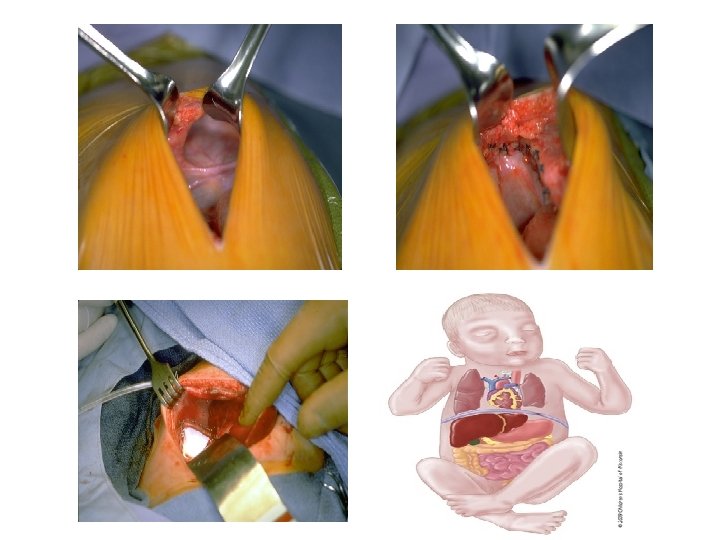
Surgical Repair
Surgical repair- options • Open (subcostal incision) / thoracoscopic repair • Abdominal/ thoracic • Primary repair after reduction of contents with interrupted non absorbable sutures (Prolene) • Reconstructive options: Pre renal fascia, surrounding ribs, Prosthetic patch, split abd. Muscle flap/ LD flap (thoracic) • Skin closure and creation of ventral hernia/ temporary silo • ICD – not required (only when bleeding/collection/air leak)
Long term outcome • GERD • Neurological outcome- CNS and Ear • Chronic lung disease (obst. /rest. Airway- 50% survivors) • Growth issues • Chest wall deformities, scoliosis
Newer Advances 1) In utero – Surgical repair - >70% mortality – Tracheal ligation or occlusion (FETO) • 27 -28 weeks GA • Causes distention and secondary hyperplasia • Good results in 40% of patients 2)Ventilation strategies – HFOV, NO, ECMO, Liquid ventilation (PFC) 3) Lung transplantation 4) Growth factors- combined TRH & steroids
MCQ’s
1) a. b. c. d. In CDH, the most common defect is Anterior morgagni Posteriolateral Bochdalek Right sided defect Pars sternalis
2) CDH is a/an a. Physiologic/ medical emergency b. Surgical emergency c. Both a and b d. OPD case
3) 2 kg newborn child Day 1, with respiratory distress, Scaphoid abdomen, Bowel sounds +nt in left chest and X-ray chest s/o bowel loops present in left chest with mediastinal shift to right. The first intervention you do in emergency a. Bag and mask b. Iv Fluids and antibiotics c. ETT and ventilation d. Ng tube insertion
4) Antenatal Usg s/o Polyhydramnios and absent stomach bubble, most prabable diagnosis s/o a. PUV b. Malrotation c. CDH d. TEF with EA
5) Day 4 Child with CDH on ventilator with high settings, ECHO s/o PDA, referred from Ni. CU for surgical opinion a. Immediate surgery b. Wait further c. ECMO d. PDA ligation
6) Child with CDH on ventilator requires a. Aggressive ventilation with high PIP b. Gentle ventilation with permissive hypercapnia c. High RR d. High tidal volume
7) CDH associated with better prognosis is a. Liver up b. Premature c. LHR > 1 d. Right sided
8) Antenatal review to pediatric surgery OPD, G 1 P 1, 30 wks pregnant with CDH child with polyhydramnios a. Termination of pregnancy b. Prenatal steroids and delivery at 34 wks c. SVD near term at hospital d. Refer to genecist
9) Surgery for CDH a. Reduction of contents and primary repair b. Primary repair after content reduction with ventral hernia creation c. Prosthetic Patch repair d. Thoracoscopic repair e. All of the above
10) Recent advances in treatment of CDH antenatally a. ECMO b. HFOV c. FETO d. NO
THANK YOU